Pelvic floor muscle training protocol for stress urinary
incontinence in women: A systematic review
MARLENE OLIVEIRA1, MARGARIDA FERREIRA2*, MARIA JOÃO AZEVEDO3, JOÃO FIRMINO-MACHADO4, PAULA CLARA SANTOS5,6
1Physiotherapist, Camélia Hotel Sénior & Homes, Guimarães, Portugal
2Visiting Professor, Physiotherapy Department, CESPU – Instituto Politécnico de Saúde do Norte, Vale do Sousa e Vale do Ave, Portugal 3MD, Assistant Physiatrist, Hospital Senhora da Oliveira, Guimarães, Portugal
4MD, Department of Public Health, Porto, Portugal
5Lecturer, Department of Physiotherapy, Escola Superior de Tecnologia e Saúde do Porto, Instituto Politécnico do Porto, Porto, Portugal 6Research Centre in Physical Activity, Health and Leisure, Faculty of Sport, Universidade do Porto, Porto, Portugal
S
UMMARYStudy conducted at the Department of Physiotherapy, Instituto Politécnico do Porto, Porto, Portugal
Article received: 12/12/2016
Accepted for publication: 3/1/2017
*Correspondence:
Departamento de Fisioterapia, Instituto Politécnico do Porto Address: Rua Dr. António Bernardino
de Almeida, 400 Porto – Portugal Postal code: 4200-072 margasufer@gmail.com
http://dx.doi.org/10.1590/1806-9282.63.07.642
Introduction: Strengthening exercises for pelvic floor muscles (SEPFM) are considered the first approach in the treatment of stress urinary incontinence (SUI). Nevertheless, there is no evidence about training parameters.
Objective: To identify the protocol and/or most effective training parameters in the treatment of female SUI.
Method: A literature research was conducted in the PubMed, Cochrane Library, PEDro, Web of Science and Lilacs databases, with publishing dates ranging from January 1992
to March 2014. The articles included consisted of English-speaking experimental studies in which SEPFM were compared with placebo treatment (usual or untreated). The sample had a diagnosis of SUI and their age ranged between 18 and 65 years. The assessment of methodological quality was performed based on the PEDro scale. Results: Seven high methodological quality articles were included in this review. The sample consisted of 331 women, mean age 44.4±5.51 years, average duration of urinary loss of 64±5.66 months and severity of SUI ranging from mild to severe. SEPFM programs included different training parameters concerning the PFM. Some studies have applied abdominal training and adjuvant techniques. Urine leakage cure rates varied from 28.6 to 80%, while the strength increase of PFM varied from 15.6 to 161.7%. Conclusion: The most effective training protocol consists of SEPFM by digital palpation combined with biofeedback monitoring and vaginal cones, including 12 week training parameters, and ten repetitions per series in different positions
compared with SEPFM alone or a lack of treatment.
Keywords: training, pelvic floor, urinary stress incontinence, women.
I
NTRODUCTIONThe International Continence Society(ICS) and the In-ternational Urogynecological Association define urinary incontinence (UI) as a symptom, namely “the complaint of any involuntary loss of urine.”1 UI is classified accord-ing to the record of signs, symptoms and results from urodynamic study (UDS).1 Stress urinary incontinence (SUI) is “the complaint of involuntary urine loss on effort or physical exertion, or on sneezing or coughing.”1
Worldwide, SUI is predominant in females, and the mean prevalence in the various studies is 25%.2,3 It can, however, range from 10% in young women3 to 45% among the elderly.3
UI has a devastating effect on women’s quality of life in the physical, social, sexual and psychological spheres.4 Women restrict or diminish their activity and social
par-ticipation, with serious implications.5
PELVICFLOORMUSCLETRAININGPROTOCOLFORSTRESSURINARYINCONTINENCEINWOMEN: A SYSTEMATICREVIEW
REV ASSOC MED BRAS 2017; 63(7):642-650 643
The recommendations of the Agency for Health Care Policy and Research suggest that the first intervention in the treatment of SUI should be conservative. Pelvic floor rehabilitation includes behavioral modifications and advice on everyday life hygiene, intravaginal manual reeducation, strengthening exercises for pelvic floor muscles (SEPFM), electrical stimulation, biofeedback and vaginal cones.10 Rehabilitation of pelvic floor muscles (PFM) may be active and/or passive, but reeducation depends on a request of voluntary muscle contraction. Active exercises include SEPFM, intravaginal manual reeducation, vaginal cones and biofeedback, while passive exercise refers to electrical stimulation.10 Investigations11-13 demonstrated similar ef-fectiveness of different SEPFM programs, but no evidence of a specific, standardized program. These investigations differ regarding the parameters used in the training pro-grams: eight14-16 to forty repetitions;17 two15 to five series;16 submaximal14,18 to maximum contractions;15,16 duration of five weeks16 to six months;14 three times a week14 to daily;19 instruction on muscle contraction using digital palpation;18 biofeedback19 or perineal ultrasound;20 individual20 or group sessions;21 supervised training14 or home practice.10,19,22 In general, SEPFM is effective in the treatment of female SUI; however, there is a great heterogeneity of programs, not allowing identification of the most effective protocol.
The objective of our review was to identify the most effective protocol and/or PFM training parameters to treat female SUI.
M
ETHODThe structural and content organization of our system-atic review was based on the recommendations of the PRISMA statement.23,24
Eligible studies were of an experimental nature com-paring SEPFM to placebo, usual treatment or lack of treat-ment. They presented high methodological expressiveness (score ≥ 5 on the PEDro scale) and were written in English. The participants were female, aged between 18 and 65 years, diagnosed with SUI based on subjective percep-tion (symptom) and/or clinical evaluapercep-tion (signal) and/or UDS (uroflowmetry and cystometry). Exclusion criteria included diagnosis of SUI triggered by factors external to the lower urinary tract (neurological pathologies, cog-nitive deficits), pregnant and postpartum women, ≥ stage 2 prolapse in the Pelvic Organ Prolapse Quantification (POP-Q), and other types of UI (mixed and urgent).
Search strategy
The search covered five databases: PubMed (Medline), CochraneLibrary, PEDro, Web of Science and Lilacs. In
addition, we conducted a manual survey from the bibliog-raphy of the articles, systematic reviews and meta-analyses included, as well as on the ICS website, in order to reduce publication bias.25 Studies included were published between January 1992 and March 2014. The Medical Subject Head-ings (MeSH) of the National Library of Medicine enabled the identification and the combination of keywords per-taining to: the pathology (urinary stress incontinence), interventions (pelvic floor muscle training; pelvic floor muscle exercise; physical therapy; program; protocol; re-habilitation), population (women; female), and study design (randomized controlled trial; controlled clinical trial; comparative study; research design).
The final search choice included the following keywords: (pelvic floor muscle) AND (“education” OR “training” OR “education”[MeSH Terms] OR “training”) OR (pelvic floor muscle exercise) AND physical therapy OR physiotherapy OR protocol OR program OR rehabilitation AND (stress urinary incontinence) AND women AND female AND (randomized controlled trial OR controlled clinical trial OR comparative study OR research design) NOT (preg-nancy OR animals).
Methodological quality
The methodological quality of the studies was analyzed by three independent researchers using the PEDro scale. This assessment tool has 11 items, with a maximum score of 10 points.26 For each criterion presented in the scale (except for the first one), a score of 1 or 0 points can be attributed.26 The PEDro scalewas created by Moseley et al. in 1999 based on the Delphi List, and was translated and adapted for the Portuguese population by Costa in 2011.
R
ESULTSSearch strategy results
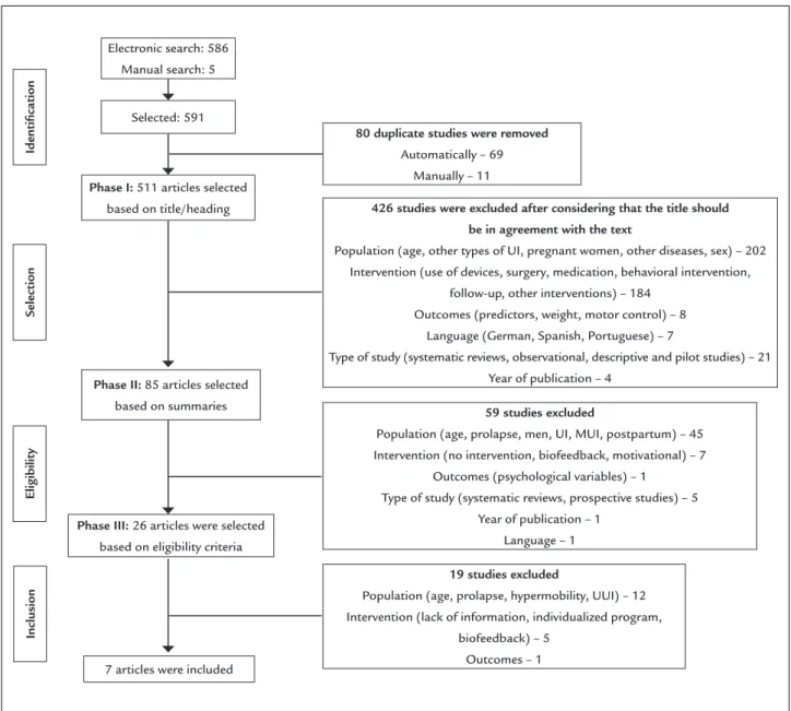
The search in the databases led to the identification of 591 potentially relevant studies (Figure 1).
Methodological quality results
The mean score for methodological quality evaluation was 5.7±1.28 (min/max: 5/8) out of 10 points (Table 1).
The items that most contributed for the decrease of the total score were the 5 (blind study regarding the participants) and 6 (blind study regarding therapists) (Table 1).
Description of the studies
TABLE 1 Classiication of the methodological quality of studies according to the PEDro scale.
Studies 1 2 3 4 5 6 7 8 9 10 11 Total
Glavind et al.30 1 1 1 1 0 0 0 1 0 1 1 6
Arvonen et al.29 1 1 0 1 0 0 0 1 0 1 1 5
Aksac et al.19 1 1 1 1 0 0 0 0 1 1 0 5
Zanetti et al.18 1 1 1 1 0 0 0 1 0 1 1 6
Felicíssimo et al.31 1 1 1 0 0 0 0 1 0 1 1 5
Sriboonreung et al.28 1 1 1 1 0 0 0 1 0 1 0 5
Kamel t al.27 1 1 1 1 0 0 1 1 1 1 1 8
Note: 1. Eligibility criteria have been speciied; 2. Participants were randomly assigned to groups; 3. The distribution into groups was blinded; 4. The groups were initially similar in relation to the most important prognostic indicators; 5. Blind study regarding the participants; 6. Blind study regarding therapists; 7. Blind study regarding evaluators who measured at least one key result; 8. Me-asurements of at least one key outcome were performed on more than 85% of participants initially allocated to groups; 9. All participants for whom outcome measures were presented received tre-atment or control intervention as planned or, whenever this was not the case, data were analyzed for at least one of the key outcomes by “intention to treat”; 10. The results of the inter-group sta-tistical comparisons were described for at least one outcome; 11. The study presents measurement points and variation measurements for at least one key result.
FIGURE 1 Study selection lowchart.
Electronic search: 586 Manual search: 5
7 articles were included
Identiication
Selection
Inclusion
Eligibility
Phase I: 511 articles selected based on title/heading
19 studies excluded
Population (age, prolapse, hypermobility, UUI) – 12 Intervention (lack of information, individualized program,
biofeedback) – 5 Outcomes – 1
Phase III: 26 articles were selected based on eligibility criteria
59 studies excluded
Population (age, prolapse, men, UI, MUI, postpartum) – 45 Intervention (no intervention, biofeedback, motivational) – 7
Outcomes (psychological variables) – 1 Type of study (systematic reviews, prospective studies) – 5
Year of publication – 1 Language – 1
426 studies were excluded after considering that the title should be in agreement with the text
Population (age, other types of UI, pregnant women, other diseases, sex) – 202 Intervention (use of devices, surgery, medication, behavioral intervention,
follow-up, other interventions) – 184 Outcomes (predictors, weight, motor control) – 8
Language (German, Spanish, Portuguese) – 7
Type of study (systematic reviews, observational, descriptive and pilot studies) – 21 Year of publication – 4
Phase II: 85 articles selected based on summaries
80 duplicate studies were removed
PELVICFLOORMUSCLETRAININGPROTOCOLFORSTRESSURINARYINCONTINENCEINWOMEN: A SYSTEMATICREVIEW
REV ASSOC MED BRAS 2017; 63(7):642-650 645
Characteristics of the studies
Sample size varied between 3027 and 6828 women, with a mean age of 48.8±5.51 years, ranging from 25 to 65 years.27-30 The mean duration of urine loss was 64±5.66 months18,29,31 with severity ranging from mild19,27 to severe (even though the definition of the severity of UI is not expressed).30
The diagnosis of SUI was demonstrated through subjec-tive evaluation/symptoms (questionnaire, interview),19,27,29,31 physical examination/signs (pad test, gynecological eval-uation)19,27-31 and/or UDS.18,19,27,31
Interventions
In most studies, the program began with instructions for contracting PFM. Methods most often used were digital palpation19,27,31 and teaching of the anatomy and function of PFM.29-31 Only one study used biofeedback,19 while two omitted the teaching of contraction.18,28
Two studies combined SEPFM and biofeedback,19,30 one combined the exercises with vaginal cones,29 two com-pared SEPFM supervised or not,18,30 and other two compared the exercises with and without the activation of abdominal muscles.27,28 SEPFM program parameters included length of contractions, which ranged from 1 s28 to 20 s29, length of rest from 1 s18 to 20 s19,27 and number of series, ranging from 227 to 40.19
Three studies used maximum contractions27,28,31 and two applied a combination of submaximal and maximum contractions.18,29 As for training positions, the one most often used was supine,18,19,27,30,31 followed by standing,18,29-31 seated18,29-31 and lateral decubitus position.31 Two studies, however, did not specify a training position.19,28
Regarding the frequency of sessions, the minimum applied was two sessions per week,30 while daily treatment was the most frequent.18,19,28,29,31
The analyzed programs lasted between 819,31 and 16 weeks,29 and most opted for a 12-week duration.18,27,28,30
Instruments used to measure outcomes
Almost all of the studies (6 out of 7) assessed the amount of urine leakage based on 1-hour and 24-hour pad tests.18,19,28-31 PFM strength was assessed by digital palpation19,29,31 and perineometry (vaginal squeeze pressure)19,27,28 while intrin-sic sphincter was assessed by UDS.27 Other outcomes in-cluded a subjective assessment based on a visual analogue scale,19 quality of life scales (QV-I-QOL, QV-ICIQ-SF)18,31 and voiding diaries.18
Cure rate results
Six studies18,19,28-31 displayed their assessments of cure rates measured by pad test ranging between < 1 g19,30 and < 2 g.18,29,31
The results of cure rate according to the type of interven-tion were: 50% (cones) versus 26% (PFM Training – PFMT);29 36.6% (supervised PFMT) versus 34.5% (unsupervised);31 58% (PFMT+biofeedback) versus 20% (PFMT);30 48% (PFMT+supervision) versus 9.5% (unsupervised);18 75% (PFMT+palpation) versus 80% (PFMT+biofeedback) ver-sus 0% (no treatment).19 For intervention periodicity, cure rates were 28.6% (daily PFMT) versus 21.2% (PFMT three times weekly) versus 20% (abdominal training)28 (Table 2).
On perineometry, PFM strength increased to 84.7% (PFMT+palpation) versus 161.7% (PFMT+biofeedback) versus 7% (no treatment);19 15.6% (SEPFM) versus 4.7% (abdominal muscle strength)27 and 63.4% (daily) versus 48.4% (three times weekly) versus 59.7% (SEPFM+abdominal, three times weekly).28 On digital palpation, PFM strength reached 37.5% (digital palpation) versus 48.9% (biofeedback) versus 0% (no treatment);19 33% (SEPFM) versus 0% (vaginal cones);29 and 50% (supervised) versus 50% (unsupervised).31 On UDS, intraurethral pressure increased 16% (abdominal muscle strength) versus 9.1% (SEPFM)27 (Table 2).
Subjective perception of cure increased from 23.818 to 75%.28
D
ISCUSSIONOur systematic review confirmed the diversity in study designs, measurement instruments, cure rate definitions, and intervention outcomes.
Zanetti et al.18 found that supervised SEPFM were more effective than unsupervised SEPFM, unlike an-other study,31 which demonstrated the equal efficacy of both. The heterogeneity of the results may derive from the different manners of measuring the pad test (24-h and 1-h) and the duration of the interventions (8 and 12 weeks), respectively.18,31 The pad test is an instrument that reveals the amount of urinary leakage in grams, in addi-tion to being inexpensive and non-invasive.32 According to Jørgensen et al.,33 the correlation coefficient varies between 0.68 and 0.93.33 The investigations are inconsis-tent regarding pad test application duration (1-h or 24-h), although some guidelines recommend the long-duration pad test (24 hours) as it allows the reproduction of urine losses during daily activities according to an individual’s bladder capacity, compared with the 1-hour pad test, which requires a standardized bladder volume and provokes urine leakage in distinct physical activities.32
O
LIV
EIR
A
M
ET
AL
.
R
EV
A
SS
O
C
M
ED
B
R
AS
2
0
1
7
; 6
3
(7
):6
4
2
-6
5
0
TABLE 2 Summary of the description of the studies according to intervention, results and conclusions.
Study Groups Severity Outcomes Results Inter -
-groups
Deinition of cure
Rate of cure
Main conclusions Pre-intervention Post-intervention
Glavind et al.30
G 1 : S E P F M + biofeedback G2: SEPFM
M i l d t o severe
Pad test 1h (g) G1: 9.0 (5-22); G2: 12.8 (9-44)
G1: 0.8 (0-4); G2: 10.0 (2-27)
p=0.02 Padtest ≤ 1 g
G1: 58% G2: 20%
Combined treatment of biofeedback with SEPFM showed a signiicant reduction of urinary loss compared to SEPFM alone.
Arvonen et al.29
G1: SEPFM G2: Vaginal cones
NR Padtest 1h (g) G1: 20; G2: 30 G1: 5; G2: 1 p=0.03 Padtest
˂ 2 g
G1: 26% G2: 50%
Treatment with vaginal cones has signiicantly reduced the amount of urinary loss compared to SEPFM. Digital palpation (0-5) G1: 3; G2: 3 G1: 3; G2: 4 p=0.05
Subjective assessment of cure (0-100%)
NR
Aksac et al.19 G1: SEPFM via
digital palpation G2: SEPFM via biofeedback G3: no treatment
Mild and moderate
Padtest 1h (g) G1: 19.9±2.5; G2: 20.5±0.7; G3: 29.1±3.2
G1: 2.1±0.4; G2: 1.2±0.2; G3: 28.2±3.7
p˂0.001 Padtest
˂ 1 g
G1: 75% G2: 80% G3: 0%
SEPFM combined with digital pal-pation or biofeedback are effec-tive compared to the untreated group.
Perineometry (cmH2O) G1: 20.3±6.2;
G2: 19.1±4.8; G3: 18.7±4.9
G1: 37.5±8.7; G2: 50.0 ±11.5; G3: 20.0±3.9
p˂0.001
Digital palpation/Oxford scale (0-5)
G1: 3.5±0.5; G2: 3.3±0.4; G3: 3.3±0.4
G1: 4.8±0.4; G2: 4.9±0.2; G3: 3.3±0.6
p˂0.001
Subjective assessment – VAS (0-10 points)
NA G1: 7.5±1.2; G2: 8.1±0.8; G3: 3.6±0.6 Zanetti et al.18 G1: Supervised
SEPFM
G2: Unsupervised SEPFM
NR Padtest 1h (g) G1: 20.1; G2: 24.7 G1: 3.2; G2: 15.0 p=0.002 Padtest
˂ 2 g
G1: 48% G2: 9.5%
The supervised SEPFM group improved signiicantly compared to the unsupervised SEPFM group. QV-I-QoL G1: 69.0; G2: 82.0 G1: 89.0; G2: 79.0 p=0.046
Voiding diary G1: 7.0; G2: G1: 1.0; G2: 10.0 p<0.0002
Subjective assessment NA G1: 66.7%; G2: 23.8%
P EL VIC FL O O R M U SC LE TR AIN IN G PR O TO CO L FO R ST R ES S U R IN AR Y IN CO N TIN EN CE IN W O M EN : A SY ST EM AT IC R EV IE W R EV A SS O C M ED B R AS 2 0 1 7 ; 6 3 (7 ):6 4 2 -6 5 0 6 4 7
TABLE 2 (cont.) Summary of the description of the studies according to intervention, results and conclusions.
Study Groups Severity Outcomes Results Inter -
-groups Deinition of cure Rate of cure Main conclusions Pre-intervention Post-intervention Felicíssimo et al.31
G1: Supervised SEPFM
G2: Unsupervised SEPFM
NR Padtest 24h G1: 4.5 (3.0-15.7); G2: 9.3 (3.3-36.1)
G1: 3.2 (1.2-8.0); G2: 2.8 (1.5-8.5)
p=0.78 Padtest
˂ 2 g
G1: 36.6% G2: 34.5%
Supervised and unsupervised SEPFMs were equally effective, with prior teaching of the correct contraction of PFM.
Digital palpation/Oxford scale (0-5)
G1: 2.0 (2.0-3.0); G2: 2.0 (2.0-3.0)
G1: 3.0 (3.0-4.0); G2: 3.0 (2.0-4.0)
p=0.20
QV-ICIQ-SF (0-21) G1: 14.0 (9-16); G2: 14.0 (10-16)
G1: 8.0 (6-12); G2: 8.0 (5-13)
p=0.76
Subjective assessment of cure (0-100%)
NA G1: 69%; G2: 70%
Sriboonreung et al.28
G1: Daily SEPFM G2: SEPFM, three times weekly G 3 : S E P F M + abdominal muscle strength, three times weekly
NR Padtest 1h (g) G1: 4.0±0.9; G2: 4.0±1.5; G3: 4.7±1.6
G1: 1.4±0.7; G2: 1.7±0.7; G3: 4.7±1.6
p>0.05 Padtest G1: 20% G2: 21.2% G3: 28.6%
Daily SEPFM signiicantly increased PFM strength compared to the three times weekly frequency group and the abdominal training group. However, all groups reduced the amount of urine leakage. Perineometry
(cmH2O)
G1: 29.0±10.2; G2: 28.7±13.1; G3: 29.0±7.4 G1: 47.4±9.6; G2: 42.6±12.4; G3: 46.3±8.2 p<0.001
Subjective assessment of cure (0-100%)
NA G1: 75%; G2: 68.4%; G3: 66.7%
Kamel et al.27 G1: Abdominal
muscle strength G2: SEPFM
Mild Perineometry (cmH2O) G1: 49.9±4.85;
G2: 50.3±6.06
G1: 57.73±6.39; G2: 52.60±7.60
p>0.05 NR NR Abdominal training signiicantly increased PFM strength compared to SEPFM.
Valsalva LPP (cmH2O) G1: 80.00±5.52;
G2: 78.00±4.49
G1: 92.80±13.57; G2: 87.33±9.07
According to Sapsford et al.,34 training of deep abdominal muscles triggers the co-contraction of PFM, causing an increase in the strength of PFM and an improvement in urinary continence. A systematic review by Kari Bø et al.35 concluded that the results are ambivalent because, to date, there is no strong clinical evidence of benefit with ab-dominal muscle training in women with UI.
In the studies included in the review, PFM training programs including adjuvant therapies such as biofeed-back, digital palpation and vaginal cones reach high rates of cure (80, 50 and 58%, respectively).19,27,31 A systematic review by Neumann et al.36 demonstrated that SEPFM combined with adjuvant therapies were effective in the treatment of SUI, reaching a cure rate of 73%. These PFM strengthening techniques allow identification, awareness of correct muscle contraction, and inhibition of syner-gistic muscles, enhancing results.37
The PFM training programs differed in the following parameters: type of muscle contraction, number of rep-etitions and series, rest time between each contraction, time of contraction and progressivity of the exercises. Nevertheless, most of the studies that were analyzed showed consistency in the repetition frequency parameter (ten initial repetitions), except for the study by Kamel et al.,27 who initiated the SEPFM program with 15 repeti-tions. This parameter corroborates the parameters of strength training to obtain muscular hypertrophy advo-cated by the American College of Sports Medicine,38,39 which recommends 8 to 12 contractions per series.
The frequency of SEPFM was predominantly intensive (one to three times per day), but the study by Sriboonreung et al.28 failed to verify significant differences in reducing the amount of urine leakage by using different frequen-cies of SEPFM. The current evidence for the principles of strength training recommends that the frequency of three times weekly is sufficient for muscle hypertrophy.38,39
In most studies,18,27,28,30 the training program duration was 12 weeks, except for two studies19,31 that applied SEPFM for 8 weeks. According to the recommendations of the American College of Sports Medicine, strength training programs should last at least 15-20 weeks.38 PFM are skeletal muscles and, therefore, the recommendations of strength training are not different from other skeletal muscles.12 In the first 8 weeks of training, the changes are essentially neural (increased number and frequency of motor unit activation), followed by muscle hypertrophy due to increased volume and number of myofibrils, es-sential for morphological or structural adaptations.36 In our systematic review, training programs of 8 to 12 weeks seem to reduce the amount of urine leakage, and/or to
increase PFM strength, inferring that short-term training is equally effective in the treatment of SUI. However, these results should be analyzed with caution, because the gain of muscular strength in this period was sustained by an increase in number and synchronism of the motor units,36 without any mention of patient follow-up after training, in addition to the fact that the studies included in the analysis used different designs, eligibility criteria and measuring instruments. Also, some of the studies28,29 in our review demonstrated that increasing the strength of PFM in this short period of time may not be related to a significant reduction in the amount of urine loss. This suggests that the increase in PFM strength and urethral resistance does not seem to guarantee the mechanism of urinary continence.28,29 According to some authors, co-ordination between early contraction of PFM and in-creased intra-abdominal pressure may be the most relevant factor in reducing urine leakage compared to the strength gain of PFM, which may justify the positive results of short training programs.7,40
We found in our review that five studies used differ-ent positions to perform the exercises, so that the most commonly applied ones were the standing, seated and lateral decubitus positions.18,27,29-31 One of the ways to promote the progression of the exercises is to create dif-ferent levels of difficulty (without and against gravity).11 According to Kari Bo et al.,41 a standing position
increas-es princreas-essure on the bladder and PFM, and may decrease the effectiveness of PFM contraction, affecting the reduc-tion of muscle strength.
According to recent studies,42,43 the PFM contraction reflex to increased intra-abdominal pressure may be inher-ent to the mechanism of urinary continence, but coordi-nation of the different patterns may be acquired as a learned behavior and is currently considered complemen-tary to SEPFM, a determining factor in any PFM reeduca-tion protocol.
The literature cites cure rates ranging from 44 to 70%.13,18,44 In our systematic review, the objective cure rate varied between 2028,30 and 75%,19 while the subjective cure rate ranged between 23.818 and 75%.28 The low cure rate can be justified by different definitions of cure using pad test (< 1 g or < 2 g). On the other hand, variations in cure rates also depend on different levels of severity of SUI,45 training program duration,22 initial PFM strength42 and patient adherence to treatment.22,46
C
ONCLUSIONPELVICFLOORMUSCLETRAININGPROTOCOLFORSTRESSURINARYINCONTINENCEINWOMEN: A SYSTEMATICREVIEW
REV ASSOC MED BRAS 2017; 63(7):642-650 649
parameters, with ten repetitions per series and in distinct positions seemed more effective to reduce the amount of urine leakage, also providing a subjective perception of cure compared with SEPFM alone or a lack of treatment. The limited number of studies and the heterogeneity of the intervention protocols did not allow us to identify the most effective PFM training protocol.
C
ONFLICT OF INTERESTThe authors declare no conflict of interest.
R
ESUMOProtocolo de treino dos músculos do pavimento pélvico em mulheres com incontinência urinária de esforço: re-visão sistemática
Introdução: Os exercícios de fortalecimento dos múscu-los do pavimento pélvico (EFMPP) são considerados a primeira intervenção no tratamento da incontinência urinária de esforço (IUE); porém, não existe evidência sobre os parâmetros de treino.
Objetivo: Identificar o protocolo e/ou os parâmetros de treino mais eficazes no tratamento da IUE feminina. Método: A pesquisa bibliográfica foi realizada entre ja-neiro de 1992 e março de 2014 nas bases de dados PubMed, Cochrane Library, PEDro, Web of Science e Lilacs. Os artigos incluídos eram de língua inglesa, estudos experi-mentais, comparando EFMPP com tratamento placebo, usual ou sem tratamento, com idade compreendida entre 18 e 65 anos e diagnóstico de IUE. A avaliação da quali-dade metodológica foi realizada por meio da escala PEDro. Resultados: Sete artigos de elevada qualidade metodo-lógica foram incluídos na presente revisão. A amostra foi constituída por 331 mulheres, com idade média de 44,4±5,51 anos, duração média das perdas urinárias de 64±5,66 meses e gravidade da IUE variando entre ligeira e grave. Os programas de EFMPP eram distintos relativa-mente aos parâmetros de treino dos MPP. Alguns estudos incluíram treino abdominal e técnicas adjuvantes. A taxa de cura da quantidade de perda urinária variou entre 28,6 e 80%, enquanto o aumento da força dos MPP variou de 15,6 a 161,7%.
Conclusão: O protocolo de treino mais eficaz consiste nos EFMPP por palpação digital e supervisão combinados com biofeedback e cones vaginais, incluindo os parâmetros de treino de 12 semanas de duração, dez repetições por série e em distintas posições comparados com os EFMPP isolados ou sem tratamento.
Palavras-chave: treinamento, assoalho pélvico, inconti-nência urinária de esforço, mulheres.
R
EFERENCES1. Haylen B, De Ridder D, Freeman R, Swift S, Berghmans B, Lee J, et al.; International Urogynecological Association; International Continence Society. An International Urogynecological Association (IUGA)/International Continence Society (ICS) joint report on the terminology for female pelvic floor dysfunction. Neurourol Urodyn. 2010;29(1):4-20.
2. Hunskaar S, Burgio K, Diokno A, Herzog A, Hjälmås K, Lapitan MC. Epidemiology and natural history of urinary incontinence in women. Urology. 2003; 62(4 Suppl 1):16-23.
3. Hunskaar S, Burgio K, Clark A, Lapitan MC, Nelson R, Sillen U, et al. Epidemiology of urinary and faecal incontinence and pelvic organ prolapse (POP). Health Publications Ltd; 2005.
4. Yip SK, Cardozo L. Psychological morbidity and female urinary incontinence. Best Pract Res Clin Obstet Gynaecol. 2007; 21(2):321-9.
5. Vigod SN, Stewart DE. Major depression in female urinary incontinence. Psychosomatics. 2006; 47(2):147-51.
6. Forte C. Incontinência urinária de esforço na mulher [dissertação]. Porto: Insti-tuto de Ciências Biomédicas Abel Salazar, Universidade do Porto, Portugal; 2011. 7. Delancey JOL, Ashton-Miller JA. Pathophysiology of adult urinary
incontinence. Gastroenterology. 2004; 126(1Suppl 1):S23-32.
8. Mangera A, Patel AK, Chapple CR. Pathophysiology of urinary incontinence. Surgery. 2011; 29(6):249-53.
9. Patel AK, Chapple CR. Pathophysiology of urinary incontinence. Surgery. 2008; 26(5):188-92.
10. Soltero GA, Campoy MP, Barrero CR, Medrano SE, Pérez PM, Rodríguez PA. Tratamiento rehabilitador en la incontinencia urinaria de esfuerzo femenina. Arch Españoles Urol. 2002; 55(9):1035-46.
11. Dumoulin C, Hay-Smith EJ. Pelvic floor muscle training versus no treatment, or inactive control treatments, for urinary incontinence in women. Cochrane Database Syst Rev. 2010; (1):CD005654.
12. Dumoulin C, Glazener C, Jenkinson D. Determining the optimal pelvic floor muscle training regimen for women with stress urinary incontinence. Neurourol Urodyn. 2011; 30(5):746-53.
13. Hay-Smith EJC, Herderschee R, Dumoulin C, Herbison GP. Comparisons of approaches to pelvic floor muscle training for urinary incontinence in women. Cochrane Database Syst Rev. 2011; (12):CD009508.
14. Castro RA, Arruda RM, Zanetti MR, Santos PD, Sartori MG, Girão MJ. Single-blind, randomized, controlled trial of pelvic floor muscle training, electrical stimulation, vaginal cones, and no active treatment in the management of stress urinary incontinence. Clinics. 2008; 63(4):465-72. 15. Parkkinen A, Karjalainen E, Vartiainen M, Penttinen J. Physiotherapy for
female stress urinary incontinence: individual therapy at the outpatient clinic versus home-based pelvic floor training: a 5-year follow-up study. Neurourol Urodyn. 2004; 23(7):643-8.
16. Turkan A, Inci Y, Fazli D. The short-term effects of physical therapy in different intensities of urodynamic stress incontinence. Gynecol Obstet Invest. 2005; 59(1):43-8.
17. Miller J, Sampselle C, Ashton-Miller J, Hong GR, DeLancey JL. Clarification and confirmation of the Knack maneuver: the effect of volitional pelvic floor muscle contraction to preempt expected stress incontinence. Int Urogynecol J. 2008; 19(6):773-82.
18. Zanetti MRD, Castro RDA, Rotta AL, Santos PD, Sartori M, Girão MJBC. Impact of supervised physiotherapeutic pelvic floor exercises for treating female stress urinary incontinence. São Paulo Med J. 2007; 125(5):265-9. 19. Aksac B, Aki S, Karan A, Yalcin O, Isikoglu M, Eskiyurt N. Biofeedback and
pelvic floor exercises for the rehabilitation of urinary stress incontinence. Gynecol Obstet Invest. 2003; 56(1):23-7.
20. Balmforth J, Cardozo LD. Trends toward less invasive treatment of female stress urinary incontinence. Urology. 2003; 62(4 Suppl 1):52-60. 21. Bø K, Talseth T, Holme I. Single blind, randomised controlled trial of pelvic floor
exercises, electrical stimulation, vaginal cones, and no treatment in manage-ment of genuine stress incontinence in women. BMJ. 1999; 318(7182):487-93. 22. Dumoulin C, Lemieux MC, Bourbonnais D, Gravel D, Bravo G, Morin M.
23. Moher D, Liberati A, Tetzlaff J, Altman DG; PRISMA Group. Preferred reporting items for systematic reviews and meta-analyses: the PRISMA Statement. J Clin Epidemiol. 2009; 62(10):1006-12.
24. Urrútia G, Bonfill X. Declaración PRISMA: una propuesta para mejorar la publicación de revisiones sistemáticas y metaanálisis. Med Clín. 2010; 135(11):507-11.
25. Margaliot Z, Chung KC. Systematic reviews: a primer for plastic surgery research. Plast Reconstruct Surg. 2007; 120(7):1834-41.
26. Costa CML. Tradução e adaptação da PEDro Scale para a cultura portu-guesa: um instrumento de avaliação de ensaios clínicos em Fisioterapia [dissertação]. Lisboa: Universidade Técnica de Lisboa, Faculdade de Mo-tricidade Humana; 2011.
27. Kamel DM, Thabet AA, Tantawy SA, Radwan MM. Effect of abdominal versus pelvic floor muscle exercises in obese Egyptian women with mild stress urinary incontinence: a randomized controlled trial. Hong Kong Physiother J. 2013; 31(1):12-8.
28. Sriboonreung T, Wongtra-ngan S, Eungpinichpong W, Laopaiboon M. Effectiveness of pelvic floor muscle training in incontinent women at Maharaj Nakorn Chiang Mai Hospital: a randomized controlled trial. J Med Assoc Thai. 2011; 94(1):1-7.
29. Arvonen T, Fianu-Jonasson A, Tyni-Lenné R. Effectiveness of two conservative modes of physical therapy in women with urinary stress incontinence. Neurourol Urodyn. 2001; 20(5):591-9.
30. Glavind K, Nøhr SB, Walter S. Biofeedback and physiotherapy versus physiotherapy alone in the treatment of genuine stress urinary incontinence. Int Urogynecol J Pelvic Floor Dysfunct. 1996; 7(6):339-43.
31. Felicíssimo M, Carneiro M, Saleme C, Pinto R, da Fonseca A, da Silva-Filho A. Intensive supervised versus unsupervised pelvic floor muscle training for the treatment of stress urinary incontinence: a randomized comparative trial. Int Urogynecol J. 2010; 21(7):835-40.
32. Ghoniem G, Stanford E, Kenton K, Achtari C, Goldberg R, Mascarenhas T, et al. Evaluation and outcome measures in the treatment of female urinary stress incontinence: Internationa Urogynecological Association (IUGA) guidelines for research and clinical practice. Int Urogynecol J Pelvic Floor Dysfunct. 2007; 19(1):5-33.
33. Jørgensen L, Lose G, Andersen J. One-hour pad-weighing test for objective assessment of female urinary incontinence. Am Coll Obstet Gynecol. 1987; 69(1): 39-42.
34. Sapsford R. The pelvic floor. A clinical model for function and rehabilitation. Physiotherapy. 2001; 87(12):620-30.
35. Bø K, Herbert R. There is not yet strong evidence that exercise regimens other than pelvic floor muscle training can reduce stress urinary incontinence in women: a systematic review. J Physiother. 2013; 59(1):159-68. 36. Neumann PB, Grimmer KA, Deenadayalan Y. Pelvic floor muscle training
and adjunctive therapies for the treatment of stress urinary incontinence in women: a systematic review. BMC Womens Health. 2006; 6:11. 37. Dannecker C, Wolf V, Raab R, Hepp H, Anthuber C. EMG-biofeedback
assisted pelvic floor muscle training is an effective therapy of stress urinary or mixed incontinence: a 7-year experience with 390 patients. Arch Gynecol Obstet. 2005; 273(2):93-7.
38. American College of Sports Medicine. American College of Sports Medicine position stand. Progression models in resistance training for healthy adults. Med Sci Sports Exerc. 2009; 41(3):687-708.
39. American College of Sports Medicine. American College of Sports Medicine health-related physical fitness assessment manual. Philadelphia: Lippincott Williams & Wilkins; 2013.
40. Bø K. Pelvic floor muscle training is effective in treatment of female stress urinary incontinence, but how does it work? Int Urogynecol J Pelvic Floor Dysfunct. 2004; 15(2):76-84.
41. Bø K, Finckenhagen B. Is there any difference in measurement of pelvic floor muscle strength in supine and standing position? Acta Obstet Gynecol Scand. 2003; 82(12):1120-4.
42. Yang JM, Yang SH, Huang WC, Tzeng CR. Factors affecting reflex pelvic floor muscle contraction patterns in women with pelvic floor disorders. Ultrasound Obstet Gynecol. 2013; 42(2):224-9.
43. Dietz HP, Erdmann M, Shek KL. Reflex contraction of the levator ani in women symptomatic for pelvic floor disorders. Ultrasound Obstet Gynecol. 2012; 40(2): 215-8.
44. Rett MT, Simoes JA, Herrmann V, Pinto CL, Marques AA, Morais SS. Management of stress urinary incontinence with surface electromyography–assisted biofeedback in women of reproductive age. Phys Ther. 2007; 87(2):136-42. 45. Hung HC, Chih SY, Lin HH, Tsauo JY. Exercise adherence to pelvic floor
muscle strengthening is not a significant predictor of symptom reduction for women with urinary incontinence. Arch Phys Med Rehabil. 2012; 93(10):1795-800.