* University of the State of São Paulo, Rio Claro-SP.
Received in 6/2/04. 2nd version received in 26/4/04. Approved in 2/5/04. Correspondence to: Gustavo Puggina Rogatto, Rua Padre Ferraz, 427 – 13970-250 – Itapira, SP. Tels.: (65) 9976-2499/(19) 3863-2328, e-mail: [email protected]
Influence of spirulina intake on
metabolism of exercised rats
*
Gustavo Puggina Rogatto, Camila Aparecida Machado de Oliveira, Júlio Wilson dos Santos,
Fúlvia de Barros Manchado, Fábio Yuzo Nakamura, Camila de Moraes, Alessandro de Moura Zagatto, Marcel Cardoso Faria, Márcia Afonso and Maria Alice Rostom de Mello
O
RIGINALA
RTICLEKey words: Spirulina. Metabolism. Exercise. Rats. Body growth.
ENGLISH VERSION
ABSTRACT
In this study we compared the metabolic response to acute ex-ercise among rats fed on a standard diet or on a spirulina diet. Young Wistar rats were divided into two groups according to diet: control (C) (standard diet) and spirulina (S) (spirulina diet). At the end of experimental period (5 weeks) rats were submitted to an acute exercise session of swimming (20 minutes, supporting a load corresponding to 5% of body weight) to determine blood lactate and serum glucose, insulin, proteins, albumin and fatty free acids (FFA). Gastrocnemius and liver samples were used to determine glycogen and lipids tenors. Both C and S groups showed increase in serum glucose and FFA, a drop in serum insulin and a decrease of muscle and liver glycogen contents after acute exercise. Blood lactate during exercise was higher in S than C rats. It was conclud-ed that the response pattern to acute exercise was similar for C and S rats. However, diet protein seemed to influence aspects of glucose metabolism.
INTRODUCTION
The protein malnutrition is a public health problem that has af-fected a large parcel of the world population for many years,
espe-cially in the developing countries(1-4). In Brazil, even regions
con-sidered as rich such as the Southeast region present significant
malnutrition prevalence (6%)(5). In states from the Northern and
Northeast regions these indexes are even higher, reaching 17% of population under 5 years of age.
The growth involvement is a consequence frequently observed in individuals in the development phase submitted to situations where the lack of nutrients is present, especially nutrients of
pro-tein origin(6). However, other harmful effects that increase the
in-fant mortality rate and impair mental development and maturation demonstrate the risk that the protein-calorie deficiency may cause
to malnourished infants(2).
Regardless the malnutrition cause, the organism generates adap-tive responses to long periods in which it is submitted to such condition. With regard to the use of energetic substrates, studies report decrease on the energy expenditure when the energy
in-take is reduced(2). This self-modulation between intake and
expen-diture is maintained until the moment where the energy expendi-ture cannot be compensated by the insufficient ingestion. Thus, the organism makes use of its fat supplies, what results in de-crease on adiposity and loss of weight. The carbohydrates and pro-teins metabolism is also influenced by the lack of protein in diet, presenting reduction on glycogen and proteins supplies in muscle
and liver(7-11). Concomitantly, higher concentrations of hepatic fats
may be observed in malnourished individuals in function of the
lack of fat transportation out of the liver(12,13). These and other
neg-ative effects indicate the need of new alternneg-ative for the control and fight against this serious problem, which is the protein-energy malnutrition (PEM).
The conventional agriculture, despite the available technology, presents limitations in its capacity of providing sufficient energy
for the world population(14). The protein malnutrition problem has
concerned public health authorities for many decades, especially the World Health Organization (WHO), which technicians have dis-cussed on new protein sources as well as on norms to be adopted for the use in human alimentation. In this context, microorganisms have received special attention as alternative sources of protein in diet.
Among the several microorganisms that have been studied, the bluish-green seaweed spirulina is considered a promising microor-ganism due to its high protein content (65 to 70% of its dried weight)
and plentifulness of vitamins and minerals(14,15).
The spirulina is a helicoidal-shaped bluish-green seaweed with
0.2-0.5 mm of length(16). The seaweed usually grows in waters of
lakes naturally alkaline localized in arid zones. Although water from these lakes cannot be used in irrigation, it may be used for the
cultivation of spirulina(17). Since this seaweed presents fast
repro-duction rate, dividing itself three times a day, an area exclusively aimed at the spirulina growth may produce 125 times more pro-tein if compared to an area of the same size aimed at the corn crop
or 70 times more protein if compared to the cattle breeding(17).
Furthermore, the spirulina presents some advantages in relation to other seaweeds among which we could mention the pleasant taste; spirulina presents no problems with digestion and no toxic-ity to humans, what does not occur to other seaweeds such as
Chlorella and Scenedesmus(14). Spirulina also presents antioxidant
and hypocholesterolemic actions(18).
Such as the malnutrition, the physical exercise is a condition where modifications on the general metabolic profile are observed, occurring mobilization on the energetic substrate supplies and
al-terations in the hormonal secretions(19,20). The acute responses to
the aerobic stimulus by means of the physical exercise have been reported in specific literature, recording maintenance or decrease on supplies of lipidic and glycidic substrates during exercise in dif-ferent experimental models (sedentariness and training, diabetes,
obesity) in humans and laboratory animals(21-23). Researches on the
METHODS
Animals
For the development of this study, Wistar male young rats (Rat-tus Norvegicus albinus, Wistar) (approximately 30 days) were used. The animals collected in the Unesp Central Bioterium – Botucatu were maintained in the Bioterium of the Physical Education De-partment Biodynamic Laboratory – Biosciences Institute – Unesp – Rio Claro. The rats were maintained in collective cages (five rats
per cage) in controlled room temperature at 25oC and photoperiod
of 12 h light/12 h dark (7 am/7 pm) and water “ad libitum”. All procedures to which animals were submitted were in agreement with norms of animal experimentation proposed by the “Interna-tional Guiding Principles for Biomedical Research Involving Ani-mals”.
Experimental groups
The animals were randomly distributed in the following experi-mental groups:
Control (C): Animals fed on standard diet (casein as source of protein) during five weeks.
Spirulina (S): Animals fed on manipulated diet (spirulina as source of protein) during five weeks.
Diet compositions are presented in table 1.
in a water tank supporting load equivalent to 5% of the body weight of each animal attached to the thorax.
The water temperature was kept between 30oC and 32oC for
being considered as thermally neutral in relation to the rat’s body
temperature(25).
During the performance of the swimming session, blood sam-ples were collected through cut at the distal extremity of tail of the resting animals and at 5, 10, 15 and 20 minutes of exercise for the determination of the lactate concentration. The lactate was deter-mined in a electrochemical analyzer YSL 2300 STAT (Yellow Springs, Inc. USA).
Animals’ sacrifice and biochemical analyses
At the end of the experimental period, the animals were sacri-ficed through decapitation without previous fasting. The sacrifice of the rats from control and spirulina groups occurred in rest condi-tions or after the performance of acute exercise session of swim-ming.
Blood samples from trunk were collected in glass tubes without anticoagulant for the evaluation of glucose (glucose-oxidase
enzy-matic method)(26), albumin, proteins, free fatty acids(27) and seric
insulin (radioimmunoassay- Kit Coat-A-Count, USA).
A median incision was performed in the back and front paw for the removal of sample from the gastrocnemius muscle. The sam-ple was weighted and used for analysis of the glycogen
concentra-tion through colorimetric method(28,29). Liver samples were used
for the dosage of glycogen(28,29) and lipids(27), also through
colorim-etry.
All biochemical determinations were performed by a specialized technician who had no access to information with regard to alloca-tion of animals to the respective groups.
Statistical analysis
The results were expressed as average ± standard deviation and statistically analyzed through t-Student test for independent samples, Analysis of Variance and post-hoc Bonferroni test. In all cases, the significance level was established in 5%.
RESULTS
Parameters evaluated during experimental period
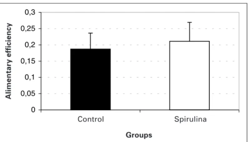
In table 1, different results of body weight, body length and food ingestion of animals from control and spirulina groups during the five weeks of treatment are found. Both groups presented signifi-cant increase on body weight after the first week of study and no differences between groups were observed. With regard to the body length, both groups presented significant weekly growth un-til the end of the experiment from the second week on. However, such as weight, the body length showed no difference between control and spirulina groups. No significant differences were ob-served in food ingestion when groups were compared. The ali-mentary efficiency of control group was also similar to the spiruli-na group (figure 1).
The blood lactate concentrations are presented in figure 2. Both animals from control and spirulina groups presented significant in-crease on this metabolite during performance of the load test. However, for animals from spirulina group, this increase was more
intense at the 20th minute (p < 0,05).
Parameters evaluated after sacrifice
In the present study, 30 animals from each group were consid-ered for the evaluation of the pre-sacrifice parameters (weight, length, ingestion and alimentary efficiency). However, in function of the experiment continuity and other evaluations, only eight ani-mals from each group were sacrificed for the attainment of biolog-ical material for the present study.
BOARD 1 Diets composition
Component 17% protein* 17% protein
(casein) (spirulina)
Spirulina (65% protein) – 280.0** Casein (84% protein) 202.0** –
Corn starch 397.0 386.0
Dextrin 130.5 130.5
Sucrose 100.0 100.0
Soy oil 070.0 070.0
Fiber 050.0 050.0
Mineral mixture* (AIN-93) 035.0 035.0 Vitamins mixture* (AIN-93) 010.0 010.0
L-cystine 003.0 003.0
Choline chlorydrate 002.5 002.5
** According to Reeves et al.(24).
** Values adjusted according to protein content in casein and spirulina.
Procedures
During all experimental period the animals were weekly weight-ed and measurweight-ed. The daily alimentary ingestion was evaluatweight-ed and the alimentary efficiency was calculated through the following equation:
AE = (WE – WB)
TF
Where:
AE: alimentary efficiency
WE: body weight at the end of the experiment (g) WB: body weight at the beginning of the experiment (g) TF: total amount of food ingested during experiment
In the last week, animals from both groups were again divided into subgroups: CR (control rest); CE (control acute exercise); SR (spirulina rest); SE (spirulina acute exercise), according to the per-formance or not of an acute session of exercise shortly before sacrifice.
Acute physical exercise
TABLE 1
Weight (g), body length (cm) and food ingestion in 24 h (g/100 g b.w) of animals during experiment
Initial 1st week 2nd week 3rd week 4th week 5th week
Weight
Control 254.5 ± 28.1 290.8 ± 32.1a 329.9 ± 29.7a,b 351.3 ± 29.7a,b,c 368.4 ± 33.5a,b,c 392.8 ± 29.0a,b,c
Spirulina 254.7 ± 21.6 286.0 ± 26.0a 315.6 ± 34.6a,b 345.8 ± 25.0a,b,c 362.9 ± 29.4a,b,c 379.1 ± 27.7a,b,c
Length
Control 20.7 ± 1.2 21.6 ± 1.2 22.5 ± 1.3a 23.1 ± 1.3a,b 23.9 ± 1.0a,b,c 24.4 ± 0.8a,b,c
Spirulina 20.7 ± 1.1 21.6 ± 1.2 22.5 ± 1.2a 23.2 ± 1.2a,b 24.1 ± 1.1a,b,c 24.6 ± 1.1a,b,c
Food ingestion
Control 11.3 ± 3.1 8.2 ± 1.1 7.4 ± 1.7a 7.6 ± 1.2a 7.0 ± 1.6a 5.0 ± 1.1a
Spirulina 09.0 ± 2.2 7.6 ± 1.1 7.5 ± 2.4a 6.8 ± 1.4a 6.7 ± 1.3a 5.1 ± 1.2a
Results expressed as average ± standard deviation, n = 30 per group a. ≠ initial; b. ≠ 1st week; c. ≠ 2nd week.
0 0,05 0,1 0,15 0,2 0,25 0,3
Fig. 1 – Alimentary efficiency (weight gain/amount of food ingested) dur-ing experiment. Results expressed as average ± standard deviation, n = 30 per group.
Alimentary efficiency
Groups
Control Spirulina
0 2 4 6 8 10 12 14 16
Repouso 5 10 15 20
*
Fig. 2 – Blood lactate in rest situation (R) and during 20 minutes of swim-ming exercise with loads corresponding to 5% of the body weight. Broken line (---) control group; constant line (—) spirulina group. * Significant differ-ence between groups in T20. N = 8 animals per group.
Time (min) Rest
Lactate (mM)
TABLE 3
Hepatic glycogen (mg/100 mg), hepatic lipids (mg/100 mg) and muscular glycogen (mg/100 mg) of animals in rest at the end of experiment
(CR and SR) and after a single session of exercise (CE and SE)
Groups Hepatic glycogenic Hepatic lipids Muscular glycogenic
(mg/100 mg) (mg/100 mg) (mg/100 mg)
CR 09.21 ± 1.30 6.64 ± 1.33 0.57 ± 0.07 CE 06.93 ± 0.97a 6.41 ± 1.45 0.29 ± 0.05a
SR 11.45 ± 2.05a 6.62 ± 1.30 0.70 ± 0.14a
SE 05.23 ± 1.48a,b,c 7.49 ± 1.11 0.18 ± 0.06a,b,c
Results expressed as average ± standard deviation, n = 8 per group. C = control; S = spirulina; R = rest; E = acute exercise. a. ≠ CR. b. ≠ SR. c. ≠ CE.
Table 2 presents the glucose seric concentrations, insulin, total proteins, albumin and free fatty acids (FFA) of animals from control and spirulina groups in conditions of rest and after a 20-minutes exercise session of swimming. An increase on the glycemia due to the acute effect of the effort was observed, being less evident in the spirulina group. The only exercise session resulted in reduc-tion of the insulin seric concentrareduc-tions in both experimental groups. No significant differences were observed in the protein concentra-tion due to both the diet acconcentra-tion and the performance of the acute exercise. At the end of the effort series, both experimental groups presented modifications in the serum albumin as follows: reduc-tion for the control group and increase for the spirulina group. With regard to the free fatty acids, the acute exercise alone caused sig-nificant alterations, resulting in increase on this lipidic substrate.
The hepatic and muscular glycogen content and the lipid con-centrations in liver are recorded in table 3. Both liver and muscle carbohydrate contents presented similar behavior due to the acute exercise performance and due to the diet modification. After the performance of the swimming series, both organs presented sig-nificant decrease in supplies of this energetic substrate and it was more intense in the spirulina group. No alterations in the hepatic lipids contents were observed.
DISCUSSION
The utilization of spirulina as food has been exhaustedly
dis-cussed in specific literature(14,30-32). Besides being a good source of
proteins, minerals and vitamins, the spirulina presents no toxicity to humans and no impairments to the development of organs and
tissue(14,31). In our study, no alterations in weight and body length
were observed in animals fed on spirulina-based diet when com-pared to control group. Furthermore, the ingestion values and ali-mentary efficiency were also similar between both nutritional con-ditions (control and spirulina), indicating that this alternative source of protein originated from biomass does no impair the growth of animals in the development phase. These results corroborate re-ports from literature that compare diets based on spirulina and
casein(14) and spirulina and “pellet”(30). The utilization of young
ani-mals (30 days at the beginning of experiment) may explain the reduction of the amount of food ingested along the study, once as animals aged, the nutrients requirements for the somatic develop-ment may have been decreased. Through evaluations performed
TABLE 2
Seric parameters of animals in rest at the end of experiment (CR and SR) and after a single session of exercise (CE and SE)
Groups Glucose Insulin Proteins Albumin AGL
(mg/dL) (µµµµµUI/mL) (g/dL) (g/dL) (mEq/mL)
CR 082.8 ± 12.8 11.0 ± 3.2 7.3 ± 0.2 6.0 ± 0.6 0.49 ± 0.11 CE 137.4 ± 31.5a 03.6 ± 1.4a 7.4 ± 0.3 5.6 ± 0.6a 0.60 ± 0.06a
SR 073.0 ± 11.7 13.1 ± 4.5 7.9 ± 0.4 5.4 ± 0.3 0.44 ± 0.07 SE 117.0 ± 23.8a,b 03.3 ± 1.1a,b 7.6 ± 0.5 6.0 ± 0.4b,c 0.56 ± 0.06b
in the present study, it was not possible to determine the reasons why animals from control group presented reduction on the amount of food ingested along the experiment before animals from spiruli-na group.
In our work, the performance of physical exercises furthered alterations on the blood lactate concentrations in both groups, in-dicating an acute metabolic stress condition. During the perfor-mance of the physical exercise, an increase on the exigency of several organic systems was observed, resulting in the activation of mechanisms related to the mobilization of energetic substrates. Such response facilitates the redistribution of these fuels in the organism aimed at the maintenance of the muscular work. When this demand exceeds the oxygen supply or its utilization rate, the energetic contribution through the anaerobic via of the carbohy-drate metabolism becomes higher, what produces an excess of hydrogen ions that combine with pyruvic acid to form lactic acid.
Later, it diffuses into the blood, being converted into lactate(33).
The lactate produced plays important role in the energy supply,
once it may be reconverted into glucose by the liver(34). The blood
lactate concentrations during the first 15 minutes of swimming (acute test) were similar between control and spirulina groups
(fig-ure 2). Only at the 20th minute, a more intense elevation was
ob-served in the group fed on spirulina-based diet. Probably, the groups exercised in different effort domains. According to Gaesser and
Poole(35), the exercise intensities in which an elevation of the blood
lactate concentration above the rest values is detected, however, with stabilization in some moment of the load, belong to the in-tense effort domain. The upper limit of this domain is the intensity corresponding to the lactate maximal stable state or anaerobic threshold. The effort intensities above the anaerobic threshold would cause a continuous increase on the blood lactate concentra-tion, hastening fatigue as result of the metabolic acidosis. These intensities are found in the effort severe domain. It is believed that
in this effort domain, the VO2 would invariably reach its maximal
value. Thus, according to results of figure 2, we could suppose that rats belonging to control group would be exercising in the ef-fort intense domain. In other words, swimming with additional overload of 5% of the body weight would not be sufficiently in-tense to cause an elevated acidosis, neither a maximal cardio
res-piratory response (inferred due to the verification of the VO2max).
Animals belonging to the spirulina group would be, during the acute test, performing severe exercise, once there is no evidence of the blood lactate stabilization, particularly at the five final minutes of effort, where the metabolite concentration suffered elevation of
2.4 mM. Some authors(36) accept that an elevation lower than 1
mM during the last 20 minutes of rectangular load of 30 minutes would be a good indicative of lactate stable state. According to this interpretation, animals fed on spirulina would be impaired on the development of their aerobic capacity if compared to animals from the control group along their growth and development, once when submitted to the same relative swimming load, they would present physiological responses that indicate higher organic over-load (acidosis).
The glycemic response during and after the performance of acute exercise in rats has varied in function of differences in the activi-ties employed. The performance of the swimming exercise with loads that ranged from 0 to 8% in relation to the body weight for 30 or 60 minutes in some cases resulted in no modifications on
the glycemic profile in acute condition(37-39). Nardo and Luciano(40)
have also observed reductions on glycemia after acute performance of aerobic effort. In these cases, the increase on the catching of glucose by peripheral tissues during exercise may be the factor
responsible for the glycemic drop(41). On the other hand, increases
on the glucose seric concentrations were verified in our work what
is in agreement with previous findings(19,20,37). This hyperglycemic
response was observed in both the control group and the spirulina group and it is possibly related to the increase on the sympathetic
activity with consequent increase on the muscular and hepatic gly-cogenolytic activity. Mechanisms involved with the neoglycogene-sis may also be contributing to the increase on the glycogen con-tent in function of elevations on the glycocorticoid hormones
secretion(42). Furthermore, several studies have reported increase
of the secretory responses of other contraregulatory hormones such as the growth hormone and catecholamines, among others
after acute exercise(43-50).
The lowering on the insulin-induced seric concentration due to effort acute performance observed in both groups of our study is in agreement with previous works that used continuous and
inter-mittent exercises(19,20,48). The sympathetic stimulation as result of
the effort acute performance may have resulted in the inhibition of the insulin release in that, in addition to the increase on the secre-tion of the contraregulatory hormones contributed to the glycemia elevation in both experimental groups.
The lipidic metabolism may also suffer interference from the physical activity, once the mobilization of this type of substrate may be found increased. In our study, the AGL concentration present significant increase after the performance of a single ses-sion of effort in both the group fed on standard diet and the group fed on spirulina. This result may indicate increase on the mobiliza-tion of triglycerides stocked in the adipocites, what is in
agree-ment with previous works(51) and may be related to elevations on
increase on the secretion of the contraregulatory hormones. The quality of the protein ingested seems not to influence the concen-tration of this lipidic substrate.
The evaluation of the total proteins and seric albumin may be used as indicative of the presence of protein malnutrition, once organisms such as Kwashiorkor present significant reduction on
these components(4). Thus, the fact that the reduction on the blood
proteins in the group fed on spirulina diet had not been verified is an evidence that this diet consists of a source of protein adequate for the development of young animals. Influences on the acute physical activity on the albumin profile were observed, which suf-fered reduction and increase in groups control and spirulina, re-spectively. A hypothesis for this type of response may be related to modifications on the organism’s catabolic activity before acute activity, generated by the ingestion of spirulina, aiming at acceler-ating the muscular and hepatic proteins mobilization with increase on the availability of albumin in circulatory system.
The hepatic steatosis or the lipids accumulation in liver is a met-abolic alteration that has also been observed in organisms
submit-ted to chronic protein-energy malnutrition(13,52). This increase of fat
concentration in liver mainly occurs in function of the increase on
the triglycerides content(13,53). In our study, no difference in the
he-patic content of lipids in both nutritional and exercise conditions was observed. In previous studies, an increase on the activity of the lipoproteic lipase enzyme in liver of animals fed on spirulina
was observed(54).
The intermediate metabolism plays important role in the main-tenance of the organic homeostasis, contributing with the supply-ing of energy in normal conditions or in situations where the met-abolic requirement is found increased. The acute physical exercise is a type of stimulus that requires higher mobilization of the ener-getic substrate from the organism, including from the carbohydrate
supplies(33,55). In our study we could observe that the hepatic and
muscular glycogen supplies presented significant reduction after acute exercise in both control diet and spirulina diet groups. This type of metabolic behavior has been frequently observed in
sever-al studies that used different forms of physicsever-al activity(16). This
de-crease on the glycogen supplies is related to the elevation of the glycogenolytic activity caused by the acute increase on the
cata-bolic hormones(56), being responsible for the increase on the
exercise, however, mechanisms related to glycogenolysis such as the activity of the glycogenolytic enzymes as well as the adrener-gic activity that might be increased in the group fed on this sea-weed, are possible explanations for this response. The muscular and hepatic glycogen supplies, significantly higher in animals in rest condition fed on spirulina if compared to animals from control group, may be very important for the maintenance of the homeo-stasis during exercise in these animals. The set of these results indicates the necessity of further researches in order to clear points yet ambiguous about the effects of the utilization of spirulina as alternative source of protein for organisms in the development phase.
We have come to the conclusion that spirulina did not cause impairments in the growth of rodents as well as in most biochem-ical parameters analyzed and presented good acceptance by ani-mals. This suggests that this seaweed may be a good alternative source of protein. However, the high blood lactate contents ob-served during effort test may induce the early fatigue in exercise, what could impair the physical performance. Further studies on the topic will be required in order to elucidate this and other ques-tions that remain answerless.
All the authors declared there is not any potential conflict of inter-ests regarding this article.
REFERENCES
1. Castro J. Geopolítica da fome. 7a ed. Rio de Janeiro: Brasiliense, 1965.
2. Torun B, Chew F. Protein energy malnutrition. In: Shils M, Olson JA, Shike M, editors. Modern nutrition in health and disease. Philadelphia: Lea & Febiger, 1994;950-76.
3. Uvin P. The state of world hunger. Nutr Res 1994;52:151-61.
4. Dâmaso A. Nutrição e exercício na prevenção de doenças. Rio de Janeiro: MED-SI, 2001.
5. Ministério da Saúde. Pesquisa nacional sobre demografia e saúde 1996: relató-rio preliminar. Brasília: Ministérelató-rio da Saúde, 1996.
6. Frank S. Metabolic adaptation in protein-energy malnutrition. J Am Coll Nutr 1986;5:371-81.
7. Alleyne GAO, Scullard GH. Alterations in carbohydrate metabolism in Jamaican children with severe malnutrition. Clin Sci 1969;37:631-42.
8. Alleyne GAO, Millward DJ, Scullard GH. Total body potassium muscle electro-lytes and glycogen in malnourished children. J Pediatr 1970;76:75-81. 9. Waterlow JC, Mendes CB. Composition of muscle in malnourished infants.
Na-ture 1957;180:1361.
10. Waterlow JC. The nature and significance of nutritional adaptation. Eur J Clin Nutr 1998;53:52-5.
11. Waterlow JC, Weisz J. The fat protein and nucleic acid of liver in malnourished human infant. J Clin Invest 1956;35:346-54.
12. Flores H, Pak N, Maccioni A, Monckeberg F. Lipid transport in kwashiorkor. Br J Nutr 1970;24:1005-11.
13. Kumar V, Deo MG, Ramalingaswami V. Mechanism of fatty liver in protein defi-cience: an experimental study in the rhesus monkey. Gastroenterology 1972; 62:445-51.
14. Contreras A, Herbert DC, Grubbs BG, Cameron IL. Blue-green alga, spirulina, as the sole dietary source of protein in sexually maturing rats. Nutr Rep Int 1979; 19:749-63.
15. Kay RA. Microalgae as food and supplement. Food Sci Nutr 1991;30:555-73. 16. Hadenshog G, Haffen AV. The ultrastructure of Spirulina plantensis. Physiol Plant
1970;23:209.
17. Furst PT. Spirulina – a nutricious alga, once a staple of Aztec diet, could feed many of the world hungry people. Human Nature 1978;3:60.
18. Rodriguez-Hernandez A, Blé-Castillo JL, Juárez-Oropeza MA, Diaz-Zagoya JC. Spirulina maxima prevents fatty liver formation in CD-1 male and female mice with experimental diabetes. Life Sciences 2001;69:1029-37.
19. Rogatto GP, Luciano E. Efeitos do treinamento físico intenso sobre o metabolis-mo de carboidratos. Revista Brasileira de Atividade Física & Saúde 2001;6:39-46.
20. Rogatto GP, Luciano E. Influência do treinamento físico intenso sobre o metab-olismo de proteínas. Motriz 2001;7:75-82.
21. Green HJ, Ball-Brunett M, Symon S, Grant S, Jamieson G. Short-term training, muscle glycogen and cycle endurance. Can J Appl Physiol 1995;20:315-24. 22. Nakamura M, Brown J, Miller WC. Glycogen depletion patterns in trained rats
adapted to a high-fat or high-carbohydrate diet. Int J Sports Med 1998;19:419-24.
23. Romijn JA, Coyle EF, Sidossis LS, Rosenblatt J, Wolfe RR. Substrate metabo-lism during different exercise intensities in endurance-trained women. J Appl Physiol 2000;88:1707-14.
24. Reeves PG, Nielsen FH, Fahey GC Jr. AIN-93 purified diets for laboratory ro-dents: final report of the American Institute of Nutrition ad hoc writing commit-tee on the reformulation of the AIN-76 rodent diet. J Nutr 1993;123:1939-51. 25. Azevedo JRM. Determinação de parâmetros bioquímicos em ratos sedentários
e treinados, durante e após exercício agudo de natação. Tese de Doutorado. Campinas: Unicamp, 1994.
26. Henry RJ, Cannon DC, Wilkeman J. Clinical chemistry, principles and techniques. 2nd ed. New York: Harper and Harper Row Publishes, 1974.
27. Nogueira DM, Strufaldi B, Hirata MH, Abdalla DSP, Hirata RDC. Métodos de bioquímica clínica: técnico-interpretação. São Paulo: Pancasat, 1990. 28. Sjörgreen B, Nordenkjold T, Holmgren H, Wollerstrom J. Bertrag zur kuntnis des
leberrhythmik. Pflügers Arch Gesamte Physiol Menschen Tiere 1938;240:247. 29. Dubois B, Gilles KA, Hamilton JK, Rebers PA. Colorimetric method for
determi-nation of sugar and related substances. Anal Chem 1965;28:350-6.
30. Tranquille N, Emeis JJ, Chambure D, Binot R, Tamponnet C. Spirulina accept-ability trials in rats. A study for the “Melissa” life-support system. Adv Space Res 1994;14:167-70.
31. Salazar M, Martinez E, Madrigal E, Ruiz LE, Chamorro GA. Subchronic toxicity study in mice fed Spirulina maxima. J Ethnophamacol 1998;62:235-41. 32. Grinstead GS, Tokach MD, Dritz SS, Goodband RD, Nelssen JL. Effects of
Spir-ulina plantensis on growth performance of weanling pigs. Anim Feed Sci Tech-nol 2000;83:237-47.
33. McArdle WD, Katch FI, Katch VL. Fisiologia do exercício: energia, nutrição e desempenho humano. Rio de Janeiro: Guanabara Koogan, 1996.
34. Brooks GA. Ácido láctico no sangue: o “vilão” dos esportes torna-se bom. Sports Science Exchange 1995;2:1-6.
35. Gaesser GA, Poole DC. The slow component of oxygen uptake kinetics in hu-mans. Exerc Sport Sci Rev 1986;24:35-70.
36. Heck H, Mader A, Hess G, Mücke S, Hollman W. Justification of the 4-mmol/l lactate threshold. Int J Sports Med 1985;6:117-30.
37. Prada FJA, Carneiro EM, Azevedo JRM, Luciano E. Respostas endócrino-meta-bólicas em ratos diabéticos: efeito estressor do exercício agudo. Revista Brasi-leira de Atividade Física & Saúde 1997;2:22-9.
38. Felício PFV, Drigo AJ, Azevedo JRM. Efeito da administração de ascorbato (vi-tamina C) associada ao exercício agudo de natação sobre os níveis de substra-tos energéticos em cobaias. Motriz 1999;5:92.
39. Luciano E, Guerino MR, Luciano EA. Influências da atividade física e da apli-cação do ultra-som sobre o reparo ósseo em ratos diabéticos. Rev Bras Cien Mov (Edição Especial) 1999;142.
40. Nardo N Jr, Luciano E. Influências do treinamento físico sobre a resposta ao estresse agudo em ratos normais e diabéticos experimentais. Anais do XXI Sim-pósio Internacional de Ciências do Esporte. p. 105. São Paulo: CELAFISCS, 1998. 41. Luciano E, Carneiro EM, Reis MAB, Peres SB, Velloso LA, Boschero AC, Saad MJA. Endurance training modulates early steps of insulin signaling in rat mus-cle. Med Sci Sports Exerc 1998;30:S24.
42. Rogatto GP, Luciano E. Hormonal and metabolic response to acute exercise: effects of high intensity training. Med Sci Sports Exerc 2000;32:S227. 43. Jurimae T, Karelson K, Smirnova T, Viru A. The effect of a single-circuit
weight-training session on the blood biochemistry of untrained university students. Eur J Appl Physiol 1990;61:344-8.
44. Tabata I, Atomi Y, Mutoh Y, Miyashita M. Effect of physical training on the re-sponses of serum adrenocorticotropic hormone during prolonged exhausting exercise. Eur J Appl Physiol 1990;61:188-92.
45. Häkkinen K, Pakarinen A. Acute hormonal response to heavy resistance loading in men and women at different ages. Int J Sports Med 1995;16:507-13. 46. Gotshalk LA, Loebel CC, Nindl BC, Putukian M, Sebastianelli WJ, Newton RU,
et al. Hormonal responses of multiset versus single-set heavy-resistance exer-cise protocols. Can J Appl Physiol 1997;22:244-55.
47. Ortega E, Rodriguez MJ, Barriga C, Forner MA. Corticosterone, prolactin and thyroid hormones as hormonal mediators of the stimulated phagocytic capacity of peritoneal macrophages after high-intensity exercise. Int J Sports Med 1996; 17:149-55.
48. Kraemer WJ, Hakkinen K, Newton RU, McCormick M, Nindl BC, Volek JS, et al. Acute hormonal responses to heavy resistance exercise in younger and older men. Eur J Appl Physiol 1998;77:206-11.
49. Viru A, Laaneots L, Karelson K, Smirnova T, Viru M. Exercise-induced hormone responses in girls at different stages of sexual maturation. Eur J Appl Physiol 1998;77:401-8.
50. Zouhal H, Rannou F, Gratas-Delamarche A, Monnier M, Bentué-Ferrer D, Delama-rche P. Adrenal medulla responsiveness to the sympathetic nervous activity in sprinters and untrained subjects during a supramaximal exercise. Int J Sports Med 1998;19:172-6.
51. Stevanato E. Efeitos do jejum sobre a interação entre o metabolismo de ácidos graxos livres e glicose em músculos esqueléticos de ratos treinados. Disserta-ção de Mestrado. Rio Claro: Unesp, 1999.
52. Ferro-Luzzi A, Spadoni MA. Protein-energy malnutrition. Prog Food Nutr Sci 1978; 2:515-41.
53. Taylor GO, Ziboh VA. Liver lipid changes in experimental protein malnutrition. Am J Clin Nutr 1972;25:285-90.
54. Iwata K, Inayama T, Kato T. Effects of Spirulina plantensis on plasma lipoprotein lipase activity in fructose-induced hyperlipidemic rats. J Nutr Sci Vitaminol 1990; 36:165-71.
55. Fox EL, Bowers RW, Foss ML. Bases fisiológicas de educação física e dos des-portos. 4a ed. Rio de Janeiro: Guanabara Koogan, 1991.
56. Guyton AC, Hall JE. Tratado de fisiologia médica. 9a ed. Rio de Janeiro: