* Study carried out at the Hospital Israelita Albert Einstein, São Paulo, Brazil. 1. Medical Student. ABC School of Medicine, Santo André, Brazil.
2. Medical Resident in General and Emergency Surgery. ABC School of Medicine, Santo André, Brazil. 3. Physician in the Department of Thoracic Surgery. Hospital Israelita Albert Einstein, São Paulo, Brazil. 4. Head of the Department of Thoracic Surgery. Hospital Israelita Albert Einstein, São Paulo, Brazil.
Correspondence to: Vanessa Assis da Silva. Rua Henrique Jacobs, 129, Artur Alvim, CEP 03566-010, São Paulo, SP, Brasil. Tel/Fax 55 11 6749-3637. E-mail: vanessa.assis@yahoo.com.br
Submitted: 28 September 2006. Accepted, after review: 20 December 2006.
Vanessa Assis da Silva1, Paula Kataguiri1, Damila Cristina Trufelli1, Leandro Luongo de Matos2,
João Carlos das Neves-Pereira3, José Ribas Milanez de Campos4
Abstract
We present the case of a 60-year-old female patient who had been in menopause for 14 years and presented a pulmonary nodule on chest X-ray diagnosed in the postoperative follow-up evaluation of breast cancer. The patient had a history of mastectomy and ipsilateral axillary lymphadenectomy for invasive ductal breast carcinoma, as well as of hormone therapy, chemotherapy, and adjuvant radiotherapy. After thoracoscopic nodulectomy, the frozen section analysis revealed a pulmonary hamartoma. Recent studies show that 75% of patients who undergo surgery for pulmonary nodules after a curative mastectomy for breast cancer present lung metastases, 11.5% present primary lung cancer, and 13.5% present benign lesions, including hamartoma.
Keywords: Hamartoma; Breast neoplasms; Lung neoplasms; Neoplasm metastasis.
Introduction
Pulmonary metastasis is a common occurrence in patients with breast cancer,(1,2) who are usually treated with
systemic chemotherapy.(1) Some clinical studies suggest
that resection of pulmonary metastases (metastasectomy) can be a treatment option.(2-5) However, the role of surgery
for metastases originating from breast cancer has yet to be fully determined.(6)
Pulmonary nodules that appear in patients who under-went mastectomy for breast cancer are usually pulmonary metastases. However, it is not uncommon to find meta-chronous lung tumors as well as some benign diseases, among which pulmonary hamartoma stands out.(6)
Case report
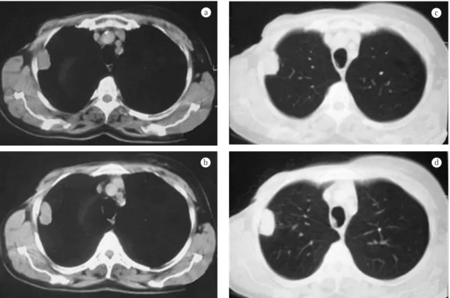
A 60-year-old Caucasian female who had been in menopause for 14 years was referred to the Department of Oncology due to the presence of a pulmonary nodule on chest X-ray (Figure 1) diagnosed in the postoperative follow-up evaluation of breast cancer.
(cyclo-interposed embryonic bronchi, compatible with pulmonary hamartoma.
The patient recovered well and was discharged on postoperative day 2. She is now under annual outpatient follow-up treatment in the Department of Oncology, having been on hormone therapy with tamoxifen for 5 years, and has presented no signs of tumor recurrence to date.
Discussion
Pulmonary hamartoma accounts for 77% of benign lung tumors(1) and for 4% of all solitary
lung nodules.(7) This lesion has been described as
a benign neoplasia of fibrous connective tissue of the bronchus surrounded by respiratory epithelium, almost always containing cartilage and fat tissue, not respecting the usual histological distribution of the lung.(2)
Ninety per cent of the hamartomas manifest as a solitary peripheral mass,(7) and rarely occur in the
form of multiple lesions.(3) They are more common
in adults, and their incidence is twice as high in males than in females,(1) with a mean population
growth of 3.2 ± 2.6 mm/year.(8)
Radiographically, the peripheral lesion is typically found in the lung bases and appears as a well-de-fined, homogeneous mass; sometimes, the margins can be lobulated or bosselated.(9) The usual size
ranges from 1 to 2 cm, although larger lesions can also be observed. There can be calcifications, usually diffuse in form, in up to 30% of the cases. Some authors(10) have also demonstrated the presence of
fat tissue in 50% of the hamartomas evaluated by computed tomography.
There is the occurrence of malignancy in hamar-tomas. Some researchers(5) have found that the
incidence of bronchial carcinoma is 6.3-fold higher in patients with hamartoma than in a normal popu-lation, suggesting the presence of an etiologic relationship.
It is common for confusion to occur when pulmonary nodules are found in the follow-up eval-uation of patients who underwent mastectomy.
According to a recent study,(6) the pathologic
diagnoses of pulmonary nodules demonstrated that 75% of the patients who underwent surgery for pulmonary nodules after a curative mastectomy for breast cancer presented lung metastases, 11.5% presented primary lung cancer (adenocarcinoma and phosphamide + methotrexate + 5- fluorouracil:
4 neoadjuvant cycles and 6 adjuvant cycles), as well as of adjuvant radiotherapy (total dose of 50 Grays for 5 weeks).
She reported no pulmonary symptoms, fever, or weight loss.
The patient then underwent computed tomog-raphy of the chest, which revealed a pleural, parenchymal pulmonary nodule measuring approxi-mately 30 × 25 mm (Figure 2) in the right lung. Therefore, a hypothetical diagnosis of pulmonary metastasis of the breast carcinoma previously removed was made, and biopsy was indicated for diagnostic confirmation.
The patient was then submitted to video-assisted thoracoscopic nodulectomy. The frozen section analysis revealed that it was a benign tumor (prob-ably a pulmonary hamartoma), and the procedure was then finished.
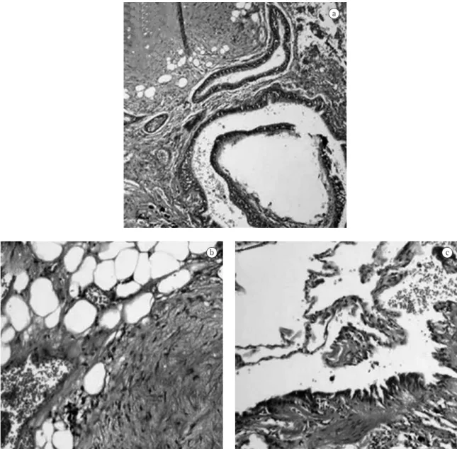
The anatomopathological examination revealed that it was a non-capsulated, regular lesion meas-uring 30 × 25 mm and presenting a firm and fibroelastic consistency. Microscopy (Figure 3) revealed vessels, adipose tissue, cartilage, well-dif-ferentiated (but disorganized) bronchial structures, and highly vascularized fibrovascular stroma with
always possible due to the stiffness of the tumor, enucleation or resection via open thoracotomy or video-assisted resection are indicated.(9,11,13)
Since breast cancer has a slow progression,(14) the
mean time between mastectomy and identification of pulmonary nodules is approximately 60 months, this time being greater in cases of primary malig-nant tumors of the lung (mean of 272 months) and benign diseases of other etiologies (mean of 132 months), including hamartoma.(6) In addition,
the incidence of multiple pulmonary nodules is significantly more frequent in patients with meta-static breast cancer.
The literature has yet to establish whether it is possible to predict the diagnosis of pulmonary metastasis in multiple pulmonary nodules that are revealed in the early follow-up evaluation (within less than five years) of patients who underwent mastectomy for breast cancer.(15)
Recently, other ways to differentiate primary malignant cancers or benign diseases from pulmo-squamous cell carcinoma), and 13.5% presented
benign lesions (pulmonary tuberculosis, sclerosing hemangioma, typical organized pneumonia), 28.6% of which were pulmonary hamartomas.
Therefore, the pulmonary nodules that appear in patients submitted to mastectomy for breast cancer are not always pulmonary metastases, and the confirmation of the histopathological diagnosis is of fundamental importance in order to define the treatment.(11)
Bronchoscopy with biopsy is indicated in endo-bronchial lesions and also in patients with pulmonary symptoms, such as cough, hemoptysis, recurrent pulmonary infections, or atelectasis.(12) Percutaneous
transthoracic aspiration biopsy can diagnose 85% of the hamartomas close to the thoracic wall, differen-tiating them from nodules of other etiologies, such as breast cancer lung metastasis. When cartilage fragments are present on cytology, the diagnosis is highly suggestive of hamartoma.(4) If a diagnosis has
not been made, since percutaneous biopsy is not a
b
c
d
nary metastases have been studied. Positron emission computed tomography using glucose marked with fluoride 18, which is an imaging method with high specificity for malignant diseases,(16) can be cited as
an example. Another equally important ally is immu-nohistochemical study, which can be performed in biopsy specimens and also differentiates those three entities; thyroid transcription factor 1 monoclonal antibody, which is one of the markers of primary lung carcinoma, is worthy of mention.(17)
a
b c
Therefore, pulmonary nodules in patients with breast cancer should only be treated after histopathological confirmation, and surgical resec-tion should be considered both as a diagnostic and as a therapeutic option.
Referências
1. Arrigoni MG, Woolner LB, Bernatz PE, Miller WE, Fontana RS. Benign tumors of the lung. A ten-year surgical experience. J Thorac Cardiovasc Surg. 1970;60(4):589-99.
10. Siegelman SS, Khouri NF, Scott WW Jr, Leo FP, Zerhouni EA. Computed tomography of the solitary pulmonary nodule. Semin Roentgenol. 1984;19(3):165-72.
11. Ramming KP. Surgery for pulmonary metastases. Surg Clin North Am. 1980;60(4):815-24.
12. Cosio BG, Villena V, Echave-Sustaeta J, de Miguel E, Alfaro J, Hernandez L, et al. Endobronchial hamartoma. Chest. 2002;122(1):202-5.
13. Friedel G, Pastorino U, Ginsberg RJ, Goldstraw P, Johnston M, Pass H, et al. Results of lung metastasectomy from breast cancer: prognostic criteria on the basis of 467 cases of the International Registry of Lung Metastases. Eur J Cardiothorac Surg. 2002;22(3):335-44.
14. Winer EP, Morrow M, Osborne CK, Harris JR. Malignant tumors of the breast. In: De Vita Jr. VT, Hellman S, Rosenberg SA, editors. Cancer. Principles and practice of oncology. Philadelphia: Lippincott Williams and Wilkins, 2001. p.1651-716.
15. Staren ED, Salerno C, Rongione A, Witt TR, Faber LP. Pulmonary resection for metastatic breast cancer. Arch Surg. 1992;127(11):1282-4.
16. Bruzzi JF, Munden RF. PET/CT imaging of lung cancer. J Thorac Imaging. 2006;21(2):123-36.
17. Su YC, Hsu YC, Chai CY. Role of TTF-1, CK20, and CK7 immunohistochemistry for diagnosis of primary and secondary lung adenocarcinoma. Kaohsiung J Med Sci. 2006;22(1):14-9.
2. Bateson EM. So-called hamartoma of the lung--a true neoplasm of fibrous connective tissue of the bronchi. Cancer. 1973;31(6):1458-67.
3. Bennett LL, Lesar MS, Tellis CJ. Multiple calcified chondrohamartomas of the lung: CT appearance. J Comput Assist Tomogr. 1985;9(1):180-2.
4. Hamper UM, Khouri NF, Stitik FP, Siegelman SS. Pulmonary hamartoma: diagnosis by transthoracic needle-aspiration biopsy. Radiology. 1985;155(1):15-8.
5. Karasik A, Modan M, Jacob CO, Lieberman Y. Increased risk of lung cancer in patients with chondromatous hamartoma. J Thorac Cardiovasc Surg. 1980;80(2):217-20.
6. Tanaka F, Li M, Hanaoka N, Bando T, Fukuse T, Hasegawa S, et al. Surgery for pulmonary nodules in breast cancer patients. Ann Thorac Surg. 2005;79(5):1711-4; discussion 1714-5.
7. Khouri NF, Meziane MA, Zerhouni EA, Fishman EK, Siegelman SS. The solitary pulmonary nodule. Assessment, diagnosis, and management. Chest. 1987;91(1):128-33. 8. Hansen CP, Holtveg H, Francis D, Rasch L, Bertelsen
S. Pulmonary hamartoma. J Thorac Cardiovasc Surg. 1992;104(3):674-8.