Signaled two-way avoidance learning
using electrical stimulation of the inferior
colliculus as negative reinforcement:
effects of visual and auditory cues as
warning stimuli
1Laboratório de Psicobiologia, Departamento de Psicologia, Faculdade de Filosofia,
Ciências e Letras de Ribeirão Preto, Universidade de São Paulo, Ribeirão Preto, SP, Brasil
2Laboratoire de Psychopathologie et Psychopharmacologie de la Cognition, U405
INSERM, Strasbourg, France A.C. Troncoso1,
G. Cirilo-Júnior1,
G. Sandner2 and
M.L. Brandão1
Abstract
The inferior colliculus is a primary relay for the processing of auditory information in the brainstem. The inferior colliculus is also part of the so-called brain aversion system as animals learn to switch off the electrical stimulation of this structure. The purpose of the present study was to determine whether associative learning occurs between aversion induced by electrical stimulation of the inferior colliculus and visual and auditory warning stimuli. Rats implanted with elec-trodes into the central nucleus of the inferior colliculus were placed inside an open-field and thresholds for the escape response to electri-cal stimulation of the inferior colliculus were determined. The rats were then placed inside a shuttle-box and submitted to a two-way avoidance paradigm. Electrical stimulation of the inferior colliculus at the escape threshold (98.12 ± 6.15 (A, peak-to-peak) was used as negative reinforcement and light or tone as the warning stimulus. Each session consisted of 50 trials and was divided into two segments of 25 trials in order to determine the learning rate of the animals during the sessions. The rats learned to avoid the inferior colliculus stimulation when light was used as the warning stimulus (13.25 ± 0.60 s and 8.63 ± 0.93 s for latencies and 12.5 ± 2.04 and 19.62 ± 1.65 for frequencies in the first and second halves of the sessions, respectively, P<0.01 in both cases). No significant changes in latencies (14.75 ± 1.63 and 12.75 ± 1.44 s) or frequencies of responses (8.75 ± 1.20 and 11.25 ± 1.13) were seen when tone was used as the warning stimulus (P>0.05 in both cases). Taken together, the present results suggest that rats learn to avoid the inferior colliculus stimulation when light is used as the warning stimulus. However, this learning process does not occur when the neutral stimulus used is an acoustic one. Electrical stimula-tion of the inferior colliculus may disturb the signal transmission of the stimulus to be conditioned from the inferior colliculus to higher brain structures such as amygdala.
Correspondence
M.L. Brandão
Laboratório de Psicobiologia Departamento de Psicologia Faculdade de Filosofia, Ciências e Letras de Ribeirão Preto, USP Av. Bandeirantes, 3900 14049-901 Ribeirão Preto, SP Brasil
Research supported by FAPESP (No. 94/5933-2). A.C. Troncoso was the recipient of a FAPESP scholarship and G. Cirilo was the recipient of a scientific initiation scholarship from CNPq. This work is part of a
Brazil-France exchange program (CAPES/COFECUB No. 164/94).
Received February 24, 1997 Accepted December 17, 1997
Key words
•Inferior colliculus
•Active avoidance
Introduction
In addition to being an important relay station for auditory information in the brain-stem, the inferior colliculus also plays an important role in the mediation of defensive behaviors together with the amygdala, me-dial hypothalamus, dorsal periaqueductal gray matter and deep layers of the superior colliculus (1,2). Evidence for the involve-ment of the inferior colliculus in the elabora-tion of aversive states has been provided by both immunohistochemical and behavioral data (3-5). C-fos immunoreactivity studies have shown that this structure is also labeled along with the amygdala, hypothalamus and dorsal periaqueductal gray matter following either electrical or chemical stimulation of the latter structure or exposure of the ani-mals to aversive environmental stimulation (3). Based on behavioral data, it has been shown that electrical or chemical stimula-tion of the inferior colliculus produces flight behavior and other fear-like responses (4-6). Furthermore, microinjection of bicuculline, a GABA-A antagonist, into the inferior col-liculus produces a behavioral activation, to-gether with autonomic responses, similar to the defense reaction induced by electrical or chemical stimulation of other sites in the brain aversion system (7). It has been sug-gested that a pathway through the inferior colliculus to the medial geniculate body is involved in tone conditioning and that the emotional processing depends upon afferent information from this thalamic region to the amygdala (8,9).
Recent evidence obtained in our labora-tory has demonstrated that pairing visual stimulation and aversive electrical stimula-tion of the inferior colliculus (10) produces associative learning. Thus, the inferior col-liculus appears to be involved in linking external stimuli and aversiveness at the brain-stem level. Since the inferior colliculus is primarily involved in the processing of audi-tory information, the present study addressed
the question of whether this property holds true regardless of the type of sensory stimu-lation (auditory or visual) used as the neutral stimulus. This approach may be important for the understanding of the processing of sensory information of different modalities and for the assessment of basic mechanisms of selective attention (11). To examine this issue, we used a two-way active avoidance procedure in which light or tone was used as a signal warning about the onset of electrical stimulation of the inferior colliculus. Thus, electrical stimulation of the inferior collicu-lus was the unconditioned stimucollicu-lus paired with the houselight or a tone as the warning stimulus.
Material and Methods
Animals
Twenty-four male Wistar rats weighing 250-300 g were housed in individual Plexiglas-walled cages under a 12:12 h dark/ light cycle (lights on at 6:00 a.m.) at 23 ±
1oC, with free access to food and water
throughout the experiment.
Surgery
The animals were anesthetized with
so-dium pentobarbital (45 mg/kg, ip) and fixed
mm (12). The electrode was fixed to the skull by means of acrylic resin and three stainless steel screws. The electrode wire was connected to an amphenol socket at the end of a flexible electrical cable and used for brain stimulation.
Apparatus and procedure
One week after surgery, the rats were placed in an open-field, which was a circular enclosure 60 cm in diameter and 50 cm high and allowed to habituate to the enclosure for 15 min. The brain was stimulated electri-cally with a sine wave stimulator (13). The stimulation current was monitored by meas-uring the voltage drop across a 1K resistor with an oscilloscope (Labo, São Paulo, Bra-zil). Brain stimulation with alternating cur-rent (AC; 60 Hz) for 15 s was presented at 1-min intervals with the current intensity in-creasing by steps of 5 µA (peak-to-peak) for measurement of the aversive thresholds. Alertness threshold was operationally de-fined as the lowest intensity producing epi-sodes of movement arrest longer than 6 s. Freezing threshold was defined as the lowest intensity producing immobility accompanied by at least two autonomic reactions such as urination, piloerection, defecation or exoph-thalmus. Escape threshold was defined as the lowest current intensity that produced running (gallop) or jumping in two succes-sive ascending series of electrical stimula-tion. Animals with an escape threshold above 200 µA (peak-to-peak) were excluded from the experiment.
The active avoidance cage consisted of a shuttle-box comprising two compartments of 30 x 25 x 25 cm with a 2.5-cm high barrier between them and was equipped with 4 pho-toelectric cells equally spaced on the back wall. This arrangement allowed the detec-tion of shuttle locomodetec-tion of the rat between the two compartments. The grid floor con-sisted of stainless steel rods spaced 1.2 cm apart. A 28-V light bulb and a loudspeaker
were centered on the rear wall of each com-partment of the chamber elevated 12 cm from the floor. The light was turned on and off noiselessly. The inferior colliculus stim-ulation was delivered by a protected wire lead that entered the conditioning chamber through a 2-cm hole located in the top wall of the chamber. The rat was placed in the shuttle-box and had its brain electrode connected to a flexible wire cable, allowing ample move-ment inside the box. The cable, in turn, was connected to the stimulator by means of a mercury swivel mounted on the top of the experimental chamber. The brain stimula-tion was applied at a current intensity 5% below the escape threshold previously deter-mined in the open-field. The adequacy of this current intensity level for the escape response was chosen on the basis of previ-ous studies from our laboratory (5,10).
dur-ing electrical stimulation of the brain (10 s) then the stimulation was automatically ter-minated (escape responses). The latencies and the number of avoidance and escape responses were individually recorded. In or-der to estimate the occurrence of learning the session was divided into two parts of 25 trials each (first and second halves). Asso-ciative learning was considered to occur when the number of avoidance responses was higher than 60% of all possible responses in the second half of the session.
A PC computer connected through an interface to the experimental chamber con-trolled the presentation and termination of the warning and unconditioned stimuli, along with all data collection.
Statistical analysis
Data are reported as mean ± SEM. Two-way ANOVA was applied to the three aver-sive thresholds - alertness, freezing and es-cape - recorded (factor 1) for the groups exposed to light or to a tone as the warning stimulus (factor 2). Latencies for avoidance and escape responses and frequency of avoid-ance responses in the present experiment were also subjected to two-way analysis of variance. Factor 1 refers to the first and second segments (25 trials each) of the ses-sions. Factor 2 corresponds to groups of pairing conditions (light or tone plus brain stimulation as well as tone plus footshock). Factors found to be significant were tested with Newman-Keuls comparisons.
Histology
Upon completion of the experiment, ani-mals were deeply anesthetized with sodium pentobarbital and perfused intracardially with saline followed by formalin solution (10%). The brains were then removed and left for three days in the formalin solution. Serial 50-µm brain sections were cut with a micro-tome and stained with neutral red in order to
localize the positions of the electrode tips according to the Paxinos and Watson atlas (12).
Results
The electrode placements for the sub-jects were mainly situated in the central nucleus of the inferior colliculus, as shown in Figure 1.
Two-way ANOVA applied to the data confirmed previous findings showing sig-nificant differences (F2,42 = 32.27, P<0.001) in the intensity of electric current applied to the inferior colliculus, in the production of alertness (47.81 ± 2.54 (A, peak-to-peak)), freezing (68.75 ± 3.80 (A)), and escape re-sponses (98.12 ± 6.15 (A)) (5,10). The groups (light or tone as a warning stimulus) did not differ in relation to the thresholds recorded (F1,42 = 1.03, P>0.05). Therefore, there was no statistically significant interaction between aversive thresholds and warning stimulus (P = 0.80).
Two-way ANOVA for repeated meas-ures applied to latencies of avoidance re-sponses revealed that the performances of the animals were significantly different de-pending on the segments of the sessions (F2,21 = 42.01, P<0.001). This analysis did not reveal significant differences among groups when performance in the whole ses-sion was considered (F2,21 = 1.39, P>0.05). However, the interaction between groups and halves barely reached significance (F2,21
= 3.43, P = 0.05). Post-hoc Newman-Keuls
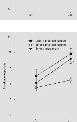
were no statistically significant interactions between groups or segments of the sessions (F2,41 = 2.41, P>0.05). Although all three groups increased their performances during the sessions the avoidance curves show a poorer performance of tone + brain stimula-tion in relastimula-tion to the other groups (Figure 3). Actually, the mean values of the number of avoidance responses in the second halves of the sessions for this latter group (11.2 ± 1.1) did not reach the criterion of learning fixed at 60% of all possible responses as did the two other groups, light + brain stimulation (19.6 ± 1.6) and tone + footshock stimula-tion (18.0 ± 1.7). Furthermore, only one out of eight animals reached this criterion in the tone + brain stimulation group while this ratio was higher for the tone + footshock stimulation (6 of eight rats) and light + brain stimulation (7/8 animals) groups.
These data are consistent with the asser-tion that pairings of 20-s illuminaasser-tion with the houselight with inferior colliculus stimu-lation caused significant avoidance responses in the signaled avoidance learning paradigm used in this study. The same does not hold
Figure 1 - Location of sites on cross-sections from the rat brain atlas of Paxinos and Watson (12). Figures represent the atlas coor-dinates in millimeters posterior to the bregma. Black circles indi-cate the inferior colliculus sites where electrical stimulation was applied. aq = Aqueduct; CG = central gray; DR = dorsal raphe nucleus; Cnf = cuneiform nu-cleus; ml = medial lemniscus; MnR = median raphe nucleus; 4V = fourth ventricle; RPn = raphe pontis nucleus.
aq
Bregma - 8.3 mm
Bregma - 8.8 mm
Bregma - 9.3 mm
4V
RPn -MnR
DR Cnf
Cnf
DR CG
true for the pairing of tone to this stimulation. Escape responses always occurred when the animals did not respond over the 20-s period of the warning stimulus. When we considered only these latencies of escape responses for the three groups tested, light + brain stimulation (5.50 ± 0.64 s), tone + brain stimulation (6.06 ± 0.68 s) and tone + footshock (4.25 ± 0.73 s) ANOVA did not reveal any statistically significant differences among them (F2,21 = 1.84, P>0.05).
Discussion
The present study provides additional evidence for the involvement of the inferior colliculus in neural circuits subserving aver-sive reactions. Electrical stimulation of the inferior colliculus shares many of the aver-sive properties of the stimulation of struc-tures classically considered as components of the brain aversion system, such as the dorsal periaqueductal gray matter, deep lay-ers of the superior colliculus, medial hypo-thalamus and amygdala (4,14-16). This is consistent with the demonstration of
ana-m -l
m -l DR
warning stimulus in the signaled two-way active avoidance paradigm used in this study. The rats increased their rate of responding in the presence of light over a number of trials. This effect may not be attributed to reflexive motor responses, since it was not observed when the neutral stimulus was presented alone, as demonstrated in a previous study from this laboratory (10). The pattern of results obtained in the present experiments with light as a warning stimulus parallels those which use a conditioned stimulus paired with stimulation of the dorsal periaqueduc-tal gray (19,20). It is likely that the inferior colliculus may be responsible for the elabo-ration of aversion and fear-like responses, together with other structures of the brain aversion system, such as dorsal periaque-ductal gray matter and amygdala. As a mat-ter of fact, there are important connections between the inferior colliculus and the dor-sal periaqueductal gray matter (17). Further-more, recent findings from this laboratory have shown that lesion of the central nucleus of the amygdala attenuates, while lesion of the basolateral complex enhances the aver-sive consequences of the electrical stimula-tion of the inferior colliculus (18).
In strong contrast to the association of light and inferior colliculus stimulation, acoustic stimulation paired with this stimu-lation does not function as a cue for rats to learn a shuttling response in order to avoid this aversive stimulation. These negative re-sults may not be attributed to rupture of the tympanic membrane during stereotaxic sur-gery or the tone parameters used in this study. Supporting this statement, an inde-pendent group of animals submitted to the same procedure, except for the pairing of tone with footshock, showed reliable learn-ing rates comparable to those from typical avoidance-escape procedures that utilize an auditory stimulus paired with electric shock as the unconditioned stimulus (21,22). One possible explanation for these surprising re-sults is that repeated electrical stimulation of
Avoidance latencies (s)
20
15
10
5
Light + brain stimulation Tone + brain stimulation Tone + footshocks
* *
1st 2nd
Figure 2 - Latencies of avoidance responses during the first and second segments of two-way active avoidance sessions for rats submitted to conditioning by pairing light or a tone with brain stimulation and a tone with footshock. Each session con-sisted of 50 trials. The results were pooled and divided into two halves for analysis. Data are reported as mean ± SEM for N = 8 rats in each group. *P<0.05 compared to 1st segment (Newman-Keuls comparisons).
Avoidance responses
25
20
15
10
5
0
Light + brain stimulation Tone + brain stimulation Tone + footshocks
*
*
1st 2nd
Figure 3 - Number of avoidance responses during the first and second segments of two-way active avoidance sessions for rats submitted to conditioning by pairing light or a tone with brain stimulation and a tone with footshock. Each session con-sisted of 50 trials. The results were pooled and divided into two halves for analysis. Data are reported as mean ± SEM for N = 8 rats in each group. *P<0.05 compared to 1st segment (Newman-Keuls comparisons).
tomical connections between the inferior colliculus and these structures (17,18).
the inferior colliculus leads to an increase of its reactivity to tone presentation. As a mat-ter of fact, local microinjections of low doses of bicuculline into the inferior colliculus, which mimic the effects of its electrical stim-ulation (7), increase the responsiveness of this structure to sound (23). In support of this possibility it has been shown that repeated stimulation of the inferior colliculus resulted in modified sensitivity of its neural network and in a chronic susceptibility to spontane-ous seizures (1). This shortcoming could lead to disturbances in the transmission of the to-be conditioned stimulus to a higher brain level, such as the amygdala, with which the inferior colliculus has an anatomical and functional relationship (18). Evidence show-ing that the steps of sensory processshow-ing are
modulated during learning procedures (24, 25) supports this possibility. Interestingly, pairing tone with stimulation of the midbrain tectum, in which several sites of stimulation were located in the superior colliculus, a relay for visual information, produces a strong con-ditioning compared to a weak one when light was used as the conditioned stimulus (19).
Taken together, the present results sug-gest that rats learn to avoid the inferior col-liculus stimulation when light is used as the warning stimulus. Since this does not occur when the neutral stimulus used is acoustic, it is likely that electrical stimulation of the inferior colliculus causes a disturbance in the transmission of auditory signals to higher brain structures, such as the amygdala, dis-turbing the conditioning process.
References
1. Bagri A, Sandner G & Di Scala G (1991). Wild running and switch-off behavior elic-ited by electrical stimulation of the infe-rior colliculus: effects of anticonvulsant drugs. Pharmacology, Biochemistry and Behavior, 39: 683-688.
2. Brandão ML, Melo LL & Cardoso SH (1993). Mechanisms of defense in the in-ferior colliculus. Behavioural Brain Re-search, 58: 49-55.
3. Silveira MCL, Sandner G & Graeff FG (1993). Induction of Fos-immunoreactivity in the brain by exposure to the elevated plus-maze. Behavioural Brain Research, 56: 115-118.
4. Brandão ML, Cardoso SH, Melo LL, Motta V & Coimbra NC (1994). Neural substrate of defensive behavior in the midbrain tec-tum. Neuroscience and Biobehavioral Re-views, 18: 339-346.
5. Melo LL, Cardoso SH & Brandão ML (1992). Antiaversive action of benzodiaz-epines on escape behavior induced by electrical stimulation of the inferior col-liculus. Physiology and Behavior, 51: 557-562.
6. Schmitt P, Carrive P, Di Scala G, Jenck F, Brandão ML, Bagri A, Moreau JL & Sandner G (1986). A neuropharmacologi-cal study of the periventricular neural sub-strate involved in flight. Behavioural Brain Research, 22: 181-190.
7. Brandão ML, Tomaz CAB, Leão Borges P
& Bagri A (1988). Defence reaction in-duced by microinjection of bicuculline into the inferior colliculus. Physiology and Be-havior, 44: 361-365.
8. Iwata J, Chida K & LeDoux JE (1987). Cardiovascular responses elicited by stim-ulation of neurons in the central amygda-loid nucleus in awake but not anaesthe-tized rats resemble conditioned emotional responses. Brain Research, 418: 183-188. 9. LeDoux JE, Sakagushi A & Reis DJ (1984). Subcortical efferent projections of the medial geniculate nucleus mediate emo-tional responses conditioned to acoustic stimuli. Journal of Neuroscience, 4: 683-698.
10. Brandão ML, Troncoso AC, Melo LL & Sandner G (1997). Active avoidance learn-ing uslearn-ing brain stimulation applied to the inferior colliculus as negative reinforce-ment in rats: Evidence for latent inhibi-tion. Neuropsychobiology, 35: 30-35. 11. MacIntosh NJ (1975). A theory of
atten-tion: variations in the associability of stimuli with reinforcement. Psychological Review, 82: 276-298.
12. Paxinos G & Watson P (1986). The Rat Brain in Stereotaxic Coordinates. Academ-ic Press, Sydney.
13. Marseillan RF (1977). A solid state sine wave stimulator. Physiology and Behav-ior, 19: 339-340.
14. Graeff FG (1990). Brain defense systems
and anxiety. In: Roth M, Burrows GD & Noyes R (Editors), Handbook of Anxiety. Vol. 3. Elsevier Science Publishers, Am-sterdam, 307-354.
15. Hunsperger RW (1956). Affektreaktionen auf Elektrische Reizung im Hirnstamm der Katze. Helvetica Physiologica et Pharma-cologica Acta, 14: 70-92.
16. Olds ME & Olds J (1963). Approach-avoid-ance analysis of rat diencephalon. Journal of Comparative Neurology, 120: 259-295. 17. Kudo M & Niimi K (1980). Ascending pro-jections of the inferior colliculus in the cat: an autoradiographic study. Journal of Comparative Neurology, 191: 545-546. 18. Maisonnette SS, Kawasaki MC, Coimbra
NC & Brandão ML (1996). Effects of le-sions of amygdaloid nuclei and substantia nigra on aversive responses induced by electrical stimulation of the inferior col-liculus. Brain Research Bulletin, 40: 93-98.
19. Di Scala G, Mana MJ, Jacobs WJ & Phillips MJ (1987). Evidence of Pavlovian conditioned fear following electrical stim-ulation of the periaqueductal grey in the rat. Physiology and Behavior, 40: 55-63. 20. Di Scala G & Sandner G (1989).
Condi-tioned place aversion produced by micro-injections of semicarbazide into the peri-aqueductal gray of the rat. Brain Research, 483: 91-97.
(1974). Effects of varying active/passive shock levels in shuttle box avoidance in rats. Journal of Comparative and Physi-ological Psychology, 86: 79-84.
22. Gray JA (1977). Drug effects on fear and frustration: possible limbic site of action of minor tranquilizers. In: Iversen LL, Iversen SD & Snyder SH (Editors), Hand-book of Psychopharmacology. Vol. 8.
Ple-num Press, New York, 433-529. 23. Bagri A, Sandner G & Di Scala G (1989).
Effects of unilateral microinjections of GABAergic drugs into the inferior collicu-lus on auditory evoked potentials and on audiogenic seizure susceptibility. Experi-mental Neurology, 104: 82-87.
24. Woody CD, Wang XF & Gruen E (1994). Response to acoustic stimuli increases in
the ventral cochlear nucleus after stimu-lus pairing. Neuroreport, 5: 513-514. 25. Yamamoto T, Shimura T, Sako N,