Morphological indicators of initial reproductive commitment in
Mustelus schmitti
(Springer 1939) (Chondrichthyes, Triakidae):
folliculogenesis and ovarian structure over the life cycle
Galíndez, EJ.
a,b*
, Díaz Andrade, M.
band Estecondo, S.
aaLaboratorio de Histología Animal, Departamento de Biología, Bioquímica y Farmacia, Universidad
Nacional del Sur – UNS, San Juan 670, 8000, Bahía Blanca, Argentina
bInstituto de Investigaciones Biológicas y Biomédicas del Sur – INBIOSUR, Consejo Nacional de
Investigaciones Científicas y Técnicas – CONICET, Bahía Blanca, Argentina
*e-mail: [email protected]
Received: November 6, 2012 – Accepted: June 5, 2013 – Distributed: November 30, 2014
(With 14 figures)
Abstract
This work provides information about the sexual commitment and the folliculogenesis of the gatuzo, Mustelus schmitti.
A total of 112 females of all maturity stages were fished in the Bahía Blanca estuary, between 2009 and 2010. The oogonia were present throughout the life cycle of the animals. The folliculogenesis follows a pattern similar to other elasmobranchs. The granulosa layer keeps monolayered throughout the folliculogenesis, but with two cell types in the vitellogenic follicle. The zona pellucida forms in the primordial follicles. The thecal system shows a connective inner layer and a glandular outer sheath. The microscopic beginning of the sexual commitment, indicated by the vitello hoarding, takes place in follicles from 500 micrometres, while the macroscopic evidence appears in follicles of 2500-3000 micrometres. The results presented in this study suggest that the fishery pressure may affect a susceptible range of sizes of the species, not previously considered and provides a biological framework for the development of fisheries policy.
Keywords: reproduction, ovary, sexual maturity, Chondrichthyans, gatuzo.
Indicadores morfológicos de compromisso reprodutivo inicial em
Mustelus
schmitti
(Springer 1939) (Chondrichthyes, Triakidae): folliculogenesis e
estrutura ovariana ao longo do ciclo de vida
Resumo
Este trabalho provê informações sobre o compromisso sexual e da foliculogênese do gatuzo, Mustelus schmitti. Um
total de 112 fêmeas de todas as fases de maturidade foram pescados no estuário Bahía Blanca, entre 2009 e 2010. O oogônias foi presentes durante todo do ciclo de vida dos animais. A foliculogênese segue um padrão semelhante a outros elasmobrânquios. A capa granulosa mantém-se simples durante toda a foliculogénese, mas com dois tipos de células no folículo vitelogênico. A zona pelúcida forma-se nos folículos primordiais. O sistema mostra uma capa tecal interior
de tecido conjuntivo e uma bainha exterior glandular. O início microscópico do compromisso sexual, indicado pela
acumulação do vitello, realiza-se em folículos de 500 micrómetros, enquanto que a evidência macroscópica aparece em folículos de 2500-3000 micrómetros. Os resultados apresentados neste estudo sugerem que a pressão da pesca pode afetar um amplo intervalo de tamanho das espécies não considerado anteriormente, e fornece uma base biológica para
o desenvolvimento de política comum da pesca.
Palavras-chave: reprodução, ovário, maturidade sexual, condrictios, gatuzo.
1. Introduction
Chondrichthyhan fisheries deal with stock problems worldwide and the SW Atlantic is not an exception, with a marked increase of fishing efforts in demersal fisheries
(Lucifora et al., 2012). Cartilaginous fishes have an extensive
fan of reproductive adaptations, the same that allow the survival of the group from the Devonian (Musick and
Ellis, 2005). Comparing them with teleosts, these fishes produce a few viable follicles in each reproductive season and the offspring is delivered in an advanced developmental stage, looking like small adults (McMillan, 2007). The
Despite this need, information about folliculogenesis in sharks is scarce and fragmented (Teshima et al.; 1976;
Andreuccetti et al., 1999; Prisco et al., 2001, 2002; Storrie, 2009; Davenport et al., 2011).
The production of a functional reproductive cell includes some general steps: the commitment of the primordial
germ cells that originate in the vitellin endoderm, their
migration to the genital ridges where they “nest”, their association with other epithelial cells to form the follicle and, finally, the accumulation of nutrients to support, at least, the initial stages of the future embryo’s development
(Kryskoet al., 2008). In elasmobranches, the ovarian
structure and the production of gametes are completely
adapted to their reproductive behaviour and the seasonality
of reproduction, if it exists. In some species this is a long process extending for a few years, concomitantly or not with gestation. On the other hand, some species show a clear annual reproductive season, with a few months for the production of gametes and delivery of the next
generation (Castro, 2009). Moreover, the function of the ovary is not restricted to the gametogenesis, but it is also a complex endocrine organ (Chieffi Baccari et al., 1992).
One of the most critical parameters for the implementation of adequate fishery management is the estimation of the reproductive status of the populations. Thence, the recognition of the maturation degree is basic. Usually, the most used indicators of maturity in females are macroscopic: the presence of yellow (vitellogenic) follicles in the ovary, as well as the enlargement of associated structures like the uterus and oviductal glands (Stehmann, 2002; Colonello et al., 2007). Despite this, commitment
in the reproductive process may occur before the onset of these characteristics; therefore fishery effort could be
targeted onto vulnerable size classes. Mustelus schmitti
Springer 1939, is a medium-small carchariniform shark that extends from Rio de Janeiro (Brazil) to Patagonia (Argentina). Due to its migratory habits, this shark is caught by different fishing fleets which affect different stages of its life cycle (Oddone et al., 2005; Molina and López Cazorla, 2011). Their stock decline, shrinking size
of captures and decrease in the LT50 is well documented
(Massa and Hozbor, 2011). In spite of their commercial
importance, there is scarce and fragmented information
about its reproductive biology and only one report exists
concerning the morphophysiology of reproductive organs
(Galíndez et al., 2010).
The knowledge of the morphology of folliculogenesis, as well as the determination of the onset of reproductive activity are important subjects, so for the physiological understanding of a transcending clade, as for a better comprehension of reproductive physiology from an
important economic resource.
2. Material and Methods
Females were caught seasonally during 2008 and 2009, by line fishing in the Bahía Blanca estuary (38°45’-39°45’ S; 61°30’-62°30’ W). Animals were sacrificed by blunt
trauma to the cranium, measured and immediately dissected.
The determination of maturity stage was made according
to Chiaramonte and Pettovello (2000) and Colonello et al.
(2007). For light microscopy, tissue samples were fixed in the Bouin’s fixative or formaldehyde 10%, both in seawater, dehydrated through a graded series of alcohols and embedded in paraffin wax. Sections of 3-5 micrometres were stained with hematoxylin-eosin, Masson’s trichromic stain, periodic acid Schiff reaction (PAS), the Alcian blue technique (AB pH 2.5) and the Sudan B technique. Sections were examined and photographed in an Olympus BX 51 microscope with an Olympus Camedia C-7070 camera. For transmission electron microscopy, small pieces were fixed in 2.5% glutaraldehyde in 0.05 M sodium cacodylate buffer with 12% sucrose (Hyder et al., 1983), for 12 hs at 4 °C
and post fixed in osmium tetroxide 1% in the same buffer for 90 minutes at 4 °C. Samples were washed in the same buffer, dehydrated in graded acetone and infiltrated in low density epoxy resin (Spurr). Semithin sections were colored with the methylen blue-azure II-basic fuchsine method and ultra-thin sections on grids were contrasted with uranyl acetate and lead citrate. Specimens were examined in an electron transmission Jeol CXII microscope.
3. Results
3.1 Microanatomy
The functional ovary of M. schmitti is the right one.
It is associated to the epigonal organs that, in mature
females, are caudally fused. In immature exemplars (24 to 57 cm of total length, n = 32), there is no evidence of ovary differentiation. In maturing females (42-68 cm TL, n = 58), the ovary develops from the cranial-ventral side of the right epigonal organ as an almond-shaped structure, filled with small translucent or white ova and separated from the granulopoietic tissue, by a slight sheet of loose connective tissue. As development continues, the ovary expands in detriment of the epigonal organ, so that in mature females (58-90 cm TL, n = 22), this organ withdraws to two minimal fused caudal projections. At this stage, the ovary occupies more than the 60% of the
general cavity and the residual space is shared by a very big
liver, the digestive tract and other organs. The remaining myeloid cells are intermingled with loose connective tissue between follicles (Figure 1). The ovarian epithelium is
simple columnar or cuboidal, with two cell types. One of them is less electrondense, shows small, apical, PAS positive vesicles, while the other type is denser and lacks of vesicles. Both cell types exhibit dilated spaces in the lateral surfaces and the basal cytoplasm retracts, leaving an arciform space (Figure 1, insert). Beneath the epithelium,
there is a false albuginea of dense connective tissue, with
smooth muscular cells.
3.2. Folliculogenesis
3.2.1. Oogonia (12.50-25.00 µm diameter)
These cells are present in all developmental stages of females. Even though the oogonia are more numerous
in pregnant females. These are medium roundish cells, with a high nucleus:cytoplasm ratio, located beneath the ovarian epithelium and normally forming nests (Figure 2).
The euchromatic nucleus shows partially condensed chromosomes and one or two evident nucleolus. The
acidophilic cytoplasm displays small vacuoles, glycogen
and lipid droplets. Inside the nests and intermingled with the oogonia, there are companion epithelial-like cells that will form the follicular envelope (Figure 3). These
cells are lightly basophilic and depict numerous RER profiles, as well as a moderate heterochromatic nucleus. At this time, in a pre-follicular stage, it is possible to find atretic structures, which show a high degree of cytoplasm vacuolization and nuclear disorganization. A thick basal
membrane (Figure 3) surrounds each nest.
3.2.2. Primordial Follicles (37.50-360 µm diameter) The first steps of follicle formation are the association of the oogonia with follicular epithelial-like cells enveloping it and a clear diminish of the nucleus:cytoplasm ratio. At this point, the follicular cells are flattened (Figure 4), with
an elongated euchromatic nucleus and one nucleolus. The oocyte nucleus shows the typical lampbrush chromosomes
(Figure 4). The cytoplasm is slightly acidophilic, with patches
of less eosinophilic reaction, corresponding to Balbiani’s
corps (Figure 4). As the follicle increases (approximately
100-150 mm diameter), both cell types show caveolae in
the plasmatic membrane (Figure 5). At their interface, the
zona pellucida originates as a deposition of an amorphous coat of glycosaminoglycans that react positive to PAS and
AB pH 2.5 (Figure 5). Likewise, the presence of microvilli
inside this caveolae evidences the beginning of the oolema folding (Figure 5). The follicular cells are surrounded by
a thick basal membrane and out of this, one or two layers of fibroblast-like cells are committed for the constitution of thecae (Figure 5).
3.2.3. Previtellogenic developing follicles (300-830 µm diameter)
This heterogeneous category includes from follicles with one simple coat of cubical or columnar follicular cells, to follicles with a tall-pseudostratified follicular
sheet (Figure 6), with wide empty spaces between cells (Figure 7). The nucleus:cytoplasm ratio of the oocyte
diminishes and their eosinophilic cytoplasm takes a foamy appearance. A morphological evidence of interaction between the oocyte and follicular cells is seen. On the one hand, the oolema folds tightly forming a refringent edge
and, on the other hand, the granulosa cells emit apical projections that stretch out through the zona pellucida and reach the oocyte (Figures 6 and 8). The zona pellucida
itself is characterised by its thickness, which is maximal in these follicles (Figures 6 and 7). In some of them, an inner
plywood-like zone is evident in the zona pellucida. Both thecas are well developed (Figures 6 and 7). The internal
one is fibrous, while the outer one comprises polyhedral glandular-like cells, which form acinar–like structures in the bigger follicles (Figures 6 and 7).
Figure 1. General view of the ovary of a sexually
mature female. Follicles at different maturity stages are intermingled with a connective stroma. The circle indicates a group of primordial follicles. The insert shows a medium magnification of the germinative epithelium, with large intercellular spaces (arrow). Pre: previtellogenic follicles; arrowheads: primary follicles; Vit: vitellate follicle. Scale
bar = 350 mm; insert Scale bar = 50 mm. Masson trichromic
stain.
Figure 2. Cist of oogonia. There are multiple oogonia
(oog), intermingled with prefollicular cells pfc). The arrow
makes point on the thick basal membrane. Semithin section, methylen blue-azure II-basic fuchsine. Scale bar = 30 mm.
3.2.4. Vitellogenic follicles (1100-2820) µm diameter Vitellogenic follicles depict many special features
(Figures 9-11). Their granulosa comprises a simple layer
of two cell types, differentiated by their electrondensity
(Figures 9-10). The cytoplasm of “dark” cells shows
granules PAS and AB 2.5 positive, while those of “light”
to Sudan B (Figures 9-10). These lipid droplets are also
located in the intercellular spaces. The zona pellucida decreases in thickness as the follicle diameter increases
(Figures 9-10). The vitello hoarding is a centripetal
phenomenon that begins by the input of small vitellin granules. The precursors reach the ooplasma from a thin vascular sinus, located immediately outside the follicular
basal membrane (Figures 9-11). These precursors cross the granulosa sheath via the enlarged intercellular spaces
Figure 3. Transmission electron microscopy image of part
of one cist. Look at the thick basal membrane (arrow)
and the close interaction between the oogonia (oog) and a
prefollicular cell (pfc), where both cells interdigitate their
membranes. The asterisk depicts one apoptotic oogonia.
Scale bar = 20 mm.
Figure 4. High magnification of a primordial follicle. Look
at the flat follicular cells (fc) and the fibroblastic-like cells
forming an incipient theca (th). Balbiani’s Corps (Bc) are
present in the ooplasm. Masson Trichromic stain.Scale bar = 30 mm. The insert depicts the thin zona pellucida
(arrow and zp). PAS reaction.Scale bar = 20 mm.
Figure 5. Transmission electron microscopy image of the
interface between the oolema (oo) and the follicular cells
in a primordial follicle. Look at the incipient formation of the zona pellucida defined by the incipient folding of the
oolema (arrowheads) facing the peripheral caveolae (cv)
of the follicular cells. The thick basal membrane is evident
(arrow). The connective cells begin the organization of the
theca (th). n: nucleus of a follicular cell. Scale bar = 5 mm.
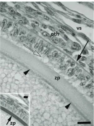
Figure 6. High magnification of envelops of one
previtellogenic follicle. The peripheral ooplasma looks foamy, indicating the coming vitellogenesis. The oolema is tightly folded (arrowheads), while follicular cells emit
apical projections (empty arrow). Between them, a thick
zona pellucida (zp) is evident. The basal membrane (arrow)
separates the follicular epithelium from the thecas. The external one shows their cells arranged in an acinar-like
structure (oth). The connective stroma is highly vascularized
(vs). Masson trichromic stain. Scale bar = 40 mm. The insert
depicts the same follicle stained with the PAS reaction, evidencing the thick zona pellucida (arrow, zp). Scale
3.2.5. Corpora lutea
When the oocyte leaves the ovary, the remaining
follicular structures transform into a corpus luteum. The first morphological hint is the invagination and folding of the granulosa sheath, with both thecas wrapping all the
structure (Figure 12). A simple layer of very tall cells lines
the central cavity. These have a vacuolated acidophilic cytoplasm with two kinds of granules: one of them, of greater size, are AB pH 2.5 and PAS positive, while the
others are small and react to the Sudan B (Figure 12,
insert). The acinar-like structures of the outer theca expand
in size (Figure 12). 3.2.6. Corpora atretica
Follicular death occurs in every stage of development.
In primordial (Figure 13) and previtellogenic phases, the atresia is indicated by nuclear apoptotic images, the
enlargement of the zona pellucida and the disorder of the granulosa. In vitellogenic follicles, some granulosa cells show evidence of hyperplasia and others become Figure 7. Semithin section of a latter previtellogenic
follicle (or an incipient vitellogenic one). The empty
intercellular spaces in the granulosa are evident (broad empty arrows). Look at the extreme development of the
zona pellucida (zp) with the inner side apposed to the folded
oolema (arrowheads). Thearrow shows the follicular basal
membrane. Both thecae are clearly developed (ith: inner
theca; oth: outer theca). Blood vessels (vs) are common in
the interstitial tissue. Methylen blue-azure II-basic fuchsine. Scale bar = 60 mm.
Figure 8. Detail of the interface between the oolema (oo)
and the zona pellucida (zp) in a vitellogenic follicle. Look at
the closed folding of the oolema. Scale Bar = 50 nm.
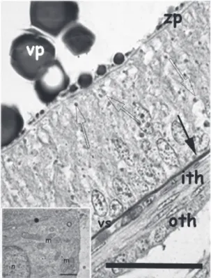
Figure 9. High magnification of a vitellogenic follicle.
Look at the clear pseudostratified epithelium. The zona
pellucida is very thin at this stage (zp). There are numerous
intercellular spaces filled with granules (empty arrows).
Both thecae are well defined and the external is composed
by polyhedral cells (oTh). One vascular sinus (vs) is
interposed between the internal theca (iTh) and the basal
membrane of the granulosa (arrow). Vp: vitelline plate.
Sudan B technique, Scale bar = 40 mm. The insert depicts an electron micrography of the cells of the outer theca. Tubular mitochondria profiles (m) and SER (O) are seen.
Cells present profuse lateral interdigitations (●).Scale Bar = 0.7 mm.
(Figure 9-10), pass through the zona pellucida and, finally,
enter into the ooplasma, by the oolema’s folds (Figure 10).
Inside the gamete, the vitellin platelets accrete to form the typical ellipsoidal structures, with an electrondense core
(Figure 10). Both thecas are well defined (Figures 9-11).
The inner one develops a vascular sinus that surrounds the follicle, while the outer is glandular-like and their cells are cuboidal, with short microvilli, mitochondria with tubular cristae, many SER profiles and developed lateral
phagocytic and transform into tall, glandular-like cells, similar to those of the corpus luteum (Figure 14). The
remaining follicle collapses. Both thecas involute and adopt the morphology of a dense connective tissue that infiltrates all the structure, transforming it in a dense scare (corpus albicans).
4. Discussion
The elamobranchs are key organisms in the evolutive context. They show many innovations in several aspects, such as the presence of jaws, placoid scales, an adaptable Figure 10. Microscopic electron image of the follicular
cells in a vitellogenic follicle. Two cellular types were seen, the dark follicular cells (dfc) and the light ones (lfc).Broad empty arrows depict the transport of vitellin precursors
in the intercellular spaces, while emptyarrowheads show
the intracytoplasmatic granules. Black arrowhead makes
point on the folding of the oolema; zp: zona pellucida, vp:
vitelline plate. Scale Bar = 4 mm.
Figure 11. Microscopic electron image of the follicular
cells in an early vitellogenic follicle. Follicular cells (fc)
are arranged in a seudostratified epithelium. One vascular
sinus (vs), plenty of blood cells, is developed between the
follicular basal membrane (black arrow) and the inner
theca (iTh). The outer theca (oTh) shows polyhedral cells.
The empty arrow makes point on granules of vitellin
precursors. Scale Bar = 5 mm.
Figure 12.Medium size image of an early corpus luteum.
The inner folding of the granulosa is clear, as well as the hypertrophy of the outer theca (th). The vascular sinus is
enlarged (vs). The insert is a magnification of the luteinic
tall cells, with evidence of phagocytosis (broadblack arrows). Masson trichromic stain. Scale Bar = 60 mm.
immune system (Haines et al., 2005) and display an adaptive
spectrum of reproductive characteristics (McMillan, 2007).
The ovary structure and the dynamics in the production of gametes agree with the manner of reproduction and the periodicity of litters. This information becomes relevant when it comes from an important economic resource.
The presence of only one functional ovary, closely associated with the epigonal organ, is the rule for the
genus (Tewinkel, 1950; Hamlett et al., 2002; Walker, 2007; Saïdi et al., 2008,2009) and M. schmitti is not an
exception. Unlike M. antarcticus Günther, 1870 (Storrie, 2004), in the gatuzo, the left epigonal organ is caudally
fused with the right one. If we add to this the fact that the mature ovary, plenty of ova, occupies more than one half of the general cavity, the laterality of the ovary in this species is unclear at naked eye.
The ovarian differentiation triggers the beginning of sexual development at the expense of the most important granulopoietic organ. This epigonal organ produces mainly granulocytes, related to the innate immunity, as well as
lymphocytes associated to the adapted immunity (Galíndez and Aggio, 2002). However, it is interesting that this fact
seems not to be crucial for the healthiness of the animals.
The gatuzo’s female reaches the sexual maturity at
2.7-2.4 years (Massa and Lasta, 2000; Colautti et al., 2010), corresponding to a total length higher than 57 cm
for the south of Brazil and the northern coasts of Argentina
and Uruguay (Oddone et al., 2005; Sidders et al., 2005)
and 79 cm for the Patagonian district (Chiaramonte and Pettovello, 2000). The microscopic evidence of the onset of vitellogenesis in animals macroscopically immature indicates, that they are already committed in the reproductive process
with an important energetic investment. That results in an indicator, nor previously considered, for the determination of the maturity stage and may contribute in the planning of specific regulations. Otherwise, it is possible that this early commitment will reflect a population disturbance, probably due to the increasing fishing pressure that is
undergoing this species.
The ovary exhibits a massive structure, with no cortex and medulla, as well as it was reported for M. asterias
Cloquet, 1819 (Capapé, 1983). It is lined by a simple epithelium, with different cell types and enlarged intercellular
and basal spaces. Some similar features were found in
M. manazo Bleeker, 1854 and M. griseus Pitschmann, 1908 (Teshima, 1981) but the presence of two cell types
has not been reported before. However, the presence of
smooth muscle in the albuginea has not been previously
reported for the genus. The occurrence of this cells might
be associated to the maximal distention attained by the
organ in mature females, due to the increment in number and size of the follicles.
The recruitment of follicles is modulated by the relationships established between the oogonia and the pre-follicular cells (Oktem and Oktay, 2008). As in other
species (Capapé, 1983; Prisco et al., 2007), the oogonia
of M. schmitti may occur isolated or forming clusters of sister cells all through the female’s life. Cortes and Massa
(2006) suggested that the gatuzo has some characteristics
that may enable a sustainable exploitation, if managed properly. If we take into account that the pools of germinal cells, as well as the atresia, define the amount of viable gametes, the results here presented support the idea of the sustainable fishery of the gatuzo.
Folliculogenesis begins with the meiotic process and the envelopment of the oocyte by the follicular cells. The interrelation between these cells and the local production of growth factors (Hutt and Albertini, 2007) define two possible routes: or development continues and the germinative cell
becomes a mature gamete, or the follicle undergoes atretia. In general, the follicle development in sharks follows
the same pattern as it does in other vertebrate species (Davenport et al., 2011). The first morphological step is
the differentiation of a primordial follicle, where a simple layer of flattened cells coats the oocyte. In M. schmitti,
these follicles are bigger than the observed in M. manazo and M. griseus (Teshima, 1981). On the other hand, the
earlier deposition of the zona pellucida, in the primordial stage from both, oocyte and follicular cells, is proper of this species and different from other fish’s pattern. Moreover, the zona pellucida of the gatuzo attempts its maximal thickness in previtellogenic stages (less than 2500 micrometers), as it does in M. canis (Mitchell, 1815)
(Davenport et al., 2011).
In M. schmitti, the onset of vitellogenesis occurs in
follicles of about 1100 - 1300 micrometers, although, at that point, it is not detectable at naked eye. As a rule, the follicular layer in this developmental step is simple in selachians, while batoids show a stratified granulosa
(Prisco et al., 2002; Díaz Andrade et al., 2009, 2011). At
early gatuzo’s vitellogenesis, the granulose increases in size and adopts the aspect of a pseudostratified epithelium,
as it does in M. antarcticus (Storrie, 2004), but different of
M. manazo and M. griseus, which depict stratified granulosa
layers as the skates (Teshima, 1981). However, despite the
diversity exhibited by chondrichthyans about the dynamics
of the follicular wall, it seems to be no consistent pattern
associated to the reproductive mode, either the oviparity
or the placentary or aplacentary viviparity. There are no previous reports about different cell types in the follicular coat of the genus Mustelus. In the gatuzo there is evidence
Figure 13. Apoptosis in incipient folliculogenesis
(primordial follicles). The asterisk depicts an apoptotic follicle. Masson trichromic stain. Scale Bar = 60 mm.
Figure 14.Medium size image of the follicular wall of an
atretic vitellogenic follicle. The is a clear transition between
the hyperplasic and the normal epithelium. In the image top there are some vitellin debris (vd) and zp indicates the disorganized zona pellucida. Th: thecas. Masson trichromic
of two different electrondense cells. However, it is not yet clear if they constitute two functionally different cells or two developmental stages of the same cellular type, so
that more studies are needed to explain this singularity and its implications.
Close to the ovulation point, the follicular cells reduce their height. These epithelial-like cells show dilated intercellular spaces filled with dense material. It is known
that vitellogenin, a lipophosphoprotein, is synthesised in the liver and transported by blood vessels to the oocyte,
through the follicular intercellular spaces (Prisco et al., 2002). Even though it is known that this protein is present in elasmobranchs (Pérez and Callard, 1989), the role of
follicular cells in their production is unclear (Prisco et al.,
2004). However, the presence of small cytoplasmic
granules that react positively to Sudan B in M. schmitti
supports the theory of the uptake of precursors from the
maternal circulation.
The maximal size registered for one follicle “at term” in this work, agrees with the range of values for the species
(Chiaramonte and Pettovello, 2000; Sidders et al., 2005). All studies Mustelus species have “at term” follicles ranged
from 15 to 28 mm (M. antarcticus, Walker, 2005; M. asterias,
Capapé, 1983; M. canis, Conrath and Musick, 2005;
M. griseus and M. manazo, Teshima et al., 1976; M. henlei
(Gill, 1863), Pérez and Sosa, 2008; M. lenticulatus Phillips,
1932, Francis and Mace, 1980; M. mustelus (Linnaeus,
1758), Capapé et al., 2006 and M. punctulatus Risso, 1827, Saïdi et al., 2009). This condition may not be related with
the reproductive mode or the mother’s size; the species referred above include placentary and aplacentary animals and comprise sharks from 95 to 185 cm of total length. If we take into account the particular structure of the zona pellucida and the interface between follicular cells and
the oocyte (Davenport et al., 2011), it is probably that the
constancy in the egg size responds to a midcourse between size, volume, resistance to deformation (flexibility) and the maximal thinness that allows fecundation.
The endocrine function of the follicle in Vertebrates
is beyond question (McMillan, 2007). There are two
known morphological ways of thecal arrangement: or the glandular tissue is part of the inner layer (Guraya, 1986), or the endocrine cells are found in the external sheet (Dodd, 1983; Prisco et al., 2002; Storrie, 2004). The
gatuzo exhibits this last model. The presence of a great amount of REL and tubular mitochondria in the thecal cells, constitute a strong evidence of the secretory nature of this external theca, since all these are characteristics of
cells involved in steroid production (Chieffi Baccari et al., 1992). M. manazo and M. griseus depict different thecas
too, but no morphological data was reported, except for a reference to a light eosinophilia in the inner one (Teshima,
1981), that might suggest a fibroblastic nature, in agreement
with the other members of the genus.
The results exposed show that atresia may occur in
any developmental stage, even in oogonia (Ricchiari et al., 2003). In early follicles, there is no major evidence than
the disorganization and resorption of the follicle. On the
other hand, in the vitellogenic ones, the hyperplasia of the follicular epithelium seen in M. schmitti has not been previously reported, so that only one histologic study exists in the genus (M. canis,Hisaw and Hisaw, 1959). Moreover,
there is much confusion refering to the identity of corpora lutea and corpora atretica in Chondrichthyes. The corpus
luteum seems to be present in all studied Mustelus species (Storrie, 2004) with similar characteristics, such as the
granulosa folding and the tall phagocytic cells. In spite of this morphological features, there are no conclusive
studies about their glandular nature.
Despite its importance from the evolutionary, ecological and commercial standpoint, fisheries have led to the elasmobranchs stocks worlwide to a critical reduction, even just to the point of commercial and biological extinction
(Oddone et al., 2005). That is why the knowledge of all
biological features, with special emphasis on reproductive traits, is necessary for the implementation of adequate management and protection policies. As mentioned before, the sharks of the genus Mustelus have some characteristics that may enable a sustainable exploitation, under adequate
management. Is in this context, it is hoped that the new data
presented here may contribute to a better understanding
and protection of this important resource.
Acknowledgements
This work was supported by SGCyT-UNS, PGI:24/B173. We thank the collaboration of the Argentine Naval Prefecture for its assistance in the sampling.
References
ANDREUCCETTI, P., IODICE, M., PRISCO, M. and GUALTIERI,
R., 1999. Intercellular bridges between granulosa cells and the
oocyte in the elasmobranchs Raya asterias. The Anatomical Record, vol. 255, no. 2, p. 180-187. http://dx.doi.org/10.1002/
(SICI)1097-0185(19990601)255:2<180::AID-AR8>3.0.CO;2-S.
CAPAPÉ, C., 1983. Nouvelles données sur la biologie de la reproduction de Mustelus asterias Cloquet, 1821 (Pisces,
Pleurotremata, Triakidae) de côtes Tunisiennes. Vie et Milieu, vol. 33, p. 143-152.
CASTRO, J., 2009. Observations of the reproductive cycles of some viviparous North American sharks. Aqua International Journal of Ichthyology, vol. 15, p. 205-222.
CHIARAMONTE, G. and PETTOVELLO, A., 2000. The
biology of Mustelus schmitti in southern Patagonia, Argentina. Journal of Fish Biology, vol. 57, no. 4, p. 930-942. http://dx.doi. org/10.1111/j.1095-8649.2000.tb02202.x.
CHIEFFI BACCARI, G., MINUCCI, S., DI MATTEO, L. and
CHIEFFI, G., 1992. Ultrastructural investigation of the corpora atretica of the electric ray, Torpedo marmorata. General and Comparative Endocrinology, vol. 86, no. 1, p. 72-80. http://dx.doi.
org/10.1016/0016-6480(92)90127-6. PMid:1505731
COLAUTTI, D., BAIGUN, C., LÓPEZ CAZORLA, A.,
LLOMPART, F., MOLINA, JM., SUQUELE, P. and CALVO,
S., 2010. Population biology and fishery characteristics of the
Fisheries Research, vol. 106, no. 3, p. 351-357. http://dx.doi.
org/10.1016/j.fishres.2010.09.004.
COLONELLO, J., CHRISTIANSEN, H. and MACCHI, G., 2007. Escala de madurez sexual para peces cartilaginosos de la Plataforma Continental Argentina. Mar del Plata: INIDEP. Informe Técnico no. 74.
CORTÉS, F. and MASSA, A., 2006. Aspectos reproductivos del gatuzo (Mustelus schmitti). Mar del Plata: INIDEP. Informe Técnico no. 81.
DAVENPORT, IR., WEAVER, AL. and WOURMS, JP., 2011. A
novel set of structures within the elasmobranch, ovarian follicle.
Journal of Morphology, vol. 272, no. 5, p. 557-565. http://dx.doi. org/10.1002/jmor.10932. PMid:21308727
DÍAZ-ANDRADE, MC., GALÍNDEZ, E. and ESTECONDO,
S., 2009. The ovary of the bignose fanskate Sympterygia acuta Garman, 1877 (Chondrichthyes, Rajidae) in the Bahía Blanca estuary, Argentina: morphology and reproductive features.
Revista Brasileira de Biologia = Brazilian Journal of Biology, vol. 69, no. 2, p. 405-413. http://dx.doi.org/10.1590/S1519-69842009000200025. PMid:19675946
DÍAZ-ANDRADE, MC., GALÍNDEZ, EJ., LÓPEZ CAZORLA, A. and ESTECONDO, S., 2011. Ovarian folliculogenesis in the
smallnose fanskate Sympterygia bonapartii (Müller and Henle,
1841) (Chondrychthyes, Rajidae). International Journal of Morphology, vol. 29, no. 1, p. 174-181. http://dx.doi.org/10.4067/ S0717-95022011000100030.
DODD, J., 1983. Reproduction in cartilaginous fishes (Chondrichthyes).
In HOAR, W., RANDALL, J. and DONALDSON, E. Fish fisiology.
New York: Academic Press. p. 31-95.
GALÍNDEZ, E. and AGGIO, M., 2002. The granulopoietic
organs of the narrownose smooth hound Mustelus schmitti
(Chondrichthyes, Triakidae): a light and electron microscopic
study. Revista Chilena de Anatomía, vol. 20, p. 49-54.
GALÍNDEZ, EJ., DÍAZ ANDRADE, MC., MOYA, AC. and
ESTECONDO, S., 2010. Morphological changes in the pregnant
uterus of the smooth hound dogfish Mustelus schmitti Springer,
1939 (gatuzo) (Chondrichthyes, Triakidae). Microscopic study and
phylogenetic reproductive implications. International Journal of Morphology, vol. 28, no. 4, p. 1003-1010. http://dx.doi.org/10.4067/ S0717-95022010000400004.
GURAYA, S., 1986. The cell and molecular biology of fish oogenesis. In SAUER, H. Monographs in Developmental Biology. Basel: Karger. p. 111-163. vol. 18.
HAINES, AN., FLAJNIK, MF., RUMFELT, LL. and WOURMS,
JP., 2005. Immunoglobulins in the eggs of the nurse shark,
Ginglymostoma cirratum. Developmental and Comparative Immunology, vol. 29, no. 5, p. 417-430. http://dx.doi.org/10.1016/j. dci.2004.08.007. PMid:15707663
HAMLETT, WC., MUSICK, JA., HYSELL, CK. and SEVER, DM., 2002. Uterine epithelial-sperm interaction, endometrial
cycle and sperm storage in the terminal zone of the oviducal gland
in the placental smoothhound, Mustelus canis. The Journal of Experimental Zoology, vol. 292, no. 2, p. 129-144. http://dx.doi. org/10.1002/jez.1149. PMid:11754029
HISAW JUNIOR, FL. and HISAW, FL., 1959. Corpora lutea of
elasmobranch fishes. The Anatomical Record, vol. 135, no. 4, p. 269-277. http://dx.doi.org/10.1002/ar.1091350405. PMid:14402031
HUTT, KJ. and ALBERTINI, DF., 2007. An oocentric view of
folliculogenesis and embryogenesis. Reproductive Biomedicine
Online, vol. 14, no. 6, p. 758-764. http://dx.doi.org/10.1016/
S1472-6483(10)60679-7. PMid:17579993
HYDER, SL., CAYER, ML. and PETTEY, CL., 1983. Cell types
in peripheral blood of the nurse shark: an approach to structure and function. Tissue & Cell, vol. 15, no. 3, p. 437-455. http://dx.doi.
org/10.1016/0040-8166(83)90075-7. PMid:6612712
KRYSKO, DV., DIEZ-FRAILE, A., CRIEL, G., SVISTUNOV, AA., VANDENABEELE, P. and D’HERDE, K., 2008. Life and
death of female gametes during oogenesis and folliculogenesis.
Apoptosis, vol. 13, no. 9, p. 1065-1087. http://dx.doi.org/10.1007/ s10495-008-0238-1. PMid:18622770
LUCIFORA, L., GARCÍA, V., MENNI, R. and WORM, B., 2012.
Spatial patterns in the diversity of sharks, rays and chimaeras (Chondrichthyes) in the southwest Atlantic. Biodiversity and Conservation, vol. 21, no. 2, p. 407-419. http://dx.doi.org/10.1007/ s10531-011-0189-7.
PRISCO, M., VALIANTE, S., ROMANO, M., RICCHIARI, L., LIGUORO, A., LAFORGIA, V., LIMATOLA, E. and
ANDREUCCETTI, P., 2004. Ovarian follicle cells in torpedo
marmorata synthesize vitellogenin. Molecular Reproduction and Development, vol. 67, no. 4, p. 424-429. http://dx.doi.org/10.1002/ mrd.20036. PMid:14991733
MASSA, A. and LASTA, C., 2000. Gatuzo (Mustelus schmitti).
In BEZZI, S., AKSELMAN, R. and BOSCHI, E. Síntesis del estado actual de las pesquerías marítimas argentinas y de la Cuenca del Plata. Años 1997-1998, con actualización de 1999, Contrib. 1129. Mar del Plata: INIDEP. p. 131-137.
MASSA, A. and HOZBOR, N., 2011. Evolución de las estimaciones de abundancia de los peces cartilaginosos demersales de mayor
valor comercial del Atlántico Sudoccidental, capturados entre 34° y 41° S, a profundidades menores de 50 m. In WOHLER, O., CEDROLA, P. and COUSSEAU, B. Contribuciones sobre biología, pesca y comercialización de tiburones en la Argentina. Aportes para la elaboración del Plan de Acción Nacional, Contrib. 1711.
Mar del Plata: INIDEP. p. 193-205.
MCMILLAN, D., 2007. Fish histology: female reproductive system. Dordrecht: Springer.
MOLINA, JM. and LÓPEZ CAZORLA, A., 2011. Trophic ecology
of Mustelus schmitti (Springer, 1939) in a nursery area of northern
Patagonia. Journal of Sea Research, vol. 65, no. 4, p. 381-389. http://dx.doi.org/10.1016/j.seares.2011.03.001.
MUSICK, J. and ELLIS, J., 2005. Reproductive evolution of
chondrichthyans. In HAMLETT, W. Reproductive biology and phylogeny of chondrichthyes: sharks, rays and chimaeras. Enfield:
Science Publishers. p. 45-71. vol. 3.
ODDONE, C., PAESCH, L. and NORBIS, W., 2005. Reproductive
biology and seasonal distribution of Mustelus schmitti (Elasmobranchii, Triakidae) in the Rio de la Plata oceanic front, south-western
Atlantic. Journal of Marine Biology, vol. 85, no. 5, p. 1193-1198. http://dx.doi.org/10.1017/S0025315405012294.
OKTEM, O. and OKTAY, K., 2008. Stem cells: a perspective on oocytes. Annals of the New York Academy of Sciences, vol. 1127, no. 1, p. 20-26. http://dx.doi.org/10.1196/annals.1434.010. PMid:18443325
PÉREZ, L. and CALLARD, I., 1989. Evidence for progesterone
inhibition of vitellogenesis in the skate, Raja erinacea. American Zoologist, vol. 29, p. 357A.
PRISCO, M., RICCHIARI, L. and ANDREUCCETTI, P., 2001.
in Torpedo marmorata. The Anatomical Record, vol. 263, no. 3, p. 239-247. http://dx.doi.org/10.1002/ar.1093. PMid:11455532 PRISCO, M., LOREDANA, R. and PIERO, A., 2002. Ultrastructural
studies on developing follicles of the spotted ray Torpedo marmorata.
Molecular Reproduction and Development, vol. 61, no. 1, p. 78-86. http://dx.doi.org/10.1002/mrd.1133. PMid:11774378
PRISCO, M., LIGUORO, A., RICCHIARI, L., DEL GIUDICE, G. and ANDREUCCETTI, P., 2007. Oogenesis in the spoted ray, Torpedo marmorata. Reviews in Fish Biology and Fisheries, vol. 17, no. 1, p. 1-10. http://dx.doi.org/10.1007/s11160-006-9013-y. RICCHIARI, L., D’ONGHIA, B., LIGUORO, A., TAMMARO, S., MOTTA, CM., FILOSA, S., ANDREUCCETTI, P. and PRISCO, M., 2003. Role of apoptosis and Fas/FasL system in
the oogenesis of the spotted ray Torpedo marmorata. Molecular Reproduction and Development, vol. 66, no. 1, p. 54-59. http:// dx.doi.org/10.1002/mrd.10324. PMid:12874799
SAÏDI, B., BRADAÏ, M. and BOUAÏN, A., 2008. Reproductive
biology of the smooth hound shark Mustelus mustelus (L.) in the Gulf of Gabès (South-Central Mediterranean Sea). Journal of Fish Biology, vol. 72, no. 6, p. 1343-1354. http://dx.doi. org/10.1111/j.1095-8649.2008.01801.x.
SAÏDI, B., NEJMEDDINE BRADAÏ, M. and BOUAÏN, A., 2009.
Reproductive biology and diet of Mustelus punctulatus (Risso,
1826) (Chondrichthyes, Triakidae) from de Gulf of Gabès, Central
Mediterranean Sea. Scientia Marina, vol. 73, no. 2, p. 249-258. http://dx.doi.org/10.3989/scimar.2009.73n2249.
SIDDERS, M., TAMINI, L., PÉREZ, J. and CHIARAMONTE,
GE., 2005. Biología reproductiva del gatuzo Mustelus schmitti Springer 1939 (Chondrichthyes, Triakidae) en el área de Puerto
Quequén, Provincia de Buenos Aires. Revista del Museo Argentino de Ciencias Naturales. Revista del Museo Argentino de Ciencias Naturales, vol. 7, no. 1, p. 89-101.
STEHMANN, M., 2002. Proposal of a maturity stages scale for oviparous and viviparous cartilaginous fishes (Pisces, Chondrichthyes). Archiv fuer Fischerei und Meeresforschung, vol. 50, p. 23-48.
STORRIE, M., 2004. Microscopic modifications of the female reproductive tissues of Mustelus antarcticus. Australia: Deakin University. 153 p. PHD Thesis.
STORRIE, MT., WALKER, TI., LAURENSON, LJ. and
HAMLETT, WC., 2009. Gestational morphogenesis of the uterine epithelium of the gummy shark (Mustelus antarcticus).
Journal of Morphology, vol. 270, no. 3, p. 319-336. http://dx.doi. org/10.1002/jmor.10693. PMid:19117062
TESHIMA, K., 1981. Studies on the reproduction of Japanese smooth dogfishes: Mustelus manazo and Mustelus griseus. Journal of the Shimonoseki University of Fisheries, vol. 29, p. 113-199.
TESHIMA, K., CHEN, C. and MIZUE, K., 1976. Studies on
sharks. IX. Ovary and oogenesis in selachians. Journal of the Shimonoseki University of Fisheries, vol. 25, p. 41-45.
TEWINKEL, LE., 1950. Notes on ovulation, ova and early development in the smooth dogfish, Mustelus canis. The Biological Bulletin, vol. 99, no. 3, p. 474-486. http://dx.doi. org/10.2307/1538478. PMid:14801013
WALKER, TI., 2007. Spatial and temporal variation in the reproductive biology of gummy shark Mustelus antarcticus