Ste ps in the fo rm atio n o f ne urite s
and synapse s studie d in culture d
le e ch ne uro ns
Departamento de Biofísica, Instituto de Fisiología Celular, Universidad Nacional Autónoma de México, DF, México F.F. De-Miguel
Abstract
Leech neurons in culture have provided novel insights into the steps in the formation of neurite outgrowth patterns, target recognition and synapse formation. Identified adult neurons from the central nervous system of the leech can be removed individually and plated in culture under well-controlled conditions, where they retain their characteris-tic physiological properties, grow neurites and form specific chemical or electrical synapses. Different identified neurons develop distinctive outgrowth patterns that depend on their identities and on the molecular composition of the substrate. On native substrates, the patterns dis-played by these neurons reproduce characteristics from the adult or the developing neurons. In addition, the substrate may induce selective directed growth between pairs of neurons that normally make contact in the ganglion. Upon contact, pairs of cultured leech neurons form chemical or electrical synapses, or both types depending on the neuronal identities. Anterograde and retrograde signals during mem-brane contact and synapse formation modify the distribution of synap-tic terminals, calcium currents, and responses to 5-hydroxytryptamine. Co rre spo nde nce
F.F. De-Miguel Departamento de Biofísica Instituto de Fisiología Celular Universidad Nacional Autónoma de México
Apartado Postal 70-253 México, 04510 DF México
Fax: + 52-5-622-5607 E-mail: ffernand@ ifisiol.unam.mx
Presented at the International Neurobiology Course, Faculdade de O dontologia de Ribeirão Preto, Universidade de São Paulo, Ribeirão Preto, SP, Brasil, May 20-27, 1998.
Research supported by CO NACYT, PAPIIT and the Human Frontiers Science Program to F.F. De-Miguel.
Received February 23, 1999 Accepted February 8, 2000
Ke y wo rds
·Regeneration ·Neurite outgrowth ·Synapse formation ·Cultured neurons ·Leech
Intro ductio n
During development and regeneration of the nervous system neurons must grow, find their targets and form the correct connec-tions. Cultured neurons from adult inverte-brates have been useful to characterize the steps from neurite outgrowth to synapse for-mation (1-5). After isolation, many inverte-brate neurons remain differentiated and con-tinue synthesizing the same transmitters, ionic channels and transmitter receptors as they normally do in the animal (4,5). Moreover, pairs or triplets of neurons plated together regrow, recognize each other and form spe-cific synapses (4-6). This review focuses on studies with cultured leech neurons, in which
many of the steps from neurite outgrowth to synapse formation have been elucidated.
of variable length from which they will re-grow and form synaptic connections with other neurons (4,10). Early experiments showed that cultured leech neurons survive for weeks, preserving their resting potentials and the distinctive shape and duration of their action potentials (11). Moreover, cells can regrow neurites displaying characteris-tic branching patterns that depend on their identities and on the substrate (7,8), and, when plated in contact with other cells, they form specific chemical or electrical synapses. Cell-to-cell contact and synapse formation induce changes in the distribution of release sites, ion channels and postsynaptic recep-tors.
Spro uting and re tracting ne urite s
Neurite outgrowth during development and regeneration of the nervous system is regulated by growth-promoting molecules, soluble factors and cell-cell interactions (12). The family of extracellular matrix (ECM) proteins provides important sources for neu-rite growth, retraction and branching of neurites in the CNS as well as in culture (13). Different ECM proteins such as laminin, tenascin and fibronectin have been identi-fied, isolated and cloned, and some of their functions regulating neurite extension or re-pulsion are known (12). Nevertheless, al-though studies analyzing attachment of neu-rons to purified molecules have provided extensive information as to some of the ef-fects of these molecules, in the CNS of ver-tebrates and inverver-tebrates ECM has a broad molecular composition, thus suggesting a complex regulation of neurite outgrowth. On the other hand, cell-cell interactions and soluble factors influence neurite outgrowth in embryos or regenerating nervous systems. How far could native ECM shape a neuronal outgrowth pattern in the absence of soluble factors and cell-cell interactions? The fol-lowing studies, conducted on two different leech neurons plated on different substrates,
have provided interesting insights as to how complex native mixtures of ECM proteins regulate neurite extension and directed growth.
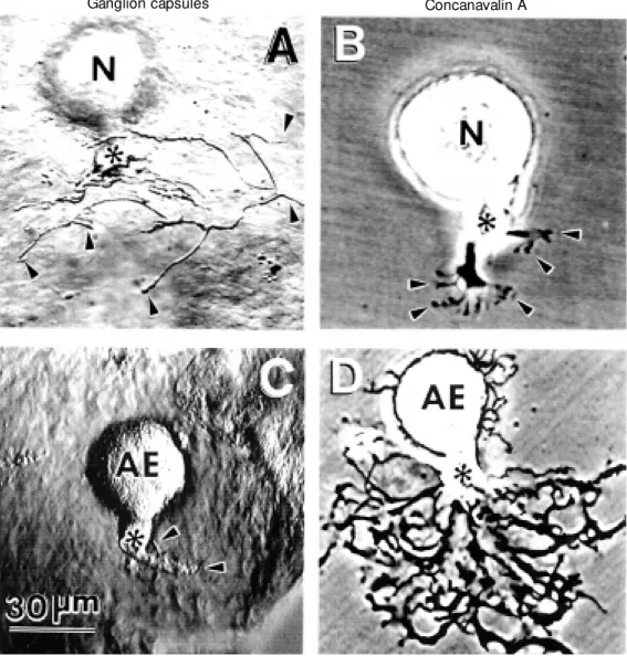
Retzius neurons, which in the animal coordinate swimming, and anterior pagoda (AP) neurons, of unknown function, start growing a few minutes after being plated in culture with characteristic patterns that de-pend on the substrate and on the neuronal type. For each cell type the outgrowth pat-terns are consistently very similar, but quite different from patterns of the same cells on different substrates (14). An interesting ex-ample has been provided by AP or Retzius neurons plated on a substrate they normally contact in the animal, i.e., the ganglion cap-sule, which is enriched in ECM proteins. AP neurons plated on capsules (Figure 1A) de-velop a T-shaped pattern consisting of two main branches in opposite directions that later bifurcate (15,16). A completely differ-ent pattern is developed by Retzius neurons plated on the same substrate, where they sprout a bundle of long and thick processes emerging from the axonal stump (Figure 1B). Plating a particular cell type on differ-ent substrates can be used to demonstrate the importance of the composition of the sub-strate in the outgrowth pattern. As can also be seen in Figure 1, the pattern of AP neu-rons on ganglion capsules is quite different from the patterns developed by the same neuronal type on other substrates. For ex-ample, AP neurons plated on leech laminin extracts extend long neurites with few branch-ing points (Figure 1C). In contrast, on the plant lectin concanavalin A (Con A) (a sub-strate they never encounter in the animal), AP neurons extend flat and curved processes with multiple branching points (Figure 1D; 14,15).
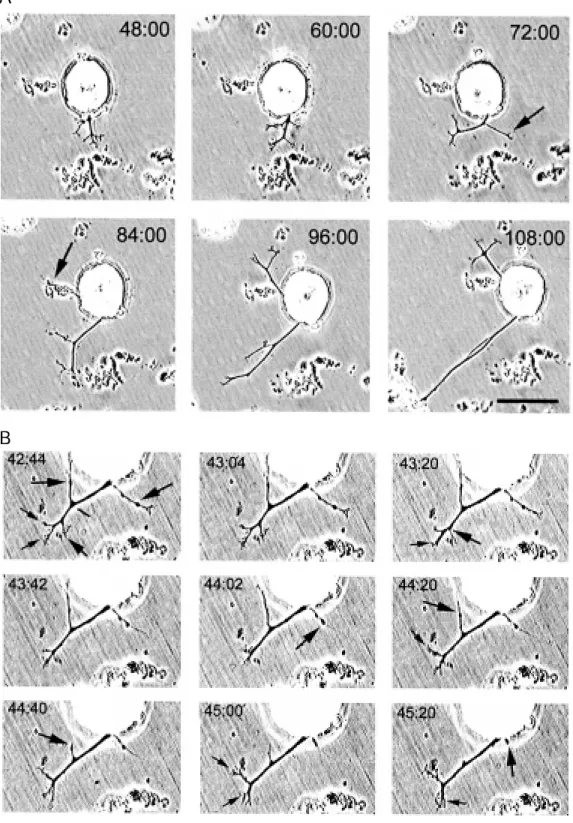
em-bryonic AP neurons at around day 9 (17). This similarity between growth of embry-onic and adult cultured neurons suggests that an ECM similar to that in the ganglion capsules may provide the initial messages for structuring the outgrowth pattern. This pattern can be quantitatively reproduced if AP neurons are plated on CNS or capsule homogenates as substrates (Figure 2), thus providing the possibility to analyze the steps of its formation. Time-lapse analysis of AP neurons growing on CNS homogenates has shown a complex dynamics of neurite elon-gation and retraction. As shown in Figure 2B, while two main neurites grow in oppo-site directions, secondary and presumably supernumerary neurites are retracted, reflect-ing competition. Indeed, after a total neuritic length of about 100 µm, growth of primary neurites occurs at the same rate as retraction of secondary neurites, suggesting an eco-nomical mechanism for selective growth plus
elimination of supernumerary neurites, re-utilizing the same precursors (De Miguel FF and Vargas J, unpublished data).
ECM proteins may regulate neurite ex-tension and retraction through a complex set of indirect mechanisms. For example, growth of neurites on leech laminin increases cal-cium currents evoked by electrical activity in cultured leech neurons (18). In addition, cal-cium injection into the cells in response to electrical activity may induce neuritic re-traction in leech neurons (19,20). Similar events have been described in different neu-ronal types during development, for example in amphibian spinal neurons in vitro,
in-creases in the intracellular concentration of calcium reduce neurite extension and pro-duce retraction (21,22). Growth cones of leech neurons collapse in response to electri-cal stimulation (19,20) or to the application of a calcium ionophore, presumably due to a disruption of microfilaments (23). Although
A
B
C
D
Figure 2 - Development of the outgrow th pattern of one ante-rior pagoda (AP) neuron plated on leech CNS homogenate. A, At 48 h in culture, one branch had grow n w ith several second-ary processes. Tw ent y-f our hours later the primary branch w as reoriented and one of the secondary branches had elon-gated (marked by the arrow ). At 84 h in culture, a second branch had grow n (marked by the ar-row ). Both branches continued grow ing for 24 h. Scale bar = 50 µm (adapted from Ref. 16). B, Time-lapse analysis of neurite ext ension and ret ract ion of neurites of the same AP cell pre-sented in A. The time represents the grow th time. The large ar-row s point at neurites that re-tracted w hile the small arrow s point at neurites that elongated. Scale bar = 40 µm.
A
B
the soma of adult AP neurons is unable to produce action potentials (24) these are produced at distant sites. Therefore, it is possible that during neurite extension
D ire cting gro wth
In addition to influencing neuritic out-growth, the substrate is also determinant for target finding. When pairs of Retzius neu-rons are plated at a distance on a ganglion capsule, neurites are sent towards each other and make contact even if the cells were originally plated orienting their processes 180o from each other. Similarly, ECM on
ganglion capsules induces directed growth in pairs of AP neurons. In contrast, directed growth failed when mixed pairs of one Retzius cell and one AP cell were plated on capsules, or when pairs of neurons were plated on Con A or leech laminin. It is note-worthy that in the animal, pairs of Retzius or AP neurons form connections, but Retzius neurons do not contact AP neurons. There-fore, directed growth on capsules again seems to reproduce a developmental phenomenon selectively induced by native ECM proteins. That directed growth fails in pairs of neurons plated on laminin or Con A underscores the importance of ECM inducing multiple ef-fects, such as the possible release of soluble attractants.
Se le cting the synaptic attribute s
After finding an appropriate target, a next step in development is the formation of cor-rect synapses. How does a neuron choose when and with what to make a chemical or an electrical synapse? Are neurite outgrowth, directed growth and synapse formation part of a cascade of events triggered by contact with an appropriate substrate? To what ex-tent extrinsic influences may determine neu-ronal connectivity? An answer to these ques-tions has again been provided by the analysis of growth and synapse formation of leech neurons on different substrates. In the leech, a monosynaptic reflex response that short-ens the animal is mediated by the activation of nociceptive (N) sensory neurons that in the ganglion make chemical connection with
annulus erector (AE) motoneurons (25). When these two neuronal types are isolated and plated in culture they form an electrical synapse instead of the chemical one (26). The difference between the connection in the animal and in culture provided an attrac-tive preparation to examine if the selection of a synapse is also influenced by the sub-strate. When growth was tested, N and AE neurons had opposite responses to the sub-strates. As shown in Figure 3, single N cells failed to grow on Con A, but grew profusely on ganglion capsules. In contrast, AE neu-rons sprouted profusely on Con A but failed to grow on ganglion capsules. Interestingly, as shown in Figure 4, pairs of N and AE neurons plated at a distance on the same capsule grew towards each other and made contact. Nevertheless, neurons did not form a chemical synapse but rather developed an electrical rectifying synapse (Figure 4C) simi-lar to that formed on Con A (27). These data provide an example that neurite outgrowth and target recognition may obey different signaling patterns than synapse formation, and suggest the contribution of multiple in-fluences being involved in every step of neuronal development and regeneration. A question that still remains to be answered is what are the signals that induce the forma-tion of the correct chemical synapse during development?
depolar-izing current spreads only from the P cell to its partner, while hyperpolarizing current spreads only in the opposite direction (1,4). From these observations one can conclude that the characteristic synapses established by a particular cell type are, at least in part, determined by their identities. What kind of synapse will a Retzius and a P cell form if plated together? Even though Retzius and P cells form electrical synapses with different targets in culture, when plated together they form purely chemical inhibitory synapses, with the Retzius cell always being presynap-tic (11).
In the nervous system, one single neuron
may establish synaptic contacts with many other neurons. Chemical synapses may be excitatory or inhibitory; other synapses may be electrical, either rectifying or non-rectify-ing. For each particular case, different mem-brane specializations are required. To what extent are different regions of the cell mem-brane pre-appointed to form chemical, elec-trical, pre- or postsynaptic specializations? Liu and Nicholls (10), plating pairs of Retzius neurons with different membrane regions making contact, have provided interesting information. Particular membrane segments of leech neurons have shown different pref-erences to form particular synaptic
modali-Figure 3 - Outgrow th patterns of nociceptive (N) sensory neurons and annulus erector (AE) moto-neurons on ganglion capsules and Con A after 48 h in culture. A, N sensory neurons on gan-glion capsules sprouted long and fine neurites. Some are pointed by the arrow s. B, On Con A, N cells did not grow and only ex-tended short lamellar grow th cones w ith fine filopodia, as pointed by the arrow s. C, AE motoneurons plated on ganglion capsules failed to grow . Only fine filopodia elongated from the remaining stump, as pointed by the arrow s. D, On Con A, AE m ot oneurons sprout ed pro-fusely. The stumps are indicated by the asterisks (adapted from Ref. 27).
ties. The tip of the stump, a remaining por-tion of the primary process, is a preferential region from which Retzius neurons can form presynaptic chemical endings. In contrast, the soma of a Retzius cell forms postsynap-tic but never presynappostsynap-tic components. In addition, either region develops electrical synapses after one or two days.
Mo dificatio ns during synapse fo rma-tio n: change s in re le ase site s and calcium curre nt distributio n
The series of events that start with neu-ronal contact continue long after synapses have formed and induce permanent changes
in pre- and postsynaptic neurons. Synapse formation requires a whole set of adapta-tions, including the incorporation of presyn-aptic vesicles and ion channels in presynap-tic terminals and transmitter receptors on the postsynaptic side, among many other com-ponents. For example, calcium channels should cluster at the presynaptic endings to regulate transmitter release, while transmit-ter receptors should be located at the post-synaptic endings (4,5). Cultured serotoner-gic synapses between two Retzius neurons or between a Retzius and a P neuron have also been useful to analyze the consequences of synapse formation in the incorporation of presynaptic terminals at the sites of contact
N-AE AE-N
100 ms
Figure 4 - Grow th and synapse formation of pairs of nocicep-tive (N) and annulus erector (AE) neurons. A, Nomarski image of a pair of neurons grow ing on a ganglion capsule. In this case bot h neurons ext ended pro-cesses that made contact. Arrow s point at processes gArrow -ing from the AE neuron. B, Only AE neurons sprouted on Con A. This phase cont rast im age show s processes in dark tones. The st um p of t he N cell is marked by the asterisk. As can be seen, no neurites are emerg-ing from this stump. C, Rectify-ing electrical synapse betw een N and AE neurons after 48 h plated on ganglion capsules. In all cases, traces from the N cell are above and those from the AE neuron are below . Low gain is used for the cell in w hich cur-rent is passed and high gain is used for the follow er cell. Depo-larization spreads only from N to AE neuron (left-hand traces), w hile hyperpolarization spreads in both directions. The charac-t erischarac-t ics of charac-t his coupling re-mained after nine days in cul-ture w ithout traces of a chemi-cal component (adapted from Ref. 27).
50 mV
5 mV
20 mV
5 mV
50 mV 5 mV
20 mV 5 mV
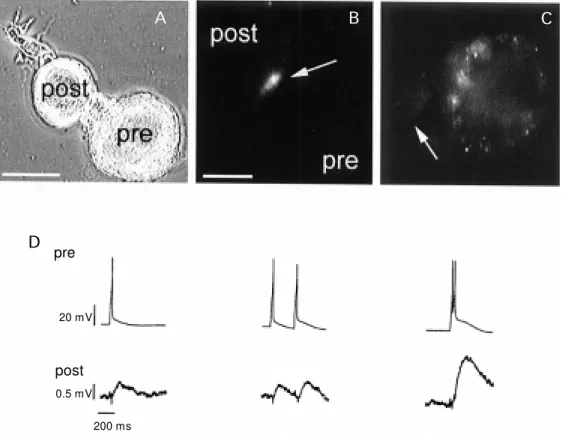
or in the distribution of calcium currents. The stump of a Retzius neuron forms presynaptic endings on a P neuron or on another Retzius neuron, with a rapid accu-mulation of newly produced release sites (Hernandez H, Trueta C and De Miguel FF, unpublished results). Figure 5 shows a pair of Retzius and P neurons making contact. Terminals shown in Figure 5B and C have been stained with the fluorescent dye FM1-43 which is taken up by synaptic vesicles upon endocytosis. Release sites shown in Figure 5B develop within a few hours right at the contact site of the presynaptic Retzius neuron. In single Retzius neurons, release sites are virtually absent from the stump of the cell. Upon synapse formation, electrical activity such as that in Figure 5D can be recorded. An action potential in a Retzius neuron produces a synaptic potential in the P neuron. In this case, the synaptic potentials have been artificially reversed by the injec-tion of chloride through the intracellular
elec-trode. The figure also shows that when pairs of impulses are produced close to each other, the synaptic potential shows facilitation, a typical property of chemical synapses (Trueta C and De Miguel FF, unpublished results).
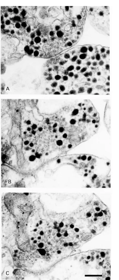
An ultrastructural study of Retzius neu-rons has shown clouds of dense-cored vesicles surrounded by clear-cored vesicles in the cytoplasm of the neuronal stump, near to but not in contact with the presynaptic membrane, suggesting that assemblies of presynaptic elements are a step preceding the formation of release sites. Figure 6 shows a typical presynaptic ending of a Retzius neuron with clear vesicles near the presyn-aptic membrane and surrounded by dense-cored vesicles (28). Some of the clear vesicles are in close apposition with the membrane. Figure 6B and C shows two serial sections of a bouton-like structure in which dense-cored vesicles are surrounding clear-cored vesicles. There is no evidence of contact of vesicles with the presynaptic membrane (Hernandez
A B C
D pre
20 mV
0.5 mV
post
200 ms Figure 5 - A, Pair of Retzius and
H, Morales M and De Miguel FF, unpub-lished results).
The establishment of chemical synapses also correlates with a redistribution of cal-cium currents (29). The large size of Retzius neurons and the possibility that they have access to very near sites and to the presynap-tic endings allowed us to use the loose-patch clamp technique (30) to record local macro-scopic currents from different membrane areas of Retzius neurons, thus providing a map of current density. The technique con-sists of having a large tip-diameter pipette (5 µm) and establishing a low resistance seal with the membrane. In this way, ion currents inside the pipette can be isolated and the pipette can be removed and introduced at another location without damaging the cell membrane. Since the same pipette is used over and over again to record from different membrane areas, the amplitude of the cur-rents at different locations provides a map of current density. Using cultured synapses be-tween pairs of Retzius neurons allowed us to compare currents between pre- and postsyn-aptic neurons of the same pair.
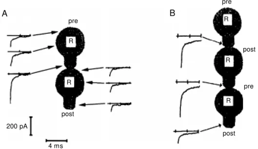
Formation of chemical synapses induces a marked reduction in postsynaptic calcium currents at the cell stump without affecting currents in other cell regions. In contrast, the characteristic amplitudes of calcium currents along presynaptic cells remain unaffected (29). Interestingly, as can be seen in Figure 7, synapse formation also has a retrograde effect on calcium currents. In triplets of cells in which one cell is purely presynaptic, the middle one is both pre- and postsynaptic and
Figure 6 - A, M orphology of a characteristic presynaptic ending in a Retzius neuron. Dense-cored vesicles sur-round clear vesicles near the cell membrane. B and C are tw o serial sections of a Retzius neuron show ing intracellular “ clouds” of clear vesicles surrounded by dense-cored vesicles but w ithout making contact w ith the cell membrane. Presumably this accumulation of vesicles could be a step preceding the formation of a presynaptic ending (Hernandez H, M orales M and De M iguel FF, unpublished results). Scale bar = 200 nm.
A
B
the third cell is purely postsynaptic. Calcium currents are reduced only in the purely post-synaptic cell, while the cell in the middle is unaffected even though it has also postsyn-aptic elements. In addition, neurite outgrowth of the postsynaptic neuron is inhibited by about 70% (31). Anterograde influences have been detected by Drapeau and his colleagues (4,5), affecting the postsynaptic responses to transmitter in P cells, in which contact with Retzius neurons induces the loss of an extrasynaptic receptor to serotonin.
Co nclusio ns and future dire ctio ns
From studies with identified leech neu-rons in culture, it has been possible to under-stand some of the events underlying neurite outgrowth and synapse formation. In addi-tion, the well-controlled conditions of the culture system have provided the chance to
analyze the mechanisms of transmitter re-lease in great detail (1). An interesting future direction will be to analyze the molecular basis for these signals. Approaches such as production of DNA libraries and injection of cloned RNA or DNA are currently being used by Blackshaw and her group (32) in studies on these large neurons. Another av-enue that has only begun to be explored is the relationship between neurite outgrowth, the molecules which support it and the speci-ficity of synapse formation. Ultimately, the mechanisms characterized with adult neu-rons in culture need to be tested with these same neurons in the developing CNS. Thus, the CNS of the leech offers the rare possibil-ity of characterizing signaling mechanisms during synapse formation and neurotrans-mission, and of determining the roles of these mechanisms during in vivo
develop-ment and regeneration.
Figure 7 - Anterograde and retro-grade effects of synapse forma-tion on calcium currents in Ret-zius (R) cells. The amplitude of calcium tail currents recorded on different spots of the neuronal membrane reflects the distribu-tion of voltage-sensitive chan-nels. A, Amplitudes of tail cur-rents in different locations of pre- and postsynaptic cells. The cell on the top is presynaptic and the cell on the bottom is post-synapt ic. The current at t he stump of the postsynaptic cell is markedly decreased relative to the same location on the presyn-aptic cell. Currents in presynap-tic cells and in the soma of post-synaptic cells remain unchanged w hen compared to single cells. B, A retrograde signal can also be detected in triplets of cells. Only currents at the stumps are show n, since the somatic cur-rents did not change. The cell in the middle (postsynaptic to the one on the top but presynaptic to the one on the bottom) does not decrease its calcium current at the stump, although it is post-synaptic to the cell on the top (adapted from Refs. 29,31).
A
B
pre
post
200 pA
4 ms
post pre
post
pre R
R
R R
R
Re fe re nce s
1. Nicholls JG (1987). The search for con-nections: Studies of regeneration in the nervous system of the leech. M agnes Lecture Series. Vol. II. Sinauer Associates Inc., Sunderland, M A.
2. Kater SB & M ills RL (1991). Regulation of grow th cone behavior by calcium. Journal
of Neuroscience,11: 891-899.
3. Dagan D & Levitan IB (1981). Isolated identified Aplysia neurons in cell culture. Journal of Neuroscience, 7: 736-740. 4. Fernández-de-M iguel F & Drapeau P
(1995). Synapse formation and function: Insights from identified leech neurons in
culture. Journal of Neurobiology, 27: 367-379.
5. Haydon PG & Drapeau P (1995). From contact to connection: Early events dur-ing synaptogenesis. Trends in Neurosci-ences,18: 196-201.
neurones isolated from the leech CNS make selective connections in culture. Na-ture,281: 67-69.
7. M asuda-Nakagaw a LM & Wiedemann C (1992). The role of matrix molecules in regeneration of leech CNS. Journal of Neurobiology, 23: 551-567.
8. Von Bernhardi R & M uller KJ (1995). Re-pair of the central nervous system: les-sons from lesions in leeches. Journal of Neurobiology, 27: 353-366.
9. Dietzel lD, Drapeau P & Nicholls JG (1986). Voltage dependence of 5-hydroxy-tryptamine release at a synapse betw een identified leech neurones in culture. Jour-nal of Physiology, 372: 191-205. 10. Liu Y & Nicholls JG (1989). Steps in the
development of chemical and electrical synapses by pairs of identified leech neu-rones in culture. Proceedings of the Royal Society of London. B, Biological Sciences, 236: 253-268.
11. Fuchs PA, Nicholls JG & Ready DF (1981). M embrane properties and selective con-nections of identified leech neurones in culture. Journal of Physiology, 316: 203-223.
12. Bixby JL & Harris WA (1991). M olecular mechanisms of axon grow th and guid-ance. Annual Review of Cell Biology,7: 117-159.
13. Sanes JR (1989). Extracellular matrix mol-ecules that influence neural development. Annual Review of Neuroscience, 12: 491-516.
14. Grumbacher-Reinert S (1989). Local influ-ence of substrate molecules in determin-ing distinctive grow th patterns of identi-fied neurons in culture. Proceedings of the National Academy of Sciences, USA, 86: 7270-7274.
15. Fernandez de M iguel F (1997). Outgrow th patterns and directed grow th of identified neurons induced by native substrates in
culture. Journal of Comparative Neurol-ogy, 380: 1-15.
16. De-M iguel FF & Vargas J (1999). Extra-cellular matrix induces a w ell-organized outgrow th pattern w ith neurite extension and retraction in cultured leech neurons. Journal of Comparative Neurology, 417: 387-398.
17. Gao WQ & M acagno ER (1987). Exten-sion and retraction of axonal projections by some developing neurons in the leech depend upon the existence of neighbor-ing homologues. II. The AP and AE cells. Journal of Neurobiology, 18: 295-313. 18. Ross WN, Arechiga H & Nicholls JG
(1987). Influence of substrate on the dis-tribution of calcium channels in identi-fied leech neurons in culture. Proceed-ings of the National Academy of Sci-ences, USA, 85: 4075-4078.
19. Grumbacher-Reinert S & Nicholls JG (1992). Influence of substrate on retrac-tion of neurites follow ing electrical activ-ity of leech Retzius cells in culture. Jour-nal of Experimental Biology, 167:1-14. 20. Neely M D (1993). Role of substrate and
calcium in neurite retraction of leech neu-rons follow ing depolarization. Journal of Neuroscience, 13: 1292-1301.
21. Holliday J & Spitzer NC (1990). Sponta-neous calcium influx and its roles in dif-ferentiation of spinal neurons in culture. Developmental Biology, 141: 13-23. 22. Gomez TM & Spitzer NC (1999). In vivo
regulation of axon extension and path-finding by grow th cone-calcium tran-sients. Nature, 397: 350-355.
23. Neely D & Gesemann M (1994). Disrup-tion of microfilaments in grow th cones follow ing depolarization and calcium in-flux. Journal of Neuroscience, 14: 7511-7520.
24. M elinek R & M uller KJ (1996). Action potential initiation site depends on
neu-ronal excitation. Journal of Neuroscience, 16: 2585-2591.
25. Nicholls JG & Purves D (1970). M onosyn-aptic chemical and electrical connections betw een sensory and motor cells in the central nervous system of the leech. Jour-nal of Physiology, 209: 647-667. 26. Vyklicky L & Nicholls JG (1988).
Specific-ity of connections formed by nociceptive cells of the leech in tissue culture. Journal of Experimental Biology, 134: 17-26. 27. De M iguel FF & Vargas J (1997). Different
determinants on grow th and synapse for-mation in cultured neurons. NeuroReport, 8: 761-765.
28. Kuffler DP, Nicholls JG & Drapeau P (1987). Transm it t er localizat ion and vesicle turnover at a serotoninergic syn-apse betw een identified leech neurons in culture. Journal of Comparative Neurol-ogy, 256: 516-526.
29. Fernández-de-M iguel F, Cooper R & Adams WB (1992). Synaptogenesis and changes in calcium current distribution in cultured leech cells. Proceedings of the Royal Societyof London. B, Biological Sci-ences,247: 215-221.
30. Stühmer W, Roberts WS & Almers W (1983). The loose pat ch clam p. In: Sakmann B & Neher E (Editors), Single Channel Recording. Plenum Press, New York, London, 123-132.
31. Cooper RL, Fernández-de-M iguel F, Adams WB & Nicholls JG (1992). Antero-grade and retroAntero-grade effects of synapse formation on calcium currents and neurite outgrow th in cultured leech neurons. Pro-ceedings of the Royal Society of London. B, Biological Sciences,249:217-222. 32. Korneev S, Blackshaw S & Davies JA