ISSN 0100-879X
BIOMEDICAL SCIENCES
AND
CLINICAL INVESTIGATION
www.bjournal.com.br
www.bjournal.com.br
Volume 44 (11) 1070-1193 November 2011
Institutional Sponsors
The Brazilian Journal of Medical and Biological Research is partially financed by
Faculdade de Medicina de Ribeirão Preto Campus
Ribeirão Preto
Ex plor e H igh - Pe r for m a n ce M S Or bit r a p Te ch n ology I n Pr ot e om ics & M e t a bolom ics
analit icaw eb.com .br S C I E N T I F I C
Braz J Med Biol Res, November 2011, Volume 44(11) 1118-1124
doi: 10.1590/S0100-879X2011007500128
Testosterone therapy delays cardiomyocyte aging via an androgen
receptor-independent pathway
Testosterone therapy delays
cardiomyocyte aging via an androgen
receptor-independent pathway
L. Zhang
1, S.Z. Wu
1, Y.J. Ruan
2, L. Hong
1, X.W. Xing
3and W.Y. Lai
41Department of Cardiology, Nangfang Hospital, Southern Medical University, Guangzhou, Guangdong, China 2Department of Cardiology, Guangzhou General Hospital, Guangzhou Military Area Command of Chinese PLA,
Guangzhou, Guangdong, China 3Laboratory of Cardiovascular Diseases, The First Affiliated Hospital of Guangzhou Medical College,
Guangzhou, Guangdong, China 4Laboratory of Cardiovascular Diseases, Nanfang Hospital, Southern Medical University,
Guangzhou, Guangdong, China
Abstract
The testicular feminized (Tfm) mouse carries a nonfunctional androgen receptor (AR) and reduced circulating testosterone levels. We used Tfm and castrated mice to determine whether testosterone modulates markers of aging in cardiomyocytes via its classic AR-dependent pathway or conversion to estradiol. Male littermates and Tfm mice were divided into 6 experimental groups. Castrated littermates (group 1) and sham-operated Tfm mice (group 2, N = 8 each) received testosterone. Sham-operated Tfm mice received testosterone in combination with the aromatase inhibitor anastrazole (group 3, N = 7). Castrated littermates (group 4) and sham-operated untreated Tfm mice (group 5) were used as controls (N = 8 and 7, respectively). An additional control group (group 6) consisted of age-matched non-castrated littermates (N = 8). Cardiomyocytes were isolated from the left ventricle, telomere length was measured by quantitative PCR and expression of p16INK4α, retinoblastoma (Rb) and p53 proteins was detected by Western blot 3 months after treatment. Compared with group 6, telomere length was short (P < 0.01) and expression of p16INK4α, Rb and p53 proteins was significantly (P < 0.05) up-regulated in groups 4 and 5. These changes were improved to nearly normal levels in groups 1 and 2 (telomere length = 0.78 ± 0.05 and 0.80 ± 0.08; p16INK4α = 0.13 ± 0.03 and 0.15 ± 0.04; Rb = 0.45 ± 0.05 and 0.39 ± 0.06; p53 = 0.16 ± 0.04 and 0.13 ± 0.03), but did not differ between these two groups. These improvements were partly inhibited in group 3 compared with group 2 (telomere length = 0.65 ± 0.08
vs 0.80 ± 0.08, P = 0.021; p16INK4α = 0.28 ± 0.05 vs 0.15 ± 0.04, P = 0.047; Rb = 0.60 ± 0.06 vs 0.39 ± 0.06, P < 0.01; p53 = 0.34 ± 0.06 vs 0.13 ± 0.03, P = 0.004). In conclusion, testosterone deficiency contributes to cardiomyocyte aging. Physiological
testosterone can delay cardiomyocyte aging via an AR-independent pathway and in part by conversion to estradiol.
Key words: Aging; Testosterone; Cardiomyocyte; Androgen receptor; Estradiol; Tfm mice
Introduction
Correspondence: S.Z. Wu, Department of Cardiology, Nanfang Hospital, Southern Medical University, Guangzhou, Guangdong, 510515, China. Fax: +86-2061-642-151. E-mail: wusaizhu@126.com
Received February 8, 2011. Accepted September 16, 2011. Available online September 30, 2011. Published November 14, 2011.
Aging per se is a risk factor for reduced cardiac function
and heart disease, even when adjusted for conventional cardiovascular risk factors. With aging, cardiac function is organically and cellularly impaired. Cardiomyocytes, the major components of the contractile apparatus, undergo a number of physiological and morphological changes with age, and these changes are thought to contribute to reduced cardiac function and heart disease (1). It is likely that modulation of cardiomyocyte aging changes the threshold for the manifestation of signs and symptoms of
heart disease.
Testosterone is the major circulating androgen and its level declines with age in men (2). It is known that testos-terone replacement therapy restores many of the adverse
pathophysiological events, which occur in androgen defi -ciency (3). Several studies have suggested a relationship between testosterone and brain aging with an effect on cognition (4-6). Also, reduced androgen levels have been associated with aging-related cardiovascular diseases and
-Testosterone therapy and cardiomyocyte aging 1119
cial effects on cardiovascular function (7). Furthermore, in experimental rats with low testosterone levels, physiological testosterone therapy can improve reduced cardiac functional capacity, probably by inhibition of tumor necrosis factor alpha
(TNF-α) (8), which is a pro-inflammatory cytokine closely
linked to the aging process (1). These data have led to the hypothesis that testosterone has a positive impact on the aging process of the cardiovascular system.
Testosterone exerts a variety of anabolic and andro-genic effects on many organs and most of these actions are mediated by the nuclear androgen receptor (AR) (9). The AR gene is also expressed in mammalian and primate cardiomyocytes, suggesting that androgens may play a role in the heart (10,11). Golden et al. (11) reported that gonadectomy slowed the contractile velocity by altering myosin heavy chain composition in isolated cardiomyocytes of adult male rats. Moreover, in castrated rats testosterone supplementation reversed the decline in myosin heavy chain content to values seen in sham-operated controls.
The same investigatorsreported that testosterone regulated
the functional expression of L-type calcium channel mRNA levels in isolated rat cardiomyocytes and this effect was
mediated bythe AR (12). In addition to the AR pathway,
the beneficial effect of long-term testosterone on cardio
-myocytes may have been due tothe conversion to estradiol
by aromatase activity (13,14). Although testosterone may
be of pathophysiological importance tothe heart, there are
no published studies, which demonstrate the role of testos-terone and AR as modulators of aging in cardiomyocytes. The testicular feminized (Tfm) mouse exhibits an X-linked, single base-pair deletion in the gene encoding the AR. This deletion results in premature termination of AR protein synthesis, with the occurrence of a nonfunctional AR (15,16). In addition, circulating levels of testosterone are reduced in the Tfm mouse, reportedly being 10-fold lower compared to male littermates (16). Cellular aging is characterized by an irreversible cell cycle arrest and loss of
specialized functions. Telomere length, p16INK4α and p53
expression are plotted as a function of age (17). Cellular
aging is also modulated by Rb. p16INK4α inhibits
cyclin-de-pendent kinases and maintains Rb in its unphosphorylated, growth-suppressive state (17). Therefore, in the present
study, we investigated whether testosterone deficiency
and/or the absence of a functional AR are associated with the changes of telomere length and of the expression of the above-mentioned genes in murine cardiomyocytes. Furthermore, we determined whether physiological tes-tosterone therapy modulates cardiomyocyte aging via the AR-dependent pathway or conversion to estradiol in Tfm and castrated male mice.
Material and Methods
Experimental animals and design
All experimental procedures and protocols used in
this investigation were reviewed and approved by the Ethics Committee of Nanfang Hospital, Southern Medical University. Male littermates and Tfm mice were bred from mice purchased from the Model Animal Research Center of
Nanjing University (female = AW-J/AW-J EdaTa-6J +/+ArTfm;
male = AW-J/AW-J EdaTa-6J +/Y). The gender of Tfm mice
was identified by PCR. Mice were kept under standard
temperature (20-26°C), humidity (40-70%), and illumination (12-h light/12-h dark cycle, lights on at 8:00 h).
At 8 weeks of age, male littermates and Tfm mice un-derwent surgical castration or sham operation, respectively. Four weeks after surgery, castrated mice were treated with (N = 8) or not (N = 8) with testosterone propionate (Guangzhou Mingxing Pharmaceutical Company, China), while sham-operated Tfm mice were also treated (N = 8) or not (N = 7) with testosterone propionate. Another 7 sham-operated Tfm mice were treated with testosterone
propi-onate in combination with 10 mg·kg-1·day-1 of the aromatase
inhibitor anastrazole (AstraZeneca, UK) in drinking water (18). Testosterone was injected intramuscularly at 3 mg/kg, diluted in sesame oil, once during each 72-h period for 3 months. Age-matched 24-week-old nonsurgical littermates were used as controls (N = 8). Before these manipulations, pharmacokinetic determination of physiological testosterone was performed.
Pharmacokinetic determination of physiological testosterone
To establish and maintain a dosing regimen of
tes-tosterone at physiological concentrations, we first had to
determine the appropriate volume and frequency of admin-istration of 25 mg/mL testosterone propionate. At 8 weeks of age, male littermate mice (N = 36) underwent surgical castration and were allowed to recover in individual cages for 4 weeks. Next, 12-week-old mice received a single 3 mg/kg intramuscular injection of testosterone propionate (the human replacement dose of testosterone propionate is 0.35 mg/kg administered 2-3 times per week according
to manufacturer instructions. Mice were sacrificed at 30
min, and at 1, 2, 4, 8, 16, 24, 32, 48, and 72 h after injec-tion, and testosterone concentrations were measured in duplicate (ELISA kits, R&D Systems, Inc., USA). To ensure reproducibility, 18 additional castrated littermates received a second-cycle injection (double administration) beginning
72 h after the first and were sacrificed at 1, 2, 4, 8, 24,
and 72 h after the second injection. At the same time, in order to determine normal testosterone concentrations,
9 non-castrated male mice were sacrificed at 0:00, 8:00,
and 16:00 h.
Isolation of cardiomyocytes
Left ventricular myocytes were enzymatically isolated
from 39 male mice as described previously (17,19). Briefly,
were supplements of modified commercial minimal essential
medium (MEM) Eagle-Joklik. HEPES-MEM contained 117
mM NaCl, 5.7 mM KCl, 4.4 mM NaHCO3, 1.5 mM KH2PO4,
17 mM MgCl2, 21.1 mM HEPES, and 11.7 mM glucose
with amino acids and vitamins, 2 mM L-glutamine, and 21 mU/mL insulin; pH was adjusted to 7.2 with NaOH. The washing solution was HEPES-MEM with the addition of 0.5 mM EGTA. The resuspension medium was HEPES-MEM supplemented with 0.5% bovine serum albumin (BSA) and
0.3 mM CaCl2. The cell isolation procedure consisted of 3
main steps: 1) for low-calcium perfusion, blood washout in the presence of EGTA was performed for about 10 min, and 0.05% collagenase (selected type I, Worthington Biochemi-cal Corp., USA) perfusion of the myocardium was carried
out at 37°C with HEPES-MEM gassed with 85% O2, 15%
N2. 2) After the heart was removed from the cannula, the
left ventricle was cut into small pieces and subsequently shaken in resuspension medium at 37°C. Supernatant cell suspensions were washed and resuspended in resuspen-sion medium. 3) Intact cells were concentrated by centrifu-gation. This procedure was repeated 4-5 times.
Quantitative PCR assay
Quantitative PCR assay was used to measure
telom-ere (T) signals and single-copy gene signals (S, β-globin
gene) in experimental DNA samples extracted from isolated cardiomyocytes. The relative T/S ratios indicated relative telomere length (20,21).
The final reaction system in the quantitative PCR con
-sisted of a final volume of 20 µL per reaction, including 10 µL 2X AllinOne™ quantitative-PCR Mix (SYBR Green I, GeneCopoeia, USA), 2 µL T primer pair telg and telc (final concentrations of 900 nM each), or 2 µL S primer pair hbgu and hbgd (final concentrations 200 nM each), and 2 µL of each experimental DNA sample (20 ng DNA). Five
concentrations of a reference DNA sample (genomic DNA extracted from any individual mouse DNA sample) ranging
from 0.16 to 100 ng/µL were prepared by serial dilution to
provide data for the generation of the standard curves used for relative quantitation.
The thermal cycling profile consisted of 10 min at 95°C,
followed by 2 cycles of 10 s at 95°C, 20 s at 49°C, and 15 s at 72°C, and 40 cycles of 10 s at 94°C, 20 s at 60°C, and 15 s at 72°C. After thermal cycling, the Bio-Rad (USA) iQ5 2.0 Standard Edition Optical System Software was used to generate two standard curves and to read cycle threshold (Ct) values of T and S signals.
T and S primers were as follows: telg, ACACTAAGGTTTG GGTTTGGGTTTGGGTTTGGGTTAGTGT and telc, TGTT AGGTATCCCTATCCCTATCCCTATCCCTATCCCTAACA. hbgu, CTGCCCTGGCTCACAAGTAC, and hbgd, AGATG CCCAAAGGTCTTCATC.
Western blotting
Freshly isolated cardiomyocytes were lysed in 250 μL
lysis buffer (50 mM Tris-HCl, pH 7.5, 5 mM EDTA, 250 mM NaCl, and 0.1% Triton X-100) containing the following
protease inhibitors: 2 mM PMSF, 1 µg/mL aprotinin, 5 mM
DTT, and 1 mM Na3VO4. Myocyte lysates were centrifuged
at 6700 g for 10 min (19). Forty micrograms protein was
separated by 10-15% SDS-PAGE and electroblotted onto
polyvinylidene difluoride membranes (Bio-Rad). After block -ing with 5% BSA for 1 h, the membranes were incubated overnight with primary antibody dilution buffer at 4°C.
Anti-p16INK4α (F-12, Santa Cruz Biotechnology, Inc., USA),
anti-p53 (FL-393, Santa Cruz), and anti-unphosphorylated Rb (Wuhan Boster Co., China) were used. HRP-conjugated goat anti-mouse IgG and goat anti-rabbit (Bio-Rad) served
as the secondary antibodies. The membranes were briefly
incubated with ECL detection reagent (Thermo, USA) to
visualize the proteins and exposed to X-ray film. β-actin
(Wuhan Boster Co.) was used as an internal control for all
the Western blotting procedures. The Western blottingdata
presented in this study were from at least 3 independent experiments.
Measurement of testosterone concentration
At the time of measurementsand the end of the study, all
animals were anesthetized and blood was collected without
anticoagulantfrom the retroorbital venous plexus. Samples
were centrifuged at 1400 g for 10 min and serum was
col-lected and stored at -20°C until assay. Total testosterone levels were measured in duplicate using commercially avail-able ELISA kits (R&D Systems, Inc.). Interassay variation was 2.9-4.0% and intra-assay variation was 5.6-6.8%.
Statistical analysis
SPSS Version 13.0 (Statistical Software for Social Sciences, USA) was used for statistical analysis. Data are reported as means ± SD and were analyzed by one-way ANOVA, with equal variances assumed with the least
significant difference (LSD) test and equal variances not
assumed with Dunnett’s T3 multiple comparisons. The level
of significance was set at P < 0.05.
Results
Determination of a doseregimen to reach
physiological testosterone levels in castrated mice
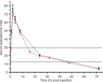
The normal range of testosterone concentrations was 12.6 to 28.7 nM. After castration, baseline levels of serum testosterone were about 6% of normal levels. The levels
rose significantly from baseline after intramuscular injection
of 3 mg/kg testosterone at 30 min, reached a peak at 2 h, and remained within the normal range between 14 and 50 h (Figure 1). The mean area under the curve (AUC) for
the first 72-h period was 20.6 nM and the mean AUC for
Testosterone therapy and cardiomyocyte aging 1121
and double administrations. Furthermore, after single and double administrations, the mean AUC were all within the normal range. These results agree with those of Nettle-ship et al. (18) and indicate a good reproducibility of the
pharmacokinetic profile and that a sufficient physiological
replacement therapy would be3 mg·kg-1·72 h-1
intramus-cular testosterone propionate in castrated male mice.
Telomere length
The Ct values of T and S were calculated with their independent standard curves. The relative T/S ratios of castrated and sham-operated Tfm mice did not differ
sig-nificantly (Figure 2, columns 2 and 4, respectively). These, however, were significantly lower than those observed in
control mice (column 1, both P < 0.01). After testosterone
therapy, an increase in the relative T/S ratios was detectedin
castrated (column 3) and sham-operated Tfm mice (column
5) compared with their untreated controls(columns 2 and
4, both P < 0.01). Moreover, the values for testosterone-treated castrated and sham-operated Tfm mice did not
differ significantly (P > 0.05). The relative T/S ratios were significantly lower in sham-operated Tfm mice receiving
testosterone in combination with anastrazole (column 6) compared with those receiving testosterone alone (column 5, P = 0.021). However, the values were still higher than those of untreated Tfm mice (column 4, P = 0.044).
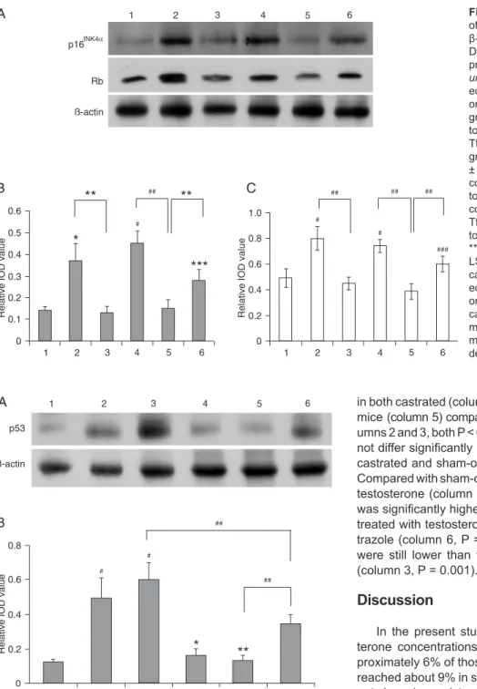
Expression of p16INK4α and Rb proteins
As shown in Figure 3, expression of p16INK4α and Rb
proteins was low in control mice. The expression levels,
however, were significantly higher in both castrated and
sham-operated Tfm mice than in control mice (p16INK4α
= both P < 0.05. Rb = both P < 0.01). There were no
sig-nificant differences between the two experimental groups (both P > 0.05). After testosterone treatment, the levels
of p16INK4α and Rb proteins were down-regulated in both
castrated and sham-operated Tfm mice compared with
untreated mice (p16INK4α = both P < 0.05. Rb = both P <
0.01). Moreover, the values did not differ significantly be -tween testosterone-treated castrated and sham-operated
Tfm mice (both P > 0.05). Expression of p16INK4α and Rb
proteins was significantly higher in sham-operated Tfm
mice receiving testosterone in combination with anastra-zole compared with those receiving testosterone alone (p16INK4α, P = 0.047; Rb, P < 0.001). However, the levels
were still lower than those of untreated Tfm mice (p16INK4α,
P = 0.045; Rb, P = 0.008).
Expression of p53 protein
Expression of p53 protein did not differ significantly (P > 0.05) between castrated (Figure 4, column 2) and
sham-operated Tfm mice (column 3). The levels, however, were
significantly higher than those observed in control mice
(column 1, both P < 0.01). After testosterone treatment, a down-regulation of p53 protein expression was detected
Figure 1. Pharmacokinetic curves of the single and double 72-h period injection of testosterone propionate in castrated littermates. Castrated mice received a single 3 mg/kg intramuscular injection of 25 mg/mL testosterone (N = 36, red solid line). An additional 3 mg/kg intramuscular injection of 25 mg/mL testosterone was
administered at 72 h after the first injection (N = 18, black broken line). Mice were sacrificed at the times indicatedafter injection and serumtestosterone concentrations were measured. There
was no statistically significant difference in peak time, mean area
under the curve, or testosterone concentrations between single and double administrations. The area between the dashed paral-lel lines indicatesthe physiological range of testosterone (12.6 to 28.7 nM).
Figure 2. Quantitative PCR assay of relative telomere length in cardiomyocytes expressed as the relative T/S ratios. Columns:
1, control group; 2, castrated group; 3, castrated + testoster-one group; 4, sham-operated Tfm group; 5, sham-operated Tfm + testosterone group; 6, sham-operated Tfm + testosterone + anastrazole group. Data are reported as means ± SD. #P < 0.01 compared to the control group; ##P < 0.05 compared to the sham-operated Tfm group; *P < 0.01; **P < 0.05 (ANOVA + Dunnett
T3 test). There were no significant differences between castrated
in both castrated (column 4) and sham-operated Tfm mice (column 5) compared with untreated mice (col-umns 2 and 3, both P < 0.01). Moreover, the values did
not differ significantly between testosterone-treated castrated and sham-operated Tfm mice (P > 0.05).
Compared with sham-operated Tfm mice treated with testosterone (column 5), expression of p53 protein
was significantly higher in sham-operated Tfm mice
treated with testosterone in combination with anas-trazole (column 6, P = 0.004). However, the levels were still lower than those of untreated Tfm mice (column 3, P = 0.001).
Discussion
In the present study, we observed that testos-terone concentrations of castrated mice were ap-proximately 6% of those of control mice. The values reached about 9% in sham-operated Tfm mice (data not shown), consistent with the study of Jones et al. (16). Left ventricular myocytes from castrated and sham-operated Tfm mice had short telomere length
and increased expression of p16INK4α, Rb and p53
proteins compared with control. Moreover, these
parameters did not differ significantly between cas -trated and sham-operated Tfm mice. These
observa-tions provide evidence that testosterone deficiency
contributes to cardiomyocyte aging. Furthermore, we observed amelioration of cardiomyocyte aging with long-term physiological testosterone therapy
Figure 3. Western blot analysis of p16INK4α and Rb proteins with
β-actin as an internal control (A). Densitometric analysis of p16INK4α protein (B) and Rb protein (C). Col-umns: 1, control group; 2, castrat-ed group; 3, castrated + testoster-one group; 4, sham-operated Tfm group; 5, sham-operated Tfm + tes-tosterone group; 6, sham-operated Tfm + testosterone + anastrazole group. Data are reported as means ± SD. *P < 0.05 compared to the control group; #P < 0.01 compared to the control group; ***P < 0.05 compared to the sham-operated Tfm group; ###P < 0.01 compared to the sham-operated Tfm group; **P < 0.05; ##P < 0.01 (ANOVA +
LSD test). There were no signifi -cant differences between castrat-ed and sham-operatcastrat-ed Tfm mice or between testosterone-treated castrated and sham-operated Tfm mice. Tfm = testicular feminized mouse; IOD = integrated optical density.
Figure 4. Western blot analysis of p53 protein with β-actin as an in -ternal control (A). Densitometric analysis of p53 protein (B). Columns:
1, control group; 2, castrated group; 3, sham-operated Tfm group; 4, castrated + testosterone group; 5, sham-operated Tfm + testosterone group; 6, sham-operated Tfm + testosterone + anastrazole group. Data are reported as means ± SD. #P < 0.01 compared to control group; *P < 0.01 compared to castrated group; **P < 0.01 compared to sham-op-erated Tfm group; ##P < 0.01 (ANOVA + LSD test). There were no
sig-nificant differences between castrated and sham-operated Tfm mice or
Testosterone therapy and cardiomyocyte aging 1123
as indicated by increased telomere length and
down-regulation of expression of p16INK4α, Rb and p53 proteins.
Surprisingly, their levels were statistically similar to those of control mice.
Senescent cardiomyocytes are characterized by an ir-reversible cell cycle arrest, which is mediated and indicated
by an up-regulation of p16INK4α and p53, and show a
re-duction in telomere length (1). Kajstura et al. (22) reported that telomeric shortening in myocytes and the fraction of p16INK4α-positive myocytes increased with age. Another study demonstrated a 39% reduction in mean telomere length in human aged hearts. Cardiomyocytes with
telom-eres equal to or shorter than 2.5 kbp were p16INK4α-positive
(23). Torella et al. (17) found that the expression of p16INK4α
and p53 increased with age in cardiomyocytes of wide-type (WT) mice. Hypophosphorylated Rb was predominant in
aging WT cardiomyocytes, reflecting the up-regulation of
p16INK4α. Cells with shorter telomeres were p16INK4α- and p53-positive. Compared to young cells (4 months), telom-ere length was reduced by approximately 35% in old WT cardiomyocytes (20-22 months). Similarly, the mRNA levels
of p16INK4α and acetylated p53 were increased in aged
rat cardiomyocytes in the study by Maejima et al. (24). In other aging animal models, telomere shortening coincides with increased expression of p53 in isolated cardiomyocyte nuclei (25).
As mentioned earlier, we observed a protective role of testosterone therapy against the development of cardio-myocyte aging. Similarly, administration of testosterone has been shown to improve brain aging in older male C57BL/6 mice and in young castrated mice (6). Testosterone also protects skeletal muscle cells and neurons from cell death (26,27). Lu et al. (28) showed that androgen down-regulated the expression of the cyclin-dependent kinase inhibitor p16INK4α gene in LNCaP-FGC cells. In a previous study conducted by Gregory et al. (29), the hypophosphorylated form of Rb was down-regulated by testosterone propionate treatment in castrated mice. Zhang et al. (30) found that expression of p53 mRNA was decreased in rat prostate glands after testosterone replacement therapy. These data are consistent with the observations of the present study. The amelioration of cardiomyocyte aging did not
differ significantly between castrated and sham-operated
Tfm mice after physiological testosterone therapy. Due to the following observations, we conclude that the positive
effects of testosterone on cardiomyocyte aging occur via an AR-independent pathway: a) Tfm mice carry nonfunctional AR and still respond to testosterone, and b) castrated mice with functional AR show signs of cardiomyocyte aging that can be cured with testosterone.
In fact, local conversion of testosterone to estradiol via aromatase has been proposed to mediate the potentially
beneficial effects of testosterone. After conversion, estra -diol elicits direct cellular effects by activation of estrogen
receptor α (ERα) and ERβ (31,32). In the present study,
increased telomere length and down-regulation of
expres-sion of p16INK4α, Rb and p53 proteins in sham-operated
Tfm mice receiving testosterone therapy were significantly
inhibited by co-treatment with the aromatase inhibitor. The degree of cardiomyocyte aging in anastrazole-treated Tfm mice, however, was still lower than in sham-operated Tfm mice. These data demonstrate that physiological testos-terone levels ameliorate cardiomyocyte aging mediated in part by aromatization to estradiol in cardiac myocytes. Michels et al. (13) reported that the long-term effect of testosterone on single T-type calcium channel was me-diated in part via the estrogen pathway in neonatal rat cardiomyocytes. Co-incubation of rat cardiac myocytes
with testosterone and the specific aromatase inhibitor
showed an inhibition of estrogen-responsive element activation but this was not observed in cells incubated with testosterone alone, thereby implicating conversion to estradiol as the mechanism of action of testosterone in cardiomyocytes (14).
Our results support the view that testosterone deficiency
contributes to cardiomyocyte aging in Tfm and castrated
male mice. Wedemonstrated thebeneficial effect of physi
-ological levels of testosterone on cardiomyocyte aging,
an action that is independent of the classical AR and is mediated in part by conversion to estradiol. Given the increasing incidence and prevalence of heart disease with aging, it will be useful to study the effect and mechanism of action of testosterone on cardiomyocyte aging. However, further investigations are needed before these results can
beextrapolated from mice to humans.
Acknowledgments
Research supported by the National Key Fundamental Research and Development Project (#2007CB507404).
References
1. Bernhard D, Laufer G. The aging cardiomyocyte: a mini-review. Gerontology 2008; 54: 24-31.
2. Araujo AB, O’Donnell AB, Brambilla DJ, Simpson WB, Long-cope C, Matsumoto AM, et al. Prevalence and incidence
of androgen deficiency in middle-aged and older men:
estimates from the Massachusetts Male Aging Study. J Clin
Endocrinol Metab 2004; 89: 5920-5926.
3. Bain J. Testosterone and the aging male: to treat or not to treat? Maturitas 2010; 66: 16-22.
2005; 64: 866-871.
5. Kritzer MF, McLaughlin PJ, Smirlis T, Robinson JK. Gonad-ectomy impairs T-maze acquisition in adult male rats. Horm
Behav 2001; 39: 167-174.
6. Frye CA, Edinger K, Sumida K. Androgen administration to aged male mice increases anti-anxiety behavior and en-hances cognitive performance. Neuropsychopharmacology
2008; 33: 1049-1061.
7. Gooren LJ. Androgens and male aging: Current evidence of
safety and efficacy. Asian J Androl 2010; 12: 136-151. 8. Li ZB, Wang J, Wang JX, Chen XM, Jiang SS. [Testosterone
therapy improves cardiac function of male rats with right heart failure]. Zhonghua Nan Ke Xue 2009; 15: 994-1000.
9. Wilbert DM, Griffin JE, Wilson JD. Characterization of the
cytosol androgen receptor of the human prostate. J Clin
Endocrinol Metab 1983; 56: 113-120.
10. Marsh JD, Lehmann MH, Ritchie RH, Gwathmey JK, Green GE, Schiebinger RJ. Androgen receptors mediate hypertro-phy in cardiac myocytes. Circulation 1998; 98: 256-261. 11. Golden KL, Marsh JD, Jiang Y, Moulden J. Gonadectomy
alters myosin heavy chain composition in isolated cardiac myocytes. Endocrine 2004; 24: 137-140.
12. Golden KL, Marsh JD, Jiang Y. Testosterone regulates mRNA levels of calcium regulatory proteins in cardiac myo-cytes. Horm Metab Res 2004; 36: 197-202.
13. Michels G, Er F, Eicks M, Herzig S, Hoppe UC. Long-term and immediate effect of testosterone on single T-type cal-cium channel in neonatal rat cardiomyocytes. Endocrinology
2006; 147: 5160-5169.
14. Grohe C, Kahlert S, Lobbert K, Vetter H. Expression of oestrogen receptor alpha and beta in rat heart: role of local oestrogen synthesis. J Endocrinol 1998; 156: R1-R7. 15. He WW, Kumar MV, Tindall DJ. A frame-shift mutation in the
androgen receptor gene causes complete androgen insen-sitivity in the testicular-feminized mouse. Nucleic Acids Res
1991; 19: 2373-2378.
16. Jones RD, Pugh PJ, Hall J, Channer KS, Jones TH. Altered circulating hormone levels, endothelial function and vascular reactivity in the testicular feminised mouse. Eur J Endocrinol
2003; 148: 111-120.
17. Torella D, Rota M, Nurzynska D, Musso E, Monsen A, Shi-raishi I, et al. Cardiac stem cell and myocyte aging, heart failure, and insulin-like growth factor-1 overexpression. Circ Res 2004; 94: 514-524.
18. Nettleship JE, Jones TH, Channer KS, Jones RD. Physiologi-cal testosterone replacement therapy attenuates fatty streak formation and improves high-density lipoprotein cholesterol in the Tfm mouse: an effect that is independent of the classic androgen receptor. Circulation 2007; 116: 2427-2434. 19. Leri A, Liu Y, Wang X, Kajstura J, Malhotra A, Meggs LG, et
al. Overexpression of insulin-like growth factor-1 attenuates
the myocyte renin-angiotensin system in transgenic mice.
Circ Res 1999; 84: 752-762.
20. Cawthon RM. Telomere length measurement by a novel monochrome multiplex quantitative PCR method. Nucleic
Acids Res 2009; 37: e21.
21. Fehrer C, Voglauer R, Wieser M, Pfister G, Brunauer R, Cio -ca D, et al. Techniques in gerontology: cell lines as standards for telomere length and telomerase activity assessment. Exp
Gerontol 2006; 41: 648-651.
22. Kajstura J, Pertoldi B, Leri A, Beltrami CA, Deptala A, Dar-zynkiewicz Z, et al. Telomere shortening is an in vivo marker of myocyte replication and aging. Am J Pathol 2000; 156: 813-819.
23. Chimenti C, Kajstura J, Torella D, Urbanek K, Heleniak H, Colussi C, et al. Senescence and death of primitive cells and myocytes lead to premature cardiac aging and heart failure.
Circ Res 2003; 93: 604-613.
24. Maejima Y, Adachi S, Ito H, Hirao K, Isobe M. Induction of premature senescence in cardiomyocytes by doxorubicin as a novel mechanism of myocardial damage. Aging Cell 2008; 7: 125-136.
25. Leri A, Franco S, Zacheo A, Barlucchi L, Chimenti S, Limana F, et al. Ablation of telomerase and telomere loss leads to cardiac dilatation and heart failure associated with p53 up-regulation. EMBO J 2003; 22: 131-139.
26. Estrada M, Espinosa A, Muller M, Jaimovich E. Testosterone stimulates intracellular calcium release and mitogen-activated protein kinases via a G protein-coupled receptor in skeletal muscle cells. Endocrinology 2003; 144: 3586-3597. 27. Nguyen TV, Yao M, Pike CJ. Androgens activate
mitogen-activated protein kinase signaling: role in neuroprotection. J
Neurochem 2005; 94: 1639-1651.
28. Lu S, Tsai SY, Tsai MJ. Regulation of androgen-dependent prostatic cancer cell growth: androgen regulation of CDK2, CDK4, and CKI p16 genes. Cancer Res 1997; 57: 4511-4516.
29. Gregory CW, Johnson RT Jr, Presnell SC, Mohler JL, French FS. Androgen receptor regulation of G1 cyclin and cyclin-dependent kinase function in the CWR22 human prostate cancer xenograft. J Androl 2001; 22: 537-548.
30. Zhang X, Colombel M, Raffo A, Buttyan R. Enhanced ex-pression of p53 mRNA and protein in the regressing rat ventral prostate gland. Biochem Biophys Res Commun
1994; 198: 1189-1194.
31. Liu PY, Death AK, Handelsman DJ. Androgens and cardio-vascular disease. Endocr Rev 2003; 24: 313-340.