Early and Delayed Effect of Functional
Endoscopic Sinus Surgery on Intraocular
Pressure
Mohammad Waheed El-Anwar
1Mohammad Abdelhady
1Hazem Saeed Amer
1Manar A. Ghali
21Department of Otorhinolaryngology - Head and Neck Surgery,
Faculty of Medicine, Zagazig University, Zagazig, Egypt
2Department of Ophthalmology, School of Medicine, Zagazig
University, Zagazig, Egypt
Int Arch Otorhinolaryngol 2016;20:359–363.
Address for correspondence Hazem Saeed Amer, MD, Department of Otorhinolaryngology, Head and Neck Surgery, School of Medicine, Zagazig University, Zagazig 0020552322665, Egypt
(e-mail: [email protected]).
Introduction
Chronic rhinosinusitis (CRS) is one of the most common diseases in the otolaryngologicfield. Functional endoscopic sinus surgery (FESS) represents the preferred surgical treat-ment for CRS.1
FESS was initially driven by Messerklinger’s work in the late 1970s.2It was then popularized and standardized in the beginning of the 1980s, particularly by Kennedy et al3and
Stammberger4with their studies regarding the philosophy of clearing the natural ostium of the diseased sinus.
Due to the close anatomical relationship between the paranasal sinuses and the orbit, involvement or injury of the orbit from procedures the paranasal sinuses may occur.5 The ocular complications after FESS include: nasolacrimal duct injury, extraocular muscle injury; periorbital, intraorbital, or retrobulbar hematoma; and even optic nerve injury. The risk of injury is correlated to the anatomical variations, the history of Keywords
►
sinusitis
►
nose
►
endoscopy
Abstract
Introduction
Due to the close anatomical relationship between the paranasal sinuses
and the orbit, involvement or injury of the orbit from paranasal sinuses procedures may
occur.
Objectives
We aimed to study the early and delayed effect of endoscopic sinus surgery
on intraocular pressure (IOP).
Methods
We included in the study 38 patients with chronic rhinosinusitis (CRS),
undergoing FESS. We performed FESS with the standard anterior to posterior approach.
We measured IOP at the same time one day before surgery as well as day 1 and 6 weeks
after surgery.
Results
One day after surgery, mean IOP in the right eye was 14.176
1.91 mm Hg
and in the left eye was 13.79
2.42 mm Hg with statistically non-signi
fi
cant difference
from preoperative values. Six weeks postoperative, the mean IOP in the right eye was
15.14
2.28 mm Hg. The difference between the mean preoperative and
postopera-tive IOP values was found to be statistically signi
fi
cant
(p
¼
0
.0012). While in the left
eye, mean postoperative IOP was 15.14
þ
2.23mm Hg. The difference between the
mean preoperative and postoperative IOP values was also found to be highly statistically
signi
fi
cant
(p
¼
0
.0005).
Conclusion
Delayed signi
fi
cant increase in IOP can occur after FESS, Thus, special
measures must be taken to reduce IOP to protect the patient
́
s eye from the risk of
increased IOP, especially in patients with glaucoma.
received
September 1, 2015
accepted
December 11, 2015
published online
February 26, 2016
DOI http://dx.doi.org/ 10.1055/s-0036-1579663.
ISSN 1809-9777.
Copyright © 2016 by Thieme Publicações Ltda, Rio de Janeiro, Brazil
previous surgery, the extent of the disease, and the skills of the surgeon.6,7
Normal intraocular pressure (IOP) is essential for normal eye structure and function.
When the balance of aqueous formation and drainage is altered, IOP changes. The IOP elevation results in corneal edema, iris atrophy, cataract and optic nerve atrophy. The common symptoms of increased IOP are blurred vision, epiphora, headache, nausea, vomiting, or a sensation of pressure in the eyes.1
Some studies have described the relationships between conventional sinus surgery and IOP.8,9A previous research1 described the early effect of FESS on IOP; however, in the current study, we aimed at detecting whether or not there is a delayed effect for this type of surgery on IOP.
Patients and Methods
Thirty-eight patients were included in this prospective study. All included patients were suffering from nasal polypi refrac-tory to medical treatment (according to Lanza and Ken-nedy10) and had been scheduled for FESS. Exclusion criteria included patients that had diabetes mellitus, hypertension, glaucoma, ocular hypertension, previous ocular trauma, his-tory of ocular surgery, defect in lamina paperatia, and patients within two days of using topical corticosteroid eye drops. Any systemic conditions, such as carotid-cavernousfistula and pulmonary hypertension, which may induce changes in episcleral venous pressure were also excluded because the
fluctuation of episcleral venous pressure results in changes in IOP.
All patients included in this study signed an informed consent. All patients underwent medical history analysis, exam-ination, diagnostic nasal endoscopy, and CT. We classified nasal polyps following the staging classification proposed by Johans-son et al11; level 0 - absent, level I - polyp in the middle meatus, level II - polyp going through the middle turbinate with clear nasalfloor, level III - polypfilling up the entire nasal cavity.
Surgical Procedure
Each nasal cavity was packed (in middle meatus) with a piece of gauze soaked with topical vasoconstrictor (with adrenaline dissolved in saline in a concentration of 1:200,000.)fifteen minutes before surgery, to be removed at the beginning of surgery.
We performed all surgeries under general anesthesia with oral endotracheal tube with the standard anterior to posterior approach for FESS (middle meatal antrostomy, anterior ethmoidectomy, posterior ethmoidectomy, sphenoidotomy, skull base clearance, and/or frontal sinus clearance). Used intranasal packing was removed on thefirst morning after surgery. We prescribed postoperative antibiotics and analgesics.
IOP was measured for both eyes in all patients using Goldmann applanation tonometry, and dilated funduscopy as baseline data one day before surgery. Then, we measured IOP on day 1 postoperatively and, lastly, at 6 weeks after surgery.
Statistical Analysis
We used the SPSS statistical software package (version 18.0; SPSS, Inc., Chicago, IL, USA) for statistical comparisons and descriptive statistics. Apvalue<0.05 was considered statistically significant.
Results
This study included 38 patients: 22 males (57.9%) males and 16 females (42.1%). Their ages ranged from 16 to 56 years with a mean of 35.3 years.
All patients underwent preoperative endoscopic nasal examination (76 nasal sides) and various grades of nasal polyposis were detected. Grade 0 was detected in 12 nasal sides (15.8%), grade 1 in 18 sides (23.7%), grade 2 in 28 sides (36.8%), and grade 3 in 18 sides (23.7%) (►Table 1).
No major complications were observed in this study; only minor complications in the form of nasal adhesions between the inferior turbinate and septum (in two cases).
Moreover, we found no significant correlation between the postoperative changes in IOP values and the grades of nasal polyposis (R value¼0.3521; R2, the coefficient of determi-nation, was 0.124).
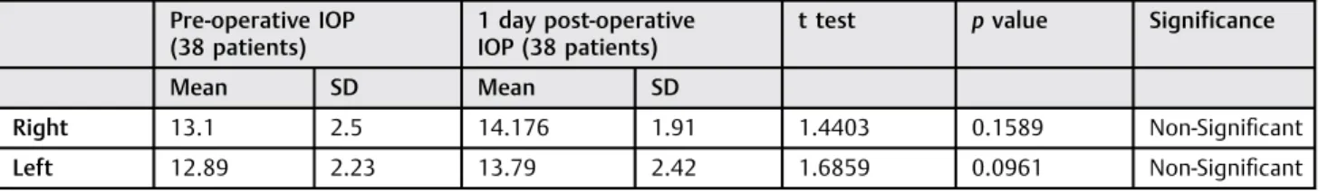
We found that the mean preoperative pressure was 13.12.5 mm Hg (range, 10 - 18 mm Hg) in the right eye and 12.892.23 mm Hg (range, 10 - 17 mm Hg) in the left eye, and the difference between the mean IOP values between the two sides was found to be statistically non-significant (p¼0.7862, t¼0.2732) (►Tables 2and3).
One day after surgery, the mean postoperative IOP in the RT eye was 14.1761.91 mm Hg (range 10 -17 mm Hg) and the difference between the mean preoperative and postoper-ative IOP values was statistically non-significant (p¼0.1589, t¼1.4403), whereas in the left eye mean postoperative IOP was 13.792.42 mm Hg (range 10 - 19 mm Hg). The differ-ence between the mean preoperative and postoperative IOP values was also statistically non-significant (p¼0.0961, t¼1.6859). Also, we observed that the difference in the postoperative IOP values between both eyes one day after surgery was statistically non-significant (p¼0.4428, t¼0.7718) (►Table 2).
Six weeks after surgery, the mean postoperative IOP in the right eye was 15.142.28 mm Hg (range 10–18 mm Hg) and the difference between the mean preoperative and postoperative IOP values was found to be statistically signifi -cant (p¼0.0012, t¼3.4027), whereas in the left eye the mean postoperative IOP was 15.142862.23 mm Hg (range 11 - 18 mm Hg). The difference between the mean
Table 1 Results of endoscopic nasal examination
Grade of nasal polyp Number of nasal sides %
0 12 15.8
1 18 23.7
2 28 36.8
preoperative and postoperative IOP values was also highly statistically significant (pvalue¼0.0005, t¼3.6808). More-over, the difference in the postoperative IOP values between right and left eyes 6 weeks after surgery was statistically non-significant.
We also performed endoscopic nasal examination during follow-up until 6 weeks after surgery for a second-look evaluation. The results presented no significantfindings.
Discussion
Functional endoscopic sinus surgery (FESS) has gained pop-ularity in the diagnosis and treatment of paranasal sinus diseases.12However, orbital negative sequels may occur due to its close anatomical relation with the paranasal sinuses.5
The incidence of serious complications of endoscopic sinus surgery reportedly reaches 0.5%. Orbital injuries can range from eye pressure, pain, retrobulbar hemorrhage, and compromised extraocular muscle movement with sequential binocular diplopia to blindness.13–16
Intraocular pressure (IOP) may be defined as the resulting balance between aqueous humor production and removal. Its increase is considered to be one of the main risk factors leading to the development of glaucoma. IOP is a variable value affected by multiple factors: age, gender, race, tobacco consumption, local ocular problems, obesity, hormonal changes, and physical exercise, among others.17–19
No clear line exists between safe and unsafe IOPs. In general, in cases in which the IOP is less than 30 mm Hg, the eye can be observed, and in cases in which the IOP is more than 40 mm Hg, a poor vision result may ensue.
The IOP can be measured clinically by various types of tonometers, including the Goldmann applanation tonometer, noncontact tonometer, and Tonopen. The Goldmann applanation tonometer is the most valid and reliable.20 That is why we used it in current study.
In the current study, the mean preoperative IOP rose 1 day postoperatively, but this increase was found to be statistically non-significant. In contrast, six weeks after surgery, we had noticed that the mean IOP increased in both eyes and this increase, surprisingly, was statistically significant in both eyes.
The postoperative results one day after surgery of this study agree with the results of Lin et al,1who found also that the postoperative increase in the average IOP values is statistically non-significant, taking into consideration that Lin et al1 studied the early effect of FESS on IOP as they measured IOP in three consecutive days after surgery.
The mechanism by which IOP is raised post-FESS whether in the early or late postoperative period is not clearly under-stood. Vascular mechanism is one of the proposed mecha-nisms for increased IOP post FESS. The eyes and paranasal sinuses are anatomically, neighboring structures. The eyeball, orbit, and parts of the sinonasal areas share the same vascular supply from the ophthalmic artery.
The venous drainage of the orbit and ocular structures is via the central retinal vein (drains into the superior ophthalmic vein) and the vortex vein (drains into the inferior ophthalmic vein). The vascular supplies of the sinuses are partial from the anterior and posterior ethmoidal arteries those are also derived from the ophthalmic artery, and the anterior and posterior ethmoidal veins drain into the superior ophthalmic vein.21
Lin et al1observed that the post-FESS symptoms of epi-phora and eye pressure were similar to the symptoms of increased IOP. Given that the eyes and sinonasal region have the same origin of vascular supply, they decided to elucidate whether FESS would result in a significant change in the IOP before and after surgery. They detected increased IOP. In the early postoperative period, in agreement with the current study, these changes were statistically insignificant.
A possible cause of increased IOP postoperatively could be excessive gauze packing and overinflated antral balloons of the Foley type, after the Caldwell-Luc operation. This may induce Table 2 Difference in IOP between preoperative and 1 day postoperative
Pre-operative IOP (38 patients)
1 day post-operative IOP (38 patients)
t test pvalue Significance
Mean SD Mean SD
Right 13.1 2.5 14.176 1.91 1.4403 0.1589 Non-Significant
Left 12.89 2.23 13.79 2.42 1.6859 0.0961 Non-Significant
Abbreviations: IOP, intraocular pressure; SD, standard deviation.
Table 3 Difference in IOP between preoperative and 6 weeks postoperative
Pre-operative IOP
(38 patients)
6 w post-operative IOP
(28 patients)
t test pvalue Significance
Mean SD Mean SD
Right 13.1 2.5 15.14 2.28 3.4027 0.0012 Significant
Left 12.89 2.23 15.14 2.34872 3.6808 0.0005 Extremely statistically significant
hypertropia and compression of the central retinal artery. Gel-man et al8reported a case of acute ocular hypertension following the use of an antral balloon after repairing fractures on thefloor of the orbit via a Caldwell-Luc antrostomy.
Another study was done by Papangelou and Christidis9to investigate whether there would be any effects on the IOP in patients who underwent a Caldwell-Luc operation for chronic sinusitis or antral polyps. They found that some changes of IOP occurred whether or not the maxillary sinus walls were denuded of mucous membrane.
They performed endoscopic grading for nasal polyposis preoperatively and observed various grades; however, when correlating the postoperative changes in IOP values with the grades of nasal polyposis, they found no correlation.
Many researchers studied the effect of endotracheal intuba-tion on IOP and found a transient increase in the IOP intra-operatively and in the early postoperative period. Malti and Geeta22reported that endotracheal intubation is associated with tachycardia, hypertension, and increase in intraocular pressure and that the latter may not have any adverse effect in patients with healthy eyes but has deleterious effects on a diseased or an injured eye. They also concluded in their study that laryngeal mask airway insertion is a suitable alternative to endotracheal intubation in patients having penetrating eye injuries, glaucoma, and strabismus in whom elevation of intraocular pressure is likely to be detrimental.
Langham et al23and Murphy24stated that endotracheal intubation can lead to adrenergic stimulation may cause vasoconstriction and an increase in central venous pressure which has a closer relationship to intraocular pressure than systemic pressure. It can produce an acute increase in intra-ocular pressure by increasing the resistance to the outflow of aqueous humor in the trabecular meshwork between the anterior chamber and the Schlemm’s canal.
In fact, these mechanisms would explain the early increase in IOP. Nonetheless, the cause of delayed increase in IOP remains unclear, so further studies are needed to explain the effect in IOP post-FESS.
To the best of our knowledge, this thefirst study to deal with the delayed effect of FEES on IOP. However, further studies are still needed to study the delayed effect of FESS on IOP in unilateral cases, as our study only included cases with bilateral sinonasal pathology.
It is important to note the risk of potential rise of IOP after surgery in patients scheduled for FESS and that had preoper-ative increase IOP, such as patients of glaucoma. Thus, preop-erative ophthalmological consultation is important to protect the patient from the hazard of possible increased IOP.
Conclusion
Delayed significant increase in IOP can occur after FESS. Therefore, special measures are necessary preoperatively to reduce IOP and protect the patient’s eye from the hazard of increased IOP, especially in patients with glaucoma. Further studies are still needed to explain the cause of this delayed significant rise in IOP post-FESS.
Conflict of Interest
The authors declare no conflict of interest.
Financial Support
The authors declare nofinancial support or interest to this study.
References
1 Lin PW, Lin HC, Chang HW, Su CY. Effects of functional endoscopic sinus surgery on intraocular pressure. Arch Otolaryngol Head Neck Surg 2007;133(9):865–869
2 Messerklinger W. [Endoscopy of the nose]. Monatsschr Ohren-heilkd Laryngorhinol 1970;104(10):451–456
3 Kennedy DW, Zinreich SJ, Rosenbaum AE, Johns ME. Functional endoscopic sinus surgery. Theory and diagnostic evaluation. Arch Otolaryngol 1985;111(9):576–582
4 Stammberger H. Endoscopic endonasal surgery—concepts in treatment of recurring rhinosinusitis. Part I. Anatomic and patho-physiologic considerations. Otolaryngol Head Neck Surg 1986; 94(2):143–147
5 Colclasure JC, Gross CW, Kountakis SE. Endoscopic sinus surgery in patients older than sixty. Otolaryngol Head Neck Surg 2004; 131(6):946–949
6 Bhatti MT, Giannoni CM, Raynor E, Monshizadeh R, Levine LM. Ocular motility complications after endoscopic sinus surgery with powered cutting instruments. Otolaryngol Head Neck Surg 2001; 125(5):501–509
7 Stankiewicz JA. Blindness and intranasal endoscopic ethmoidec-tomy: prevention and management. Otolaryngol Head Neck Surg 1989;101(3):320–329
8 Gelman HK, Janzen WR, Skolnik EM. Ocular hypertension following use of an antral balloon. Arch Otolaryngol 1974; 99(6):449–450
9 Papangelou L, Christidis M. Positive intramaxillary sinus pressure and intraocular pressure. J Laryngol Otol 1978; 92(12):1071–1074
10 Lanza DC, Kennedy DW. Adult rhinosinusitis defined. Otolaryngol Head Neck Surg 1997;117(3 Pt 2):S1–S7
11 Johansson L, Akerlund A, Holmberg K, Melén I, Stierna P, Bende M. Evaluation of methods for endoscopic staging of nasal polyposis. Acta Otolaryngol 2000;120(1):72–76
12 Weber R, Draf W, Keerl R, Schick B, Saha A. Endonasal micro-endoscopic pansinusoperation in chronic sinusitis. II. Results and complications. Am J Otolaryngol 1997;18(4):247–253
13 Cumberworth VL, Sudderick RM, Mackay IS. Major complications of functional endoscopic sinus surgery. Clin Otolaryngol Allied Sci 1994;19(3):248–253
14 Graham SM, Nerad JA. Orbital complications in endoscopic sinus surgery using powered instrumentation. Laryngoscope 2003; 113(5):874–878
15 Huang CM, Meyer DR, Patrinely JR, et al. Medial rectus muscle injuries associated with functional endoscopic sinus surgery: characterization and management. Ophthal Plast Reconstr Surg 2003;19(1):25–37
16 Stankiewicz JA. Complications in endoscopic intranasal ethmoi-dectomy: an update. Laryngoscope 1989;99(7 Pt 1):686–690 17 Fernández PC. Glaucoma. Medicine 1998;7:4770–4777.1987; 97:
1270–1273
18 Qureshi IA. Intraocular pressure: a comparative analysis in two sexes. Clin Physiol 1997;17(3):247–255
20 Liesegang TJ, Skuta GL, Cantor LB. Basic and Clinical Science Course, Section 10 Glaucoma: Intraocular Pressure and Aqueous Humor Dynamics. San Francisco, CA: American Academy of Oph-thalmology; 2003:14–24
21 Harris A, Jonescu-Cuypers CP, Kagemann L, Ciulla TA, Krieglstein GK. Atlas of Ocular Blood Flow: Vascular Anatomy, Pathophysiology, and Metabolism. Philadelphia, PA: Butterworth Heinemann; 2003
22 Malti PJ, Agarwal Geeta A. Comparative study of intraocular pressure changes with laryngeal mask airway and endotracheal tube. National Journal of Community Medicine 2012;3(2):279–282
23 Langham ME, Kitazawa Y, Hart RW. Adrenergic responses in the human eye. J Pharmacol Exp Ther 1971;179(1):47–55