Incidence of sepsis following transrectal ultrasound guided
prostate biopsy at a tertiary-care medical center in Lebanon
_______________________________________________
Mohammed Shahait
1, Jad Degheili
1, Fadi El-Merhi
2, Hani Tamim
3, Rami Nasr
11 Department of Surgery, American University of Beirut Medical Center, Beirut, Lebanon; 2 Department
of Radiology, American University of Beirut Medical Center, Beirut, Lebanon; 3 Department of Internal
Medicine and Clinical Research Institute, American University of Beirut Medical Center, Beirut, Lebanon
ABSTRACT
ARTICLE
INFO
______________________________________________________________ ______________________
Background: Urosepsis is a rare but life-threatening complication following transrectal ultrasound (TRUS) guided needle prostate biopsy. Despite the technological and phar-macological improvements, the problem of bacterial urosepsis after prostate biopsy remains. A strategy for preventing urosepsis following TRUS prostate biopsy in areas with high prevalence of resistant strains or patients presenting risk factors is lacking.
Objectives: The aim of this study was to assess the prevalence of urosepsis, as well its predictors, following TRUS guided needle biopsy of the prostate in a tertiary care medical center in Lebanon.
Materials and Methods: We carried out a retrospective study on all patients who un-derwent TRUS prostate biopsy at the American University of Beirut Medical Center between January 1, 2011 and June 31, 2013. Patients’ hospital charts were reviewed. Data collected included demographic information, pre-procedure disease specific in-formation, as well as post-procedure information. Predictors of urosepsis following TRUS were assessed.
Results: In total, 265 patients were included in this study, where the prevalence of urosepsis following TRUS prostate biopsy was found to be 9.4%. The significant in-dependent predictors of urosepsis were found to be: age with an OR=0.93 (95% CI: 0.88–1.00, p-value=0.03), and hypertension comorbidity with an OR=3.25 (95% CI: 1.19–8.85, p-value=0.02).
Conclusion: We found a high prevalence of urosepsis among patients who have un-dergone TRUS prostate biopsy, and identified two significant risk factors. The results of this study highlight the importance of implementing strategies for prevention of urosepsis following TRUS prostate biopsy.
Key words: Prostate; Biopsy; Ultrasonography; Sepsis; Neoplasms
Int Braz J Urol. 2016; 42: 60-8
_____________________
Submitted for publication: November 21, 2014
_____________________
Accepted after revision: March 26, 2015
INTRODUCTION
Prostate cancer is the second most common-ly diagnosed cancer in men and represents a signi-ficant health problem (1). A total of 233,000 new cases of prostate cancer and 29,480 deaths from the disease are anticipated in the United States in 2014 (2). The highest incidence rates f prostate cancer are
Transrectal ultrasound (TRUS)–guided prostate biopsy remains the gold standard techni-que to confirm the diagnosis of prostate cancer (5). According to recent estimates, approximately more than 1 million TRUS biopsies are performed per year in Europe and the United States (6). Although TRUS biopsy is generally considered to be a rela-tively low-risk outpatient procedure, post-biopsy complications and hospital admissions have incre-ased at alarming rates during the last decade due to an increasing rate of infection related compli-cations (7). TRUS biopsy complication rates are re-ported in up to 50% of cases and range from minor complications, such as hematuria, hematospermia or rectal bleeding, acute urine retention to much more severe complications, such as anemia, fain-ting, febrile urinary infections, syncope, and even septic shock. The infectious complications, which range from bacteriuria to sepsis, affect 1-4% of the patients who undergo this procedure (8). One stu-dy from Ontario, Canada reported that the hospital admission rate for infection-related complications within 30 days of the procedure increased from 1.0% in 1996 to 4.1% in 2005 (9). The reported in-cidence of urinary tract infections (UTI) after TRUS biopsy typically ranges between 2% and 6% with approximately 30%-50% of these patients having accompanying bacteremia (10). Severe sepsis has been described in 0.1%-3.5% of cases after TRUS biopsy (9). The proposed mechanism of infection is likely the introduction of bacteria into the bladder and bloodstream from the rectum (11). The most common organism responsible for these infectious complications is E. coli (7, 12). Moreover, the spre-ad of multiresistant E. coli is of particular concern (13). Resistant bacteria are more prevalent in some countries. Antibiotic overuse or misuse has been blamed, but a wider dissemination of resistant or-ganisms resulting from globalization and interna-tional travel may also be a factor (13, 14).
Factors that may predict which men are at greatest risk of infectious complications are: un-derlying medical comorbidities, particularly dia-betes mellitus, presence of urethral catheter, and recent hospitalization (15, 16).
Infectious complications can be serious, requiring effective preventative strategies and prompt management. Different methods have
been studied for reducing the rate of infection following TRUS guided biopsy such as the use of prophylactic antibiotic, bowel cleansing ene-ma, and using disposable instruments. Antibiotic prophylaxis is the only measure that has been shown to reduce the rate of infection post TRUS in randomized controlled trial setting, (17, 18). The American Urological Association (AUA) and the European Association of Urology guidelines for antibacterial prophylaxis for TRUS prostate biopsies recommend fluoroquinolones as agents of first choice due to their broad spectrum of ac-tivity, excellent penetration into prostatic tissue, and their prolonged post-antibiotic effect (19). The AUA guidelines also recommend aminoglycosides or aztreonam with metronidazole or clindamycin as alternatives to fluoroquinolones (17).
Fluoroquinolone-resistant E.coli is emer-ging globally (20). This poses a clinical challen-ge to the urologists to tailor the prophylactic regimen according to the resistance pattern in their hospitals. Multiple studies pointed toward the feasibility of using rectal swab culture to guide the prophylactic antibiotic regimen (21). In a survey of 3355 urologists in United States of America, Joel et al. reported 14 different du-ration of treatment using 10 different classes of antibiotic (22).
The aim of this study was to assess the prevalence of urosepsis following transrectal ul-trasound guided needle biopsy of the prostate, as well as its predictors in a tertiary-care medical center in Lebanon.
MATERIALS AND METHODS
Study design and setting
We carried out a retrospective chart re-view on all patients who underwent TRUS pros-tate biopsy at the American University of Beirut Medical Center between January 1, 2011 and June 31, 2013.
Inclusion/exclusion criteria
Ethical considerations
The institutional review board at the Ame-rican University of Beirut Medical Center appro-ved the study.
Procedure
During the study period, TRUS prostate biopsy was performed by four urologists in the Radiology Department at the American Universi-ty of Beirut Medical Center. All patients received prophylactic antibiotics. The adopted regimen by all urologists consisted of a flouroquinolone orally to be started one day prior to the procedure and gentamicin IV or IM injection 30 minutes before the procedure. The procedure was performed while the patient was in the left lateral decubitus po-sition. The anus and perineum were wiped with iodine swabs. A 5 to 9 MHz probe covered by a sterile condom and sterile K-Y Gel was introduced into the rectum and used to measure the prostate size and guide the local anesthesia injection and needle biopsies. An 18 French disposable gun and needle were used, in comparison to previous ye-ars, where we used a re-sterilizable automatic gun with a disposable needle. A standard sextant set of biopsies were taken, with the addition of targeted cores as needed to any suspicious lesion.
Outcome
The endpoint in our study was the deve-lopment of urosepsis after TRUS prostate biopsy. Urosepsis was defined as urinary symptoms, leu-kocytosis, and/or fever more than 38.0°C orally.
Data collection
Other information collected in this study included demographic information (such as age), lifestyle information (such as smoking), comorbi-dities (such as cardiac, hypertension, and diabe-tes), and pre-procedure disease specific informa-tion (including prostate size, post-void residual (PVR), PSA value and ratio, presenting urinary symptoms, recent treatment with antibiotic, po-sitive urine analysis, bowel preparation, and pre-vious biopsies). Moreover, post-procedure infor-mation were also collected, and included: positive urine analyses, urine culture and the bacteriology, as well as days to develop urosepsis.
Statistical analyses
Data entry and statistical analyses were performed using the Statistical Package for Social Sciences (SPSS), version 21.0. Descriptive analyses were carried out by reporting the number and per-cent for categorical variables, whereas the mean and standard deviation were calculated for con-tinuous ones. Associations between the different risk factors and the development of urinary tract infection were assessed using the chi-square test for categorical variables or the student’s t-test for continuous ones.
To account for the potential confounding effect of the different risk factors on the develo-pment of the outcome, multivariate logistic re-gression analyses were carried out. Included in the model were the multiple variables that could affect urinary infection post biopsy. These varia-bles included: age, smoking status, co-morbidities (such as hypertension and diabetes), prostate size, previous urine analysis, antibiotics use, and me-chanical bowel prep.
RESULTS
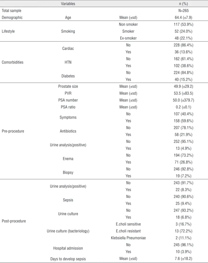
Table 1 - Baseline characteristics of the population.
Variables n (%)
Total sample N=265
Demographic Age Mean (±sd) 64.4 (±7.9)
Lifestyle Smoking
Non smoker 117 (53.9%)
Smoker 52 (24.0%)
Ex-smoker 48 (22.1%)
Comorbidities
Cardiac No 228 (86.4%)
Yes 36 (13.6%)
HTN No 162 (61.4%)
Yes 102 (38.6%)
Diabetes No 224 (84.8%)
Yes 40 (15.2%)
Pre-procedure
Prostate size Mean (±sd) 49.9 (±29.2)
PVR Mean (±sd) 53.5 (±83.5)
PSA number Mean (±sd) 50.0 (±379.7)
PSA ratio Mean (±sd) 0.2 (±0.1)
Symptoms No 107 (40.4%)
Yes 158 (59.6%)
Antibiotics No 207 (78.1%)
Yes 58 (21.9%)
Urine analysis(positive) No 252 (95.1%)
Yes 13 (4.9%)
Enema No 194 (73.2%)
Yes 71 (26.8%)
Biopsy No 246 (92.8%)
Yes 19 (7.2%)
Post-procedure
Urine analysis(positive) No 243 (91.7%)
Yes 22 (8.3%)
Sepsis No 240 (90.6%)
Yes 25 (9.4%)
Urine culture No 247 (93.2%)
Yes 18 (6.8%)
Urine culture (bacteriology)
E.choli sensitive 3 (16.7%)
E.choli resistant 13 (72.2%)
Klebsiella Pneumoniae 2 (11.1%)
Hospital admission No 245 (96.1%)
Yes 10 (3.9%)
Table-2 summarizes the association be-tween the different patient characteristics and the development of urosepsis. Patients who developed urosepsis were younger (mean=62.8 years, sd=6.3 versus non-septic patients whose age was 64.5, sd=8.0), and more likely to be smokers (33.3% versus non-septic patients, 2.8%), although the association was not significant for both charac-teristics, p-value=0.31 and 0.51, respectively. Pa-tients who developed urosepsis were more likely to be hypertensive (64.0%) compared to the non--septic ones (36%), with a p-value of 0.006.
Simi-larly, uroseptic patients were more likely to be dia-betic (32.0%) as compared to non-septic patients (13.4%), p-value=0.03. As for the pre-procedure information, none of the assessed characteristics was found to be significantly different between the uroseptic and non-septic patients. For ins-tance, uroseptic patients were less likely to have had bowel preparation (16.0%) as compared to the non-septic ones (27.9%), p-value=0.2.
Finally, Table-3 summarizes the results of the multivariate analyses carried out to iden-tify the predictors of urosepsis among patients
Table 2 - Association between different factors and urosepsis
Variables All
n (%)
No sepsis n (%)
Sepsis n (%)
P value
Total sample N=265 N=240 N=25
Demographic Age Mean (±sd) 64.4 (±7.9) 64.5 (8.0) 62.8 (6.3) 0.31
Lifestyle Smoking
Non smoker 117 (53.9%) 106 (54.9%) 11 (45.8%)
0.51
Smoker 52 (24.0%) 44 (2.8%) 8 (33.3%)
Ex-smoker 48 (22.1%) 43 (22.3%) 5 (20.8%)
Comorbidities
Cardiac No 228 (86.4%) 207 (86.6%) 21 (84.0%) 0.76
Yes 36 (13.6%) 32 (13.4%) 4 (16.0%)
HTN No 162 (61.4%) 153 (64.0%) 9 (36.0%) 0.006
Yes 102 (38.6%) 86 (36.0%) 16 (64.0%)
Diabetes No 224 (84.8%) 207 (86.6%) 17 (68.0%) 0.03
Yes 40 (15.2%) 32 (13.4%) 8 (32.0%)
Pre-procedure
Prostate size Mean (±sd) 49.9 (±29.2) 49.3 (±27.9) 54.8 (±39.8) 0.39
PVR Mean (±sd) 53.5 (±83.5) 54.8 (±88.4) 45.0 (±36.2) 0.60
PSA number Mean (±sd) 50.0 (±379.7) 50.7 (±396.0) 44.4 (±179.2) 0.94
PSA ratio Mean (±sd) 0.2 (±0.1) 0.2 (±0.1) 0.2 (±0.1) 0.14
Symptoms No 107 (40.4%) 101 (42.1%) 6 (24.0%) 0.08
Yes 158 (59.6%) 139 (57.9%) 19 (76.0%)
Antibiotics No 207 (78.1%) 188 (78.3%) 19 (76.0%) 0.79
Yes 58 (21.9%) 52 (21.7%) 6 (24.0%)
Urine analysis No 252 (95.1%) 228 (95.0%) 24 (96.0%) 1.00
Yes 13 (4.9%) 12 (5.0%) 1 (4.0%)
Enema No 194 (73.2%) 173 (72.15) 21 (84.0%) 0.20
Yes 71 (26.8%) 67 (27.9%) 4 (16.0%)
Biopsy (previous)
No 246 (92.8%) 222 (92.5%) 24 (96.0%)
1.00
who underwent TRUS prostate biopsy. Age was found to be a significant predictor, where ol-der patients are less likely to have urosepsis (OR=0.93, 95% CI: 0.88-1.00, p-value=0.03). Hypertension status was also found to be sig-nificantly associated with urosepsis (OR=3.25, 95% CI: 1.19-8.85, p-value=0.02). Smokers, diabetics, and patients with symptomatic pre-sentation were at higher chance of developing urosepsis. On the other hand, patients who had bowel preparation or cardiac disease were less likely to develop urosepsis; but none of these associations was statistically significant.
All patients who developed urosepsis were admitted to the hospital for intravenous antibiotic and monitoring. All patient received carbapenem antibiotic for 14 days at least. The average num-ber of days of hospitalization was 5.
DISCUSSION
TRUS prostate biopsy is the standard test to diagnose prostate cancer after a sus-picious digital rectal exam and elevated PSA. Occasionally this procedure is associated with significant morbidity. In this retrospective stu-dy, charts of patients who underwent TRUS prostate biopsy at the American University of Beirut Medical Center between January 1, 2011 and June 31, 2013 were reviewed to assess the prevalence of urosepsis following transrectal
ultrasound (TRUS) prostate biopsy, as well as its predictors. We found that the prevalence of (TRUS) prostate biopsy urosepsis to be 9.4%. Multivariate analysis identified age and hyper-tension comorbidity to be significantly associa-ted with an increased risk of developing uro-sepsis following TRUS prostate biopsy.
The prevalence of urosepsis following TRUS prostate biopsy found in our study (9.4%) was higher than that reported in other studies. The frequency of urosepsis varied among those studies between 0.2% and 3.06%. The lowest ra-tes of urosepsis were reported by Zaytoun et al. in a North American cohort, and Raaijmakers et al. in a European Randomized Study, who re-ported urosepsis prevalence rates of 0.2%, and 0.5%, respectively (17, 23). However, other se-ries of studies carried out by Carmignani et al., Akduman et al., and Simsir et al. in reported hi-gher rates of urosepsis (2.2%, 3.0%, and 3.06%, respectively) (16, 24, 25). Only one Asian study conducted by Raheem et al. in 2012 reported no septic complications (26). This variation in ra-tes of sepsis among different studies arises from differences in biopsy techniques, prophylactic protocols, consistent reporting, and the defini-tion of urosepsis used. The prophylactic regimes preventing infectious complications may differ with respect to the use of an antibiotic (type of antibiotic used, dose, and method of adminis-tration and duration of the therapy), as well as
Table 3 - Multivariate analysis for the predictors of urosepsis (Cox regression model).
Predictors Adjusted OR (95%CI) P value
Age 0.93 (0.88 – 1.00) 0.03
Smoking
Non smoker Reference Reference
Smoker 1.56 (0.55-4.45) 0.40
Ex-smoker 0.95 (0.30-3.07) 0.93
Cardiac 0.92 (0.27-3.19) 0.90
HTN 3.25 (1.19-8.85) 0.02
Diabetes 2.18 (0.79-6.01) 0.13
Prostate size 1.00 (0.99-1.02) 0.71
Symptoms 1.83 (0.66-5.04) 0.24
whether a cleansing rectal enema was used or not (24). Other factors that may increase the antibiotic resistance and thus increasing the rate of sepsis by an antibiotic resistant strain are past history of hospitalization, a past his-tory of exposure to antibiotics, a past hishis-tory of catheterization, and a past history of urogenital surgery (27, 28). The proponents of the high sepsis rate in our study are cited as following: the high prevalence of E. coli resistant to fluo-roquinolone in Lebanon which was noted in a recent review of the patterns and trends of bac-terial resistance to antimicrobial agents over the last decade in our center, self-medication with antibiotics which is a frequent problem in Beirut area, polypharmacy in patients with co-morbidities which may affect the antibiotic efficacy (29, 30).
As for the risk factors significantly as-sociated with urosepsis following TRUS pros-tate biopsy, age was found to be a significant risk factor OR=0.93 (95% CI: 0.88–1.00, p-va-lue=0.03), where older patients were less likely to have urosepsis. One possible reason may be that younger patients are more likely to self--report complications compared to older pa-tients. Few studies in the literature reported on the effect of age on sepsis after TRUS prostate biopsy. In a study carried out by Lee et al. be-tween 2003 and 2006 reported no significant difference in the urosepsis rate in relation to age (p-value=0.82) after TRUS prostate biopsy (29). However, several studies reported on hi-gher incidence of general complications after TRUS biopsy in younger patients (30).
Another factor found to be significantly associated with urosepsis following TRUS prostate biopsy in our study was hypertension comorbidity with an OR=3.25 (95% CI: 1.19-8.85, p-value=0.02). The study carried out by Lee et al. did not report any significant association between hypertension and sepsis following TRUS prostate biopsy (p-va-lue=0.18) (29). Further comparison of this associa-tion with the literature is hard due to the limited availability of studies assessing this association.
Although many urologists intuitively assume that increased post void residue would predispose to development of post TRUS
infec-tion, clear evidence is lacking as well. Our data did not show any significant difference in the rate of urosepsis among patients with post void residue verses those with no significant residue.
Finally, we should emphasize the limi-tations of this study, including the small num-ber of patients. In addition, the clinical results were analyzed retrospectively based on charts review.
Notwithstanding those limitations, this study elucidates of the impact of increasing bacteria resistance prevalence on the rate of post TRUS urosepsis.
CONCLUSIONS
Urosepsis after TRUS biopsy represents a great challenge for urologists; sometimes its risks are more important than its benefits. It tips the balance between the risk and the be-nefit of prostate screening. Implementation of new strategies to prevent urosepsis and early treatment is required; especially in areas where bacterial resistance is endemic. We are conside-ring revisiting our prophylactic regimen in our institution, and developing a follow-up system in which the patients will be contacted every 48 hours by a physician assistant to be screened for early urosepsis symptoms. Larger studies that explore the risks of urosepsis after TRUS biopsy are required. Moreover, identifying biomarkers that are associated with developing urosepsis after TRUS biopsy represents a unique research opportunity.
CONFLICT OF INTEREST
None declared.
REFERENCES
1. National Cancer Institute. Genetics of Prostate Cancer (PDQ®). 2014.2. available at http://www.cancer.gov/ types/prostate/hp/prostate-genetics-pdq
3. Ferlay J, Shin HR, Bray F, Forman D, Mathers C, Parkin DM. Estimates of worldwide burden of cancer in 2008: GLOBOCAN 2008. Int J Cancer. 2010;127:2893-7. 4. Shamseddine A, Saleh A, Charafeddine M, Seoud M,
Mukherji D, Temraz S, et al. Cancer trends in Lebanon: a review of incidence rates for the period of 2003-2008 and projections until 2018. Popul Health Metr. 2014;12:4. 5. American Cancer Society. Prostate cancer: early
detection, 2009. available at http://onlinelibrary.wiley. com/doi/10.3322/caac.20066/pdf
6. Loeb S, Carter HB, Berndt SI, Ricker W, Schaeffer EM. Complications after prostate biopsy: data from SEER-Medicare. J Urol. 2011;186:1830-4.
7. Ng CF, Chan SY. Re: The incidence of fluoroquinolone resistant infections after prostate biopsy-are fluoroquinolones still effective prophylaxis? J.Feliciano, E. Teper, M. Ferrandino, R. J. Macchia, W. Blank, I. Grunberger and I. Colon J Urol 2008; 179:952-955. J Urol. 2008;180:1570-1; author reply 1571.
8. Pinkhasov GI, Lin YK, Palmerola R, Smith P, Mahon F, Kaag MG, et al. Complications following prostate needle biopsy requiring hospital admission or emergency department visits-experience from 1000 consecutive cases. BJU Int. 2012;110:369-74.
9. Nam RK, Saskin R, Lee Y, Liu Y, Law C, Klotz LH, et al. Increasing hospital admission rates for urological complications after transrectal ultrasound guided prostate biopsy. J Urol. 2013;189:S12-7; discussion S17-8.
10. Zaytoun OM, Vargo EH, Rajan R, Berglund R, Gordon S, Jones JS. Emergence of fluoroquinolone-resistant Escherichia coli as cause of postprostate biopsy infection: implications for prophylaxis and treatment. Urology. 2011;77:1035-41.
11. Liss MA, Chang A, Santos R, Nakama-Peeples A, Peterson EM, Osann K, et al. Prevalence and significance of fluoroquinolone resistant Escherichia coli in patients undergoing transrectal ultrasound guided prostate needle biopsy. J Urol. 2011;185:1283-8.
12. Ozden E, Bostanci Y, Yakupoglu KY, Akdeniz E, Yilmaz AF, Tulek N, et al. Incidence of acute prostatitis caused by extended-spectrum beta-lactamase-producing Escherichia coli after transrectal prostate biopsy. Urology. 2009;74:119-23.
13. So A, Furlong M, Heddini A. Globalisation and antibiotic resistance. BMJ. 2010;341:c5116.
14. Patel U, Dasgupta P, Amoroso P, Challacombe B, Pilcher J, Kirby R. Infection after transrectal ultrasonography-guided prostate biopsy: increased relative risks after recent international travel or antibiotic use. BJU Int. 2012;109:1781-5.
15. Carignan A, Roussy JF, Lapointe V, Valiquette L, Sabbagh R, Pépin J. Increasing risk of infectious complications after transrectal ultrasound-guided prostate biopsies: time to reassess antimicrobial prophylaxis? Eur Urol. 2012;62:453-9. 16. Simsir A, Kismali E, Mammadov R, Gunaydin G, Cal C. Is it
possible to predict sepsis, the most serious complication in prostate biopsy? Urol Int. 2010;84:395-9.
17. Zani EL, Clark OA, Rodrigues Netto N Jr. Antibiotic prophylaxis for transrectal prostate biopsy. Cochrane Database Syst Rev. 2011;CD006576.
18. Zaytoun OM, Anil T, Moussa AS, Jianbo L, Fareed K, Jones JS. Morbidity of prostate biopsy after simplified versus complex preparation protocols: assessment of risk factors. Urology. 2011;77:910-4.
19. Heidenreich A, Bellmunt J, Bolla M, Joniau S, Mason M, Matveev V, et al. European Association of Urology. EAU guidelines on prostate cancer. Part 1: screening, diagnosis, and treatment of clinically localised disease. Eur Urol. 2011;59:61-71.
20. Dalhoff A. Global fluoroquinolone resistance epidemiology and implictions for clinical use. Interdiscip Perspect Infect Dis. 2012;2012:976273.
21. Duplessis CA, Bavaro M, Simons MP, Marguet C, Santomauro M, Auge B, et al. Rectal cultures before transrectal ultrasound-guided prostate biopsy reduce post-prostatic biopsy infection rates. Urology. 2012;79:556-61.
22. Hillelsohn JH, Duty B, Blute ML Jr, Okhunov Z, Kashan M, Moldwin R, et al. Variability of transrectal ultrasound-guided prostate biopsy prophylactic measures. Can J Urol. 2012;19:6573-7.
23. Raaijmakers R, Kirkels WJ, Roobol MJ, Wildhagen MF, Schrder FH. Complication rates and risk factors of 5802 transrectal ultrasound-guided sextant biopsies of the prostate within a population-based screening program. Urology. 2002;60:826-30. 24. Carmignani L, Picozzi S, Spinelli M, Di Pierro S, Mombelli
G, Negri E, et al. Bacterial sepsis following prostatic biopsy. Int Urol Nephrol. 2012;44:1055-63.
25. Akduman B, Akduman D, Tokgöz H, Erol B, Türker T, Ayoğlu F, et al. Long-term fluoroquinolone use before the prostate biopsy may increase the risk of sepsis caused by resistant microorganisms. Urology. 2011;78:250-5. 26. Raheem OA, Casey RG, Galvin DJ, Manecksha RP,
Varadaraj H, McDermott T, et al. Discontinuation of anticoagulant or antiplatelet therapy for transrectal ultrasound-guided prostate biopsies: a single-center experience. Korean J Urol. 2012;53:234-9.
28. Azap OK, Arslan H, Serefhanoğlu K, Colakoğlu S, Erdoğan H, Timurkaynak F, et al. Risk factors for extended-spectrum beta-lactamase positivity in uropathogenic Escherichia coli isolated from community-acquired urinary tract infections. Clin Microbiol Infect. 2010;16:147-51. 29. Lee SH, Chen SM, Ho CR, Chang PL, Chen CL, Tsui
KH. Risk factors associated with transrectal ultrasound guided prostate needle biopsy in patients with prostate cancer. Chang Gung Med J. 2009;32:623-7.
30. Wu MW, Sevilla EM, Raman L, Consigliere D, Siow WY, Tiong HY. Incidence of complications after transrectal ultrasonography-guided biopsy of the prostate in a local tertiary institution. Singapore Med J. 2011;52:752-7.