Licenciado sob uma Licença Creative Commons DO): http://dx.doi.org. . / - . . .AO
[T]
Histomorphometrical analysis on the effects of two therapeutic
ultrasound intensities on fracture healing in aged rats
[)]
Estudo histomorfométrico comparativo dos efeitos
de duas intensidades de ultrassom terapêuticos na
consolidação de fratura da tíbia em ratos idosos
[A]
Jeronimo Rafael Skau[a], Bruno Rodrigues[b], Felipe Oliveira Rosa[c], Rubens Correa Araujo[d], Renata Gabriel Fontinele[e], Romeu Rodrigues de Souza[f]
[a] MSc, Universidade São Judas Tadeu, Departamento de Fisioterapia, São Paulo, SP - Brazil, e-mail: [email protected] [b] PhD, Universidade São Judas Tadeu, Departamento de Fisioterapia, São Paulo, SP - Brazil, e-mail: [email protected] [c] MSc, Universidade de São Paulo, Departamento de Anatomia, São Paulo, SP - Brazil, e-mail: [email protected]
[d] PhD, Universidade de Taubaté, Departamento de Fisioterapia, Taubaté, SP - Brazil, e-mail: [email protected]
[e] PhD, Universidade de São Paulo, Departamento de Anatomia, São Paulo, SP - Brazil, e-mail: [email protected] [f] PhD, Universidade São Judas Tadeu, Universidade de São Paulo, Departamento de Anatomia, São Paulo, SP - Brazil, e-mail:
[R]
Abstract
Introduction: Experimental studies conducted in young animals show that therapeutic ultrasound TUS
has been successfully used to shorten the healing time of bone fractures. (owever, they were not found in the literature, studies comparing the effect of different intensities of UST in aged animals. Objective: To test
the efficacy of intensity . W/cm² and of . W/cm² in the consolidation of experimental fracture of the tibia from aged Wistar rats. Materials and methods: Three groups of month old rats were submitted
The results showed that fractures treated with ultrasound at . W/cm healed significantly faster than did the fractures treated with ultrasound at . W/cm and the control.
[P]
Keywords: Ultrasound. Wound healing. Bone fracture. Aging.
[B] Resumo
Introdução: Trabalhos experimentais realizados em animais jovens mostram que o ultrassom terapêutico (UST) tem sido utilizado com sucesso para abreviar o tempo de consolidação de fraturas ósseas. Entretanto, não foram encontrados na literatura, trabalhos comparando o efeito de diferentes intensidades de UST em animais idosos. Objetivo: Comparar a eficácia de duas intensidades de UST, de 0,5 W/cm² e de 1,0 W/cm² na consolidação de fratura experimental da tíbia de ratos Wistar idosos. Materiais e métodos: Três grupos de ratos com 15 meses de idade foram submetidos a osteotomia da diáfise da tíbia e a seguir o membro inferior foi imobilizado com uma barra de metal, tornando imóveis as articulações do joelho e do tornozelo. Um grupo de animais (L) recebeu UST a 0,5 W/cm²; o outro grupo (I) foi submetido ao UST de 1,0 W/cm². Um grupo controle (C) não recebeu aplicação de UST. Quinze animais (cinco por grupo) foram sacrificados após uma semana e outros quinze animais (cinco por grupo) após três semanas de imobilização. O progresso de consolidação da fratura foi obtido para cada grupo através de avaliação morfométrica de cortes histológicos da região da fra-tura. Resultados e conclusão: Os resultados obtidos mostram que, em ratos idosos, as fraturas tratadas com UST de 1,0 W/cm2 consolidaram mais rapidamente que as tratadas com o UST de 0,5 W/cm2 e as do grupo C. [K]
Palavras-chave: Ultrasom. Cicatrização. Fratura óssea. Envelhecimento.
Introduction
One of the most common kinds of fractures, both in the young and elderly is that of the middle third of the tibia . )n the repair process of such kind of fracture stable bone fracture , after the removal of the blood clot by cells of the connective tissue, there is an initial proliferation of osteogenic cells , that is followed by the appearance of minuscule bone and cartilaginous strands and fibrous tissue in the area. As a result, im-mature bone tissue is formed, and then the lamellated
bone tissue appear constituting the bone callus .
This entire process generally takes between and weeks in humans and weeks in young rats .
Several works have been carried out searching for methods to abbreviate the time for fracture con-solidation , . Methods that have been used to accelerate bone healing include electrical stimula-tion, therapeutic ultrasound TUS and precocious mobilization, among others. Low-intensity pulsed ultrasound has been shown to accelerate fracture healing in both animal models and clinical trials ,
, , , , , . The effectiveness of this resource
has been studied using different degrees of intensity. The values that showed positive results are that of
low intensity: . W/cm² and . W/ cm² , . The
use of high intensities such as those of . W/cm² proved to be detrimental to the bone tissue. (owever, investigations showing the effects of values between . and . W/cm² are scarce and have been carried
out in young animals , . )n this study, the time
for fracture consolidation of the middle third of the tibia using two different intensities . W/cm² and . W/cm² of pulsating ultrasound were compared by histomorphometric method in aged rats.
Materials and methods
Surgical proceeding
)n this work, fifteen-mo-old Wistar male rats were used. All the animals were submitted to peridural anesthetics Xylocayne % and Epinefrine, :
175
bone tissue were counted and then the Vv bone was
calculated by the formula: the hind limb was immobilized by a metal splint and
plaster of Paris, wrapping the knee and ankle joints. Animals were divided into three groups with rats each: Control group C ; Low intensity TUS group L and )ntermediary intensity TUS group ) . C Group was submitted only to the immobilization. L Group received . M(z TUS Sonopulse ))) )bramed, Amparo, São Paulo, Brazil in pulsed mode at . W/cm² for min-utes daily, times a week; animals received TUS for week and for weeks. ) Group received . M(z TUS in pulsed mode at . W/ cm², for minutes daily, times a week; animals received TUS for week and for weeks. The application of the TUS was performed through a water balloon. A thin layer of gel was placed on the area of the lesion and on top of the balloon. Then, the balloon was placed over the wound surface area, and the TUS probe was placed over it. The rats were allowed to move freely in their cages. At the end of the first week rats, being from each group were
killed and at the end of the rd week the last rats,
being from each group were also killed. The segment of the tibia with the fracture trace was collected and processed for examination. All procedures to which the animals were submitted received the approval of the Commission of Ethics in the Use of Animals of the Faculdade de Medicina Veterinária of Universidade de São Paulo, which follows the )nternational Guiding Principles for Biomedical Research )nvolving Animals,
under protocol nº / .
Histomorphometry
From each animal, the segment of the tibia con-taining the fracture trace was removed, fixed in form-aldehyde in . M phosphate buffer p( . and decalcified with % EDTA Sigma-Aldrich, St. Louis, MO, USA and % formaldehyde in . M phosphate buffer p( . at °C. The blocks were washed in the phosphate buffer and dehydrated, cleared and embedded in Paraplast. Sections micrometers μm thick were performed parallel to the long axis of the tibia. Every third section of a total of sections was then stained with hematoxylin and eosin (E and
analysed in a digital image analysis system KS- ,
Carl Zeiss, São Paulo, Brazil .
To estimate the volume density % of neoformed
bone Vv bone , a test-system with points was
superimposed to each image over the monitor screen in the video-microscopic system. The points hitting
Vv bone =Pp (bone)
PT
Where: PP bone are the number of points hitting the bone, and PT are the total number of points
of the test-system Figure . The results are
expressed as means ± SEM.
Statistical analysis
Data were compared by analysis of variance ANOVA , and Tukey’s test for multiple comparisons,
as appropriate. The significance level was set at P < . .
Results
Histomorphometric analysis
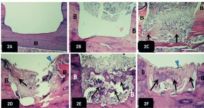
Qualitatively, at the first week, the C group has an empty space, corresponding to the area of the lesion, showing that the consolidation process has not yet begun Figure A . At third week, proliferation of osteogenic cells can be observed in the lesion area. )n the deep part of the lesion, there are recently formed bony trabeculae. Much more osseous tissue was observed in the callus Figure D . )n L Group Figure B , at the first week, osteogenic cells fill in the lesion space, indicating that the formation of bone tissue will be initiated. )n the third week, newly formed bone fill almost entirely the space of the lesion Figure E . At the first week of the ) Group Figure C , newly formed immature bony trabeculae fill the region of the fracture and osteogenic cells can be observed in the lesion space indicating that begin-ning of primary ossification takes place in the space of lesion. At the third week, bone trabeculae arrows can be seen filling almost all space of the fracture
Figure F . )n conclusion, on day st Figures D, E,
F , fractures treated with US at . W/cm² Figure F healed more rapidly than did that treated with . W/cm² Figure E or the controls Figure D .
Quantitatively, significant differences on the vol-ume density % of newly formed bone tissue were
There is disagreement among authors, related to the best intensity of TUS to be applied in the case
of fractures . Several authors , obtained
favorable results in fractures of rats treated with . W/cm² for two minutes every days or with . W/cm for ten minutes over five consecutive days . According to others, intensities from . W/
cm² to . W/cm are the most efficient . The
present study showed that fractures treated with
ultrasound at . W/cm healed significantly faster
than that treated with ultrasound at . W/cm and
the control. The fracture healing has been acceler-ated in TUS . W/cm group than in the . W/cm
group, both at the first week but mainly at the rd
week p < . .
)n the present study, tibia was choose due the easy surgical access to this bone, because of its su-perficial location and because the fracture in the middle third of the tibia is one of the most frequent
, .
p < . . The percent of the new bone tissue was
higher in ) % than in the C % and L % group
at the first week P < . . At the third week, the
percent of the newly formed bone tissue was higher
in L % than in the C % group and in ) %
than in L and C group P < . Figure .
Discussion
The consolidation time for the bone fractures that frequently occur both in sports and in aged sedentary people obliges the individuals to remain distant from
their normal activities , , . Application of TUS
is among the resources suggested with the objective of recovering the individuals in the shortest possible time. This resource have been used by several authors
obtaining positive effects , , , , , , although
some have not obtained favorable results in humans
with its application .
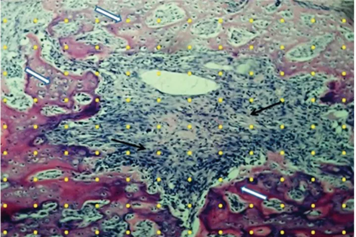
Figure 1 - Test-system with 132-points (yellow points) superimposed at a histological section from the fracture trace, used to quantify the newly formed bone (white arrows) in the four groups of rats
177
2A 2B 2C
2F 2E
2D
Figure 2 - Light micrographs of longitudinal sections of callus tissue stained with HE
Note: A, D–C Group; B, E–L Group; C, F–I Group. 2 A, B, C – At the end of the fi rst week; 2 D, E, F – At the end of the third week. New bone formation can be seen at the end of the fi rst week (7th day) in I group (C) but not in the C (A) or in L Group (B). At the end of the twenty
fi rst week fractures treated with US of 1.0 W/cm² (F) healed more rapidly than did that treated with US of 0.5 W/cm² (E) or the controls (D). Arrowheads – Periosteum. Arrows – Newly formed bone; B– Bone tissue. HE staining. Bar: 0.5mm.
Source: Research data.
Percentage
Week
80
60
C
L
I 40
20
0
First Third
Figure 3 - Volume densities of newly formed bone in the callus tissue of C, L, and I groups of rats at the end of the 1st and 3rd week
Note: The use of ultrasound accelerated the consolidation process of the fracture, compared to the C group in both weeks. * = Signifi cant vs. L and C group at the fi rst week; ** = Signifi cant
vs. L and C group at the third week; # = Signifi cant vs. C group at the third week; P < 0.05.
Source: Research data.
Assessment of the results of TUS application for the study of fracture healing with experimental ani-mals has generally been done through X-ray or bone
densitometry , , , , , . )n the present
work, results were obtained through histological and morphometric analysis of the fracture area. Only a few authors have used histological and quantitative
methods for this purpose , , and none used
histological methods associated with morphomet-ric methods.
The pulsed TUS mode was used because it pres-ents less thermal effect and because it has a proved
action on the inflammatory reaction . (owever,
some care must be taken when the TUS is used, as for example, to avoid direct contact of the head of the equipment with the surgical wound. )n this study we used the application with the water balloon, because of the impossibility of immersion of the fractured area in the water, due to the limb immobilization through the plaster of Paris.
. Matheus JP, Oliveira FB, Gomide LB, Milani JG, Volpon JB, Shimano AC. Effects of therapeutic ultrasound on the mechanical properties of skeletal muscles after contusion. Rev Bras Fisioter. ; : - . . Chan CW, Qin L, Lee KM, Zhang M, Cheng JC, Leung
KS. Low intensity pulsed ultrasound accelerated bone remodeling during consolidation stage of distraction osteogenesis. J Orthop Res. ; : - . . Malizos KN, Papachristos AA, Protopappas VC,
Fo-tiadis D). Transosseous application of low-intensity ultrasound for the enhancement and monitoring of fracture healing process in a sheep osteotomy model. Bone. ; : - .
. Yang RS, Lin WL, Chen YZ, Tang C(, (uang T(, Lu BY, et al. Regulation by ultrasound treatment on the integrin expression and differentiation of osteoblasts. Bone. ; : - .
. (antes ME, Mavrodontidis AN, Zalavras CG, Karan-tanas A(, Karachalios T, Malizos KN. Low-intensity transosseous ultrasound accelerates osteotomy heal-ing in a sheep fracture model. J Bone Joint Surg Am.
; : - .
. Azuma Y, )to M, (arada Y, Takagi (, Ohta T, Jingushi S. Low-intensity pulsed ultrasound accelerates rat femo-ral fracture healing by acting on the various cellular reactions in the fracture callus. )nt J Bone Miner Res.
; : - .
. Tsai CL, Chang W(, Liu TK. Preliminary studies of duration and intensity of ultrasonic treatments on fracture repair. Chin J Physiol. ; : - . . Fontes-Pereira AJ, Teixeira RC, Oliveira AJB, Pontes
RWF; Barros RSM; Negrão JNC. The effect of low-in-tensity therapeutic ultrasound in induced fracture of rat tibiae. Acta ortop bras. ; : - . . Gundersen (J, Kroustrup JP, Vaeth M. Stereological
analysis of three-dimensional structure organization of surfaces in multiphase specimens: statistical meth-ods and model-inferences. J Microsc. ; Pt
: - .
. Englund U, Nordström P, Nilsson J, Bucht G, Björnstig U, (allmans G, et al. Physical activity in middle-aged women and hip fracture risk: the UFO study. Osteo-poros )nt. ; : - .
activity directly by mechanical deformation of the cell membrane or the extracellular matrix or indirectly
by an electrical effect caused by cell deformation ,
. Low intensity pulsed ultrasound in isolated cell systems produced significant multifunctional effects of direct relevance to bone formation and resorption such as increasing calcium uptake and modulating adenylate cyclase activity, transforming growth fac-tor beta synthesis, bone morphogenic protein
ef-fects, and parathyroid hormone response , .
Furthermore, ultrasound stimulation increases the mechanical properties of the healing fracture callus by stimulating earlier synthesis of extracellular ma-trix proteins in cartilage, possibly altering chondro-cyte maturation and endochondral bone formation
, . Others studies suggested that TUS acts on
some cellular reactions involved in each phase of the healing process such as inflammatory reaction, angio-genesis, chondroangio-genesis, intramembranous ossifica-tion, endochondral ossificaossifica-tion, and bone remodeling
, . )n summary, the application of ultrasonic
waves of . W/cm promoted a more rapid healing of experimental bone fractures than . W/cm in Wistar rats.
References
. Lirani APR, Larzaretti-Castro M. Evidências da ação de agentes físicos sobre o metabolismo do tecido ósseo e seus potenciais usos clínicos. Arq Bras Endocrinol Metab. ; : - .
. Alvarenga EC, Rodrigues R, Caricati-Neto A, Silva-Filho FC, Paredes-Gamero EJ, Ferreira AT. Low-intensity pulsed ultrasound-dependent osteoblast prolifera-tion occurs by via activaprolifera-tion of the P Y receptor: role of the P Y receptor. Bone. ; : - . . Bazin S, Kitchen S. Eletroterapia de Clayton. São Paulo:
Manole; .
. Takikawa S, Matsui N, Kokubu T, Tsunoda M, Fujioka (, Mizuno K, et al. Low-intensity pulsed ultrasound initiates bone healing in rat nonunion fracture model. J Ultrasound Med. ; : - .
179 . Sousa VL, Alvarenga J, Padilha Filho JG, Canola JC,
Fer-rigno CRA, Alves JM, et al. Ultra-som pulsado de baixa intensidade em fraturas diafisárias: aplicação clínica em cães. Ciênc Rural. ; : - .
. Fréz AR, Ariza D, Ferreira JRL, Alves ÉPB, Breda GR, Centenaro LA, et al. Efeito do ultra-som terapêutico contínuo em placas epifisárias de coelhos. Rev Bras Med Esporte. ; : - .
. Ryaby JT, Matthew J, Duarte-Alves P. Low intensity pulsed ultrasound affects adenylate cyclase activity and TGF-β synthesis in osteoblastic cells. Trans Or-thop Res Soc. ; : .
. Naito K, Watari T, Muta T, Furuhata A, )wase (, )garashi M, et al. Low-intensity pulsed ultrasound L)PUS in-creases the articular cartilage type )) collagen in a rat osteoarthritis model. J Orthop Res. ; : - .
Received: / /
Recebido: / /
Approved: / /
Aprovado: / /
. Emami A, Petrén-Mallmin M, Larsson S. No effect of low-intensity ultrasound on healing time of intra-medullary fixed tibial fractures. J Orthop Trauma.
; : - .
. Yang K(, Parvizi J, Wang SJ, Lewallen DG, Kinnick RR, Greenleaf JF, et al. Exposure to low intensity ultra-sound increases aggrecan gene expression in a rat fe-mur fracture model. J Orthop Res. ; : - . . Rennó ACM, Faganello, FR, Navega MT, Carvalho DCL. The osteogenic effect of low intensity ultrasound. Fi-sioter Mov. ; : - .
. Kumagai K, Takeuchi R, )shikawa (, Yamaguchi Y, Fu-jisawa T, Kuniya T, et al. Low-intensity pulsed ultra-sound accelerates fracture healing by stimulation of recruitment of both local and circulating osteogenic progenitors. J Orthop Res. ; : - . . (oppenfeld S, Vasantha L. Tratamento e reabilitação
de fraturas. São Paulo: Manole; .
. (eybeli N, Yeşildag A, Oyar O, Gülsoy UK, Tekinsoy MA, Mumcu EF. Diagnostic ultrasound treatment in-creases the bone fracture-healing rate in an internally fixed rat femoral osteotomy model. Ultrasound Med.
; : - .
. Sato W, Matsushita T, Nakamura K. Acceleration of increase in bone mineral content by low-intensity ultrasound energy in leg lengthening. J Ultrasound
Med. ; : - .