mTOR Pathway Overactivation in BRAF Mutated
Papillary Thyroid Carcinoma
Alexandra Faustino,* Joana P. Couto,* Helena Po´pulo, Ana Sofia Rocha, Fernando Pardal, Jose´ Manuel Cameselle-Teijeiro, Jose´ Manuel Lopes, Manuel Sobrinho-Simo˜es, and Paula Soares
Institute of Molecular Pathology and Immunology of the University of Porto, Cancer Biology (A.F., J.P.C., H.P., A.S.R., J.M.L., M.S.-S., P.S.), 4200-465 Porto, Portugal; Medical Faculty of the University of Porto (J.P.C., H.P., J.M.L., M.S.-S., P.S.), 4200-465 Porto, Portugal; Department of Pathology (F.P.), Hospital de Sa˜o Marcos, 4701-965 Braga, Portugal; Department of Anatomic Pathology (J.M.C.-T.), Clinical University Hospital, SERGAS, University of Santiago de Compostela, 15705 Santiago de Compostela, Spain; and Department of Pathology (J.M.L., M.S.-S.), Hospital Sa˜o Joa˜o, 4200-319 Porto, Portugal
Context:There are several genetic and molecular evidences suggesting dysregulation of the mam-malian target of rapamycin (mTOR) pathway in thyroid neoplasia. Activation of the phosphati-dylinositol-3-kinase/AKT pathway byRET/PTCand mutantRAShas already been demonstrated, but no data have been reported for theBRAFV600E
mutation.
Objective:The aim of this study was to evaluate the activation pattern of the mTOR pathway in malignant thyroid lesions and whether it may be correlated with known genetic alterations, as well as to explore the mechanisms underlying mTOR pathway activation in these neoplasias.
Results:We observed, by immunohistochemical evaluation, an up-regulation/activation of the mTOR pathway proteins in thyroid cancer, particularly in conventional papillary thyroid carcinoma (cPTC). Overactivation of the mTOR signaling was particularly evident in cPTC samples harboring theBRAFV600E
mutation. Transfection assays with BRAF expression vectors as well as BRAF knock-down by small interfering RNA revealed a positive association between BRAF expression and mTOR pathway activation, which appears to be mediated by pLKB1 Ser428, and emerged as a possible mechanism contributing to the association betweenBRAFmutation and mTOR pathway up-reg-ulation. When we evaluated the rapamycin in the growth of thyroid cancer cell lines, we detected that cell lines with activating mutations in the MAPK pathway show a higher sensitivity to this drug.
Conclusions:We determined that the AKT/mTOR pathway is particularly overactivated in human cPTC harboring theBRAFV600E
mutation. Moreover, our results suggest that the mTOR pathway could be a good target to enhance therapy effects in certain types of thyroid carcinoma, namely in those harboring theBRAFV600E
mutation.(J Clin Endocrinol Metab97: E1139 –E1149, 2012)
A
bnormalities in the mammalian target of rapamycin (mTOR) signaling pathway have emerged as com-mon events observed in different types of cancer (1). mTOR is a serine/threonine kinase that functions as a cen-tral regulator of cell growth-related processes (2) and canbe found in two distinct complexes: mTORC1 and mTORC2. mTOR and its associated proteins mLST8/ GL, PRAS40, and raptor constitute the mTORC1, whose kinase activity is sensitive to rapamycin. S6 kinase 1 and eIF4E-binding protein (4EBP1) are the main
ISSN Print 0021-972X ISSN Online 1945-7197 Printed in U.S.A.
Copyright © 2012 by The Endocrine Society
doi: 10.1210/jc.2011-2748 Received October 4, 2011. Accepted April 11, 2012. First Published Online May 1, 2012
* A.F. and J.P.C. contributed equally to this work.
Abbreviations: AMPK, AMP kinase; cPTC, conventional PTC; 4EBP1, eIF4E-binding protein; FBS, fetal bovine serum; FCT, Portuguese Foundation for Science and Technology; FFPE, formalin-fixed paraffin-embedded; FTC, follicular thyroid carcinoma; FVPTC, follicular vari-ant of PTC; HEK293, human embryonic kidney (cells); LKB1, liver kinase B1; mTOR, mam-malian target of rapamycin; NIS, Na⫹/I⫺symporter; PI3K, phosphatidylinositol-3-kinase; pmTOR, phosphorylated mTOR; PTC, papillary thyroid carcinoma; PTEN, phosphatase and tensin homolog; siRNA, small interfering RNA; SRB, sulforhodamine B.
H o t T o p i c s i n T r a n s l a t i o n a l E n d o c r i n o l o g y — E n d o c r i n e R e s e a r c h
mTORC1 effectors (3). mTORC2, composed of mTOR, mLST8/GL, mSIN1, protor, and rictor, is insensitive to acute rapamycin treatment and is involved in the regula-tion of cell proliferaregula-tion and survival through phosphor-ylation of AKT (at Ser473) (4).
mTOR regulates cell growth by integrating signals de-rived from different inputs (2). Regulation of mTOR by growth factors classically occurs through the phosphati-dylinositol-3-kinase (PI3K)/AKT pathway, which is coun-teracted by the phosphatase and tensin homolog (PTEN). The cellular energy status is connected to mTORC1 through AMP-activated protein kinase (AMPK), which is activated in response to energy stress (5). The tumor sup-pressor liver kinase B1 (LKB1) has been identified as the main upstream kinase of AMPK (6). LKB1 binds and phosphorylates AMPK on its Thr172, activating this ki-nase in case of energy deprivation, which in turn inhibits mTORC1 signaling (7). Recent data reported thatBRAF
mutant melanoma cells have a dysfunctional LKB1-AMPK-mTOR energy stress sensor (8, 9). InBRAFV600E -transformed melanoma cells, LKB1 is phosphorylated by the BRAFV600Edownstream kinases ERK and p90RSK, compromising its ability to bind and activate AMPK (8). Mutations or rearrangement of genes codifying for components of the MAPK pathway seem to be crucial for the transformation process in thyroid (10), and thyroid tumors show a strong genotype-phenotype relationship (11). Papillary thyroid carcinoma (PTC) is the most com-mon thyroid malignancy [85–90% (10)], being genetically characterized by a high incidence of mutations in the
BRAFgene (particularly BRAFV600E) and by RET/PTC
rearrangements (12, 13).
In thyroid lesions, information on mTOR expression is not available, to the best of our knowledge. Despite this, PI3K/AKT pathway genetic alterations have been re-ported in follicular thyroid carcinoma (FTC) and PTC, namely PTENpromoter hypermethylation (14), PTEN
loss of heterozygosity (15, 16), andPI3KCAamplification (17, 18), suggesting a role for mTOR signaling in thyroid neoplasia. Furthermore, previous data demonstrated that
RET/PTCrearrangements andRASmutations can lead to the activation of the PI3K/AKT pathway (19 –23), whereas no data have been reported for theBRAFV600E
mutation.
The aims of this study were to evaluate the activation pattern of the mTOR signaling pathway in malignant thy-roid lesions, to verify whether it may be correlated with known genetic alterations, and to explore the mechanisms underlying mTOR pathway activation in these neoplasias.
Materials and Methods
Thyroid samples and tissue microarray construction
Thyroid tissue specimens [formalin-fixed paraffin-embedded (FFPE)] of 133 patients were collected from the files of the Hos-pital Sa˜o Joa˜o/Medical Faculty of the University of Porto, Por-tugal; the Institute of Molecular Pathology and Immunology of the University of Porto (IPATIMUP), Portugal; Clinical Univer-sity Hospital (CHUS), UniverUniver-sity of Santiago de Compostela, Spain; and Hospital S Marcos, Portugal. Cases were histologi-cally classified by experienced pathologists (J.M.C.-T., F.P., and M.S.-S.) in FTC (n⫽20), follicular variant of PTC (FVPTC; n⫽ 22), conventional PTC (cPTC; n⫽60), and cPTC metastases (n⫽21), and representative areas were selected. Areas of adja-cent normal thyroid (n⫽34) were also selected. Duplicate tissue cores 2.0 mm in diameter were extracted from the selected areas and arrayed on a recipient paraffin block. All procedures with samples were in accordance with the institutional and national ethical rules.
Immunohistochemistry
The following antibodies were used: rabbit monoclonal an-tibodies specific for PTEN (1:75), phospho-AKT Ser473 (1:50), phospho-AKT Thr308 (1:50), mTOR (1:50), phospho-S6 Ser235/236 (1:75), phospho-4EBP1 Thr37/46 (1:200), raptor (1:75), and phospho- LKB1 Ser428 (1:50), all from Cell Signal-ing Technology (Danvers, MA); rabbit polyclonal antibody spe-cific for phospho-mTOR Ser2448 (1:50; Cell Signaling Tech-nology); and antirictor mouse monoclonal antibody (1:500; Abnova, Jhongli City, Taiwan). The antibodies were validated in previous work of our group for immunohistochemical analysis in FFPE samples (24, 25).
Sections were subjected to heat-induced antigen retrieval in 1 mMEDTA (pH 8.0) for the phospho-S6 Ser235/236
anti-body or in 10 mMsodium citrate buffer (pH 6.0) for the
remain-ing antibodies, followed by blockremain-ing of endogenous peroxidase activity (3% of H2O2) and of nonspecific binding with Large
Volume Ultra V Block reagent (Thermo Scientific/Lab Vision, Fremont, CA). Sections were incubated with the primary anti-body overnight at 4 C. Negative controls were carried out by omitting the primary antibody.
The detection was performed with a labeled streptavidin-bi-otin immunoperoxidase detection system (Thermo Scientific/ Lab Vision) followed by 3,3⬘-diaminobenzidine incubation. For the anti-phospho-AKT Thr308 antibody, the immunostaining was performed with an alkaline phosphatase kit (Dako, Glostrup, Denmark), and the samples were developed with a permanent red chromogen.
The immunostaining was blindly semiquantitatively evalu-ated in terms of staining intensity (0, negative; 1, weak; 2, in-termediate; 3, strong) and percentage of stained cells (0,⬍5%; 1, 5 to 25%; 2, 25 to 50%; 3, 50 to 75%; 4,⬎75%). An im-munohistochemical score was calculated by multiplying the pro-portion of positive cells by the intensity of the staining, with 12 being the maximum score. The cellular localization was also evaluated as membranar and/or cytoplasmic and/or nuclear.
DNA extraction and mutational analysis ofBRAF
based on salting-out technology (CitogeneDNA Purification Kits; Citomed, Lisbon, Portugal).
Screening for BRAFmutations was restricted to exon 15, which was amplified by PCR and sequenced on an ABI Prism 3130xlAutomatic sequencer (Perkin-Elmer, Foster City, CA).
Cell lines,BRAFvectors, and transfection assays
T243, TPC1, K1, 8505C, C643, T241, and XTC-1 thyroid cancer cell lines used in this study were already characterized at the molecular and genotypic level (Supplemental Table 5) (26, 27). All cell lines were maintained in RPMI supplemented with 10% (vol/vol) fetal bovine serum (FBS) and antibiotics [1% (vol/ vol) Pen Strep and 0.5% (vol/vol) fungizone; all from GIBCO, Invitrogen, Carlsbad, CA], except XTC-1, which was cultured in DMEM/F12 (GIBCO, Invitrogen) supplemented with 10% (vol/ vol) FBS, insulin at 10g/ml, TSH at 10mU/ml (Sigma-Aldrich, St. Louis, MO), and antibiotics. Human embryonic kidney (HEK293) cells used in the transfection assays were cultured in DMEM supplemented with 10% (vol/vol) FBS and antibiotics. All cells were grown in a humidified incubator with 5% CO2at
37 C.
The expression vectors for BRAF were constructed in-house by insertion of the coding sequences ofBRAFWTandBRAFV600E
into the multiple cloning site of pCMV empty vector (pCMV-BRAFWTand pCMV-BRAFV600E). After transformation, the
clones were subjected to automated sequencing to verify the in-tegrity of theBRAFsequence.
For transfection assays, HEK293 cells were transiently trans-fected by the calcium phosphate coprecipitation method with 5
g of DNA, including 100 ng of expression plasmid (pCMV-empty vector, pCMV-BRAFwt, or pCMV-BRAFV600E), 100 ng
of pEGFP-C1 (Clontech, Mountain View, CA) to monitor trans-fection efficiency, and 4.8g of “carrier DNA”- pUC18. Con-firmation of BRAF expression as well as of pERK 1/2 (as a read-out of BRAF activity) was done by Western blotting.
The small interfering RNA (siRNA) assays were performed as previously reported (28), using 50 nMof oligo BRAF (BRAF-C2)
and 25 nM of LKB1 siRNA (Smartpool from Dharmacon;
Thermo Scientific). Cells were seeded and transfected with Li-pofectamine 2000, and cell lysates were obtained after 48 h. BRAF and LKB1 down-regulation was confirmed by Western blotting.
Growth inhibition assay
The growth of thyroid cancer cell lines (3⫻103C643 and K1
cells; 5⫻1038505C and TPC1 cells; 6
⫻103XTC-1 and T243
cells; and 8⫻103T241 cells) was assessed after 48 h of
rapa-mycin treatment (range, 0.1 to 1000 nM) by the sulforhodamine
B (SRB) assay. Cells were fixed with 50% (wt/vol) trichloroacetic acid and stained with a 0.1% (wt/vol) SRB solution. SRB was solubilized in a 10 mMTris-base buffer, and the absorbance was
read at 560 nm.
Western blotting
Cells were lysed in RIPA buffer supplemented with phospha-tase and protease inhibitors. Proteins were resolved by SDS-PAGE and transferred onto nitrocellulose membranes (GE Healthcare, Little Chalfont, UK). The primary antibodies were referred in the immunohistochemistry section and used at a 1:1000 dilution. Anti-phospho-44/42 MAPK (pERK 1/2) (Thr202/Tyr204) (1:1000; Cell Signaling Technology) and
anti-BRAF (1:500; Santa Cruz Biotechnology, Santa Cruz, CA) pri-mary antibodies were also used.
Protein was detected using a horseradish peroxidase-conju-gated secondary antibody (Santa Cruz Biotechnology) and a lu-minescence system (Perkin-Elmer). For protein-loading control, membranes were reprobed with an anti-␣-Tubulin (Sigma-Al-drich) or an anti-actin (Santa Cruz Biotechnology) antibody. Protein expression was quantified using the Bio-Rad Quantity One 1-D Analysis software (Bio-Rad Laboratories, Inc., Hercu-les, CA) and normalized by the levels of actin or␣-Tubulin.
Statistical analysis
Statistical analysis was done using StatView version 5.0 soft-ware (SAS Institute Inc., Cary, NC). All data are expressed as mean⫾SEM. Differences between groups were examined by the
unpaired Student’sttest and the Mann-WhitneyUtest.Pvalues ⬍0.05 were considered statistically significant.
Results
Overexpression of mTOR pathway proteins in malignant thyroid lesions
The proportion of immunopositive cases observed in the different histological types and the respective immunohisto-chemical scores are summarized in Fig. 1 and in Supplemen-tal Tables 1 and 2 (published on The Endocrine Society’s Journals Online web site at http://jcem.endojournals.org). Information relative to the cellular localization of the stain-ing is provided in Supplemental Table 3.
PTEN expression was observed in all malignant lesions with significantly higher staining scores than in normal thyroid (Pⱕ0.0001 to 0.0026; Fig. 1). Additionally, an increase in cytoplasmic PTEN and a decrease in the pro-portion of cells with nuclear PTEN were observed, par-ticularly in cPTC (Supplemental Table 3).
A statistically significant increased expression of pAKT Ser473, relative to normal tissue, was observed in cPTC and cPTC metastases (P⬍0.0001 and P⫽0.0082, re-spectively; Fig. 1). The pAKT Thr308 protein was ob-served in almost all the cases of thyroid carcinoma with a significantly elevated expression when compared with normal thyroid tissue (P⬍0.0001; Fig. 1).
Enhanced expression of mTOR and phosphorylated mTOR (pmTOR) Ser2448 was detected in FTC, FVPTC, and cPTC, when compared with normal thyroid tissue (Pⱕ0.0001 to 0.0022; Fig. 1). Total mTOR expression was significantly higher in cPTC, relative to the other car-cinoma histotypes (Pⱕ0.0001 to 0.0022). The levels of pmTOR in cPTC were significantly higher than in FVPTC and cPTC metastases (P⫽0.0108 and P⫽0.0005, re-spectively) and were borderline higher than in FTC (P⫽ 0.0589).
tissue. In cPTC, rictor score was significantly higher than in FTC and FVPTC (P ⫽ 0.0094 and P ⫽ 0.0023, respectively).
A significant increase in the expression of mTORC1 downstream targets, pS6 Ser235/236 and p4EBP1 Thr37/ 46, relative to normal thyroid tissue, was observed only in cPTC samples (P⫽0.0180 andP⬍0.0001, respectively; Fig. 1).
In primary cPTC, we found significantly higher expres-sion of PTEN (P⫽0.0086), pAKT Ser473 (P⫽0.0477), mTOR (P⬍0.0001), pmTOR (P⫽0.0005), and p4EBP1 Thr37/46 (P⫽0.0011) than in cPTC metastases.
These results show that, in thyroid cancer, particularly in cPTC, mTOR pathway proteins are overexpressed.
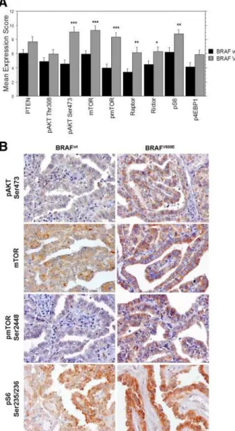
Overexpression of mTOR pathway proteins in cPTC withBRAFV600E
mutation
Because we observed distinct expression of mTOR pathway proteins across the different histotypes, with par-ticular overactivation in cPTC, and knowing the close ge-notype- phenotype correlation in thyroid tumors, we ver-ified whether the expression of mTOR pathway proteins in cPTC was correlated with the presence of the BRAF
mutation.BRAFV600Emutation was present in 23 of the
60 (38.3%) cPTC cases.
We observed a significantly higher expression of pAKT Ser473 (P ⬍ 0.0001), mTOR (P ⬍ 0.0001), pmTOR Ser2448 (P⬍0.0001), raptor (P⫽0.0037), rictor (P⫽ 0.0323), and pS6 Ser235/236 (P ⫽ 0.0084) in cPTC-BRAFV600Ethan in cPTC-BRAFwt (Fig. 2, A and B, and
Supplemental Table 4).
These observations suggest that BRAFV600E-mutated
cPTC is associated to a higher activation of the mTOR pathway.
Regulation of mTOR pathway activation by BRAF
To verify the possible effects of BRAFV600Ein the
ac-tivation of the mTOR pathway, we transiently transfected HEK293 cells with control pCMV, pCMV-BRAFwt, or
pCMV-BRAFV600E vectors. Additionally, we down-reg-ulated BRAF by siRNA in thyroid cancer cell lines and analyzed the alterations on the levels of mTOR pathway effectors.
As observed in Fig. 3A, after transfection with
pCMV-BRAFwtand pCMV-BRAFV600E, an increase in the mean
fold expression of mTOR (2.8⫾0.9 and 3.3⫾ 1.1, re-spectively), pmTOR Ser2448 (2.5⫾0.6 and 2.2⫾ 0.2, respectively), raptor (1.8 ⫾ 0.4 and 2.1 ⫾ 0.7, respec-tively), rictor (1.9⫾0.6 and 2.4⫾1.1, respectively), and pS6 Ser235/236 (1.9⫾ 0.5 and 2.0⫾ 0.3, respectively) was observed relative to pCMV-empty vector. The pCMV-BRAFV600Eexpression led to significant enhanced pmTOR Ser2448 and pS6 Ser235/236 levels (P⫽0.0023 andP⫽0.0284, respectively; Fig. 3, A and B).
The levels of mTOR pathway effectors were evaluated in K1, 8505C, TPC1, C643, and T241 thyroid cancer cell lines after BRAF down-regulation by siRNA. As observed in Fig. 4A, BRAF-C2 siRNA led to down-regulation of BRAF expression in all cell lines, although less efficiently in TPC1. Decrease of pERK1/2 was observed in all the cell lines, except in C643. In K1, such a difference was statis-tically significant (P⫽0.0002). In K1, 8505C, and C643,
a significant decrease of pmTOR Ser2448 was observed upon BRAF silencing (P ⫽ 0.0039 to 0.0127; Fig. 4B), whereas this treatment led to significant reduced expres-sion of pS6 Ser235/236 in the five cell lines (P⫽0.0004 to 0.0252; Fig. 4B).
Thesein vitroresults reveal a positive association be-tween BRAF expression and mTOR pathway activation and support the correlation between BRAFV600E and
overactivation of the mTOR pathway observed in cPTC cases, particularly the overexpression of pmTOR Ser2448 and pS6 Ser235/236.
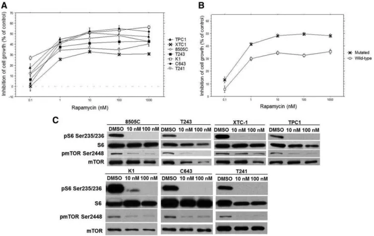
Oncogenic alterations in the ERK/MAPK pathway predict higher sensitivity of thyroid cancer cell lines to rapamycin
Basal activation of mTOR pathway was observed in the seven studied cell lines (Supplemental Fig. 1).
The sensitivity of thyroid cancer cell lines to rapa-mycin was compared. As observed in Fig. 5, A and B, rapamycin inhibited the growth of the seven cell lines, which was particularly evident at 10 and 100 nM con-centrations (Supplemental Table 5). 8505C, K1, T243,
FIG. 2. Expression of mTOR pathway proteins in cPTC with and withoutBRAFV600E
mutation. A, The immunostaining of PTEN, pAKT Thr308, pAKT Ser473, mTOR, pmTOR Ser2448, raptor, rictor, pS6 Ser235/236 and p4EBP1 Thr37/46 was evaluated. The expression score was obtained by multiplying the value attributed to the staining intensity by the value attributed to the percentage of stained cells. Results are shown as mean⫾SEM. *,P⬍0.05; **,P⬍0.01; ***,P⬍ 0.001 (unpaired Student’sttest and Mann-WhitneyUtest). B, Microphotographs representative of the immunohistochemical staining of pAKT Ser473, mTOR, pmTOR Ser2448, and pS6 Ser235/236 in cPTC-BRAFwt
and cPTC-BRAFV600E
cases. For all the evaluated proteins, the immunohistochemical staining was performed with 3,3⬘
-diaminobenzidine. Microphotographs were obtained at a 400⫻ magnification.
FIG. 3. mTOR pathway activation in transfected cells expressing exogenousBRAFwtorBRAFV600E. A, Mean fold change of protein
expression observed in HEK293 cells transfected with the expressing vectors pCMV-BRAFwtand pCMV-BRAFV600Ein comparison to cells
transfected with a pCMV-empty vector. The levels of PTEN, pAKT Thr308, pAKT Ser473, mTOR, pmTOR Ser2448, raptor, rictor, pS6 Ser235/236, and p4EBP1 Thr37/46 were quantified and normalized by the levels of control protein (actin or␣-Tubulin). Results are shown as mean expression value⫾SEMof at least three independent
experiments. *,P⬍0.05; **,P⬍0.01 (unpaired Student’sttest). B, Representative Western blot analysis of pmTOR Ser2448, mTOR, pS6 Ser235/236, and S6 expression levels in protein extracts of HEK293 cells transiently transfected with pCMV-empty vector and with the expressing plasmids pCMV-BRAFwtor pCMV-BRAFV600E. The levels of
TPC1, and C643 showed significantly higher growth inhibition than XTC1 and T241. Altogether, at 1 to 1000 nM doses, a lower sensitivity to rapamycin was observed in cell lines without MAPK alterations, com-pared with the ones harboring MAPK oncogenic alter-ations (P⬍0.0001; Fig. 5B).
In all seven cell lines, rapamycin remarkably reduced the levels of pS6 Ser235/236 (Fig. 5C), at 10 and 100 nM. Upon drug treatment, a decrease of pmTOR Ser2448 was also observed, except for the XTC-1 cell line.
These results indicate that oncogenic mutations in the MAPK pathway may confer higher sensitivity of thyroid cancer cell lines to rapamycin treatment.
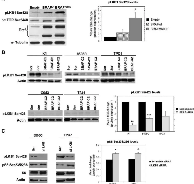
BRAFV600Eexpression regulates the levels of pLKB1 Ser428
To assess a possible mechanism underlying the link be-tweenBRAFV600Emutation and mTOR pathway
activa-tion observed in cPTC, we evaluated the levels of pLKB1 Ser428 in HEK293 cells transfected with pCMV-empty vector, pCMV-BRAFwt, or pCMV-BRAFV600Eas well as in thyroid cancer cell lines with BRAF knockdown by siRNA.
Increased expression of pLKB1 Ser428 was observed upon transfection with both BRAF expression vectors
compared to cells transfected with the empty vector: 2.2⫾ 0.8-fold for pCMV-BRAFwt, and 6.0 ⫾ 1.5-fold for pCMV-BRAFV600E (Fig. 6A); the 6-fold increase of
pLKB1 Ser428 expression observed in pCMV-BRAFV600E
transfected cells was statistically significant (P⫽0.0313; Fig. 6A).
At basal levels, pLKB1 Ser428 was barely detectable in C643 and T241. A decrease in pLKB1 Ser428 expression was observed in K1 (6.0⫾0.06-fold), 8505C (2.3⫾ 0.03-fold), and TPC1 (2.5⫾0.2-fold) upon BRAF-C2 siRNA (Fig. 6B), which was only significant in theBRAFV600Ecell lines, K1 and 8505C (P ⫽0.0019 and P⫽ 0.0009, re-spectively). We down-regulated LKB1 in 8505C, TPC1, and K1 by siRNA. pLKB1 Ser 428 expression was con-firmed to be decreased in 8505C and TPC1 (Fig. 6C), whereas it failed to be efficiently down-regulated in K1 (data not shown). In parallel, a significant up-regulation of pS6 Ser235/236 was detected in 8505C and TPC1 cell lines (P⫽0.03 and 0.02, respectively; Fig. 6C).
We analyzed the expression of phospho- LKB1 in our series of primary PTC characterized forBRAFV600E
muta-tion. We observed that BRAFV600Epositive cases had higher
phospho- LKB1 levels compared withBRAFwtcases (mean scores, 7.1vs.6.4, respectively), although the difference did not reach statistical significance (ttest,P⫽0.33).
FIG. 4. mTOR pathway expression in thyroid cancer cells lines with BRAF silencing. A, Western blot analysis of pmTOR Ser2448, mTOR, pS6 Ser235/236, and S6 expression levels in protein extracts of K1, 8505C, TPC1, C643, and T241 thyroid cancer cell lines treated with BRAF-C2 siRNA. The levels of BRAF were analyzed for control of silencing efficiency, and the levels of pERK 1/2 as readout of BRAF activity. Representative actin expression pattern is shown. Protein level, in scramble treated cell lines, was evaluated in duplicate (Scr), whereas in BRAF siRNA-treated cell lines, it was analyzed in triplicate. B, Mean fold change of protein expression observed in K1, 8505C, TPC1, C643, and T241 thyroid cancer cell lines treated with BRAF-C2 siRNA in comparison to cells treated with scramble siRNA. The levels of pmTOR Ser2448 and pS6 Ser235/ 236 were quantified and normalized by the levels of control protein (actin or␣-Tubulin). Results are shown as mean expression value⫾SEM. *,
Taken together, these results suggest that increased phosphorylation of pLKB1 Ser428 can be a mechanism contributing to the mTOR pathway overactivation ob-served inBRAFV600Ethyroid cancer cells.
Discussion
In the present study, we observed a particularly evident overactivation of AKT/mTOR pathway in cPTC. A sig-nificantly higher expression of mTOR pathway proteins was detected in cPTC compared with normal tissue and with the other types of differentiated thyroid carcinoma. Our immunohistochemical results fit with the previously reported increased expression of phospho-p70S6K and pAKT Ser473 in PTC tissues (29, 30). We observed a sig-nificantly higher up-regulation of the mTOR pathway in
BRAFV600Emutated cPTC compared withBRAFwtcPTC.
As far as we are aware, this is the first time that a
corre-lation betweenBRAFV600E mutation and overactivation
of the mTOR pathway is demonstrated in cPTC. In a pre-vious work of our group,BRAFV600E-mutated cutaneous
melanomas also showed up-regulation of mTOR pathway (25), suggesting that this effect is not tumor-type specific. Paradoxically, PTEN levels were also higher in cPTC, although with a preferential cytoplasmic localization. In fact, a decrease in PTEN nuclear localization was detected in these cases, which is in agreement with previous data showing reduced intensity of nuclear PTEN staining in PTC (16). Recent evidence showed thatPTEN posttrans-lational regulation (e.g. ubiquitination, oxidation, or phosphorylation) can have a role in cancer susceptibility by leading to inactive cytoplasmic forms and/or to defi-cient PTEN nuclear import (31–33).
In cPTC metastases, the levels of PTEN, pAKT Ser473, mTOR, pmTOR, and p4EBP1 were significantly lower relative to primary lesions. These results suggest that the
mTORC1 pathway might be less active in cPTC metasta-ses than in the primary lesions. Further work and a larger cohort are needed to determine whether the discrepancy observed between primary lesions and cPTC metastases reflects true biological heterogeneity or biologically mean-ingless quantitative differences.
The connection observed in cPTC between BRAFV600E
expression and mTOR pathway overactivation was fur-ther supported by ourin vitroresults: BRAF overexpres-sion led to enhanced pmTOR and pS6 expresoverexpres-sion, whereas BRAF down-regulation led to reduced expression of these proteins of the mTOR pathway. The up-regulation of
FIG. 6. Regulation of pLKB1 Ser428 levels by BRAFV600E
expression. A, Western blot analysis of pLKB1 Ser428 expression levels in protein extracts of HEK293 cells transiently transfected with pCMV-empty vector and with the expressing plasmids pCMV-BRAFwt
or pCMV-BRAFV600E
. Representative␣ -tubulin expression pattern is shown. The mean fold change of pLKB1 Ser428 levels in HEK293 cells transfected with the expressing vectors pCMV-BRAFwt
and pCMV-BRAFV600E
mTOR signaling in BRAF-transformed HEK293 cells was not exclusively associated with the mutated form of
BRAF, suggesting that overexpression of the wild-type BRAF protein likely mimics the situation created by the constitutional activation of BRAFV600E. Changes in
mTOR pathway uponBRAFsiRNA were also observed in cells harboring RASmutation and RET/PTC. This finding, together with the reported activation of the PI3K/AKT/mTOR pathway in cells harboring those ge-netic alterations or RETpoint mutation (19 –23, 34), led us to speculate that BRAF may (at least partially) modulate mTOR pathway activation in thyroid cells with genetic alterations in the MAPK pathway. In line with these observations, when we compared the sensitivity of thyroid cancer cell lines to rapamycin, a significantly higher growth inhibition was observed in thyroid cancer cell lines with oncogenic alterations in the MAPK pathway (BRAFV600E,RAS, andRET/PTC1).
Our results show for the first time that increased BRAF signaling leads to mTOR pathway activation and suggest that inhibition of both MAPK and mTOR pathways could be a good strategy in therapeutic targeting ofBRAFV600E -positive tumors. In fact, previous data have shown an in-creased or synergistic activity of BRAF (RAF265) or MEK inhibitors (RDEA119, AZD6244) and mTOR inhibitors (BEZ-235, temsirolimus, or rapamycin) in the growth in-hibition of thyroid cancer cell lines and xenograft tumors (35–37).
Additionally, we suggest a possible mechanism con-tributing to the effect of oncogenic BRAF on mTOR path-way activation. Specifically, we observed a positive cor-relation between BRAFV600Eexpression and pLKB1 levels
in vitro. Moreover, LKB1 down-regulation by siRNA led to activation of the mTOR pathway, as seen by increased levels of pS6 inBRAFV600Eas well as inRET/PTC
-har-boring thyroid cancer cell lines. As previously reported, LKB1 activity can be negatively regulated by Ser428 phos-phorylation induced by the RAF-MEK-ERK signaling cas-cade as well as by p90-RSK (9). Therefore, it is plausible that BRAFV600E mutations and RET/PTC
rearrange-ments, both activators of the ERK/MAPK pathway, can induce LKB1 Ser428 phosphorylation, compromising its ability to bind AMPK. In our series of human PTC, we observed a tendency for BRAFV600E-harboring cases to
display increased pLKB1Ser428 levels, although it is un-likely that mutant BRAF is the only factor regulating LKB1 phosphorylation in these lesionsin vivo.
Nonetheless, our results suggest that oncogenic BRAF-induced inactivation of LKB1 by Ser428 phosphorylation might underlie the mTOR pathway overactivation ob-served in cell lines and in human tumors harboring the
BRAFV600Emutation.
These data further suggest that AMPK could be a good target in BRAF-mutated thyroid cancer, which is in ac-cordance with a recent report showing that AICAR (AMPK activator) treatment inhibited the proliferation of
BRAFV600Emutant thyroid cancer cell lines more strongly
than of wild-type cell lines (38).
In PTC,BRAFV600Ehas been associated to a higher risk
of recurrence and radioactive iodine treatment resistance, particularly due to decreased expression of Na⫹/I⫺ sym-porter (NIS) and its impaired targeting to the membrane (39). A recent study demonstrated that rapamycin leads to an in-crease in NIS expression and iodine uptake in insulin- and TSH-stimulated thyroid PCCL3 cells (40). These previous data and our finding ofBRAFV600Eassociation with over-activation of mTOR pathway suggest that, inBRAFV600E
cPTC cases, the up-regulation of the mTOR pathway may lead to a decrease in NIS expression, and therefore, the use of mTOR inhibitors could be a strategy to increase the radio-iodine response ofBRAFV600E-cPTC.
In conclusion, we demonstrated that the AKT/mTOR pathway is particularly overactivated in human cPTC har-boring theBRAFV600Emutation. The association between
BRAFV600Emutation and mTOR pathway activation was
supported byin vitroresults. BRAF-induced phosphory-lation of LKB1Ser428 emerged as a possible mechanism contributing to such association, possibly through uncou-pling of the LKB1-AMPK-mTOR energy stress sensor.
Our results suggest and reinforce previous observations showing that targeting the mTOR pathway (as well as in combination with MAPK pathway/AMPK inhibition) could be a good strategy to improve therapy effect in thy-roid carcinomas harboring theBRAFV600Emutation.
Acknowledgments
We thank Vitor Trovisco for the BRAFwt and BRAFV600E
vectors.
Address all correspondence and requests for reprints to: Paula Soares, Institute of Molecular Pathology and Immunology of the University of Porto (IPATIMUP), Cancer Biology, Rua Dr. Ro-berto Frias, s/n, 4200-465 Porto, Portugal. E-mail: psoares@ ipatimup.pt.
This study was supported by the Portuguese Foundation for Science and Technology (FCT) through the project grant (PTDC/ SAU-OBD/69787/2006) and by Grant PS09/02050-FEDER from the Ministry of Science and Innovation (Instituto de Salud Carlos III), Spain. IPATIMUP is an Associate Laboratory of the Portuguese Ministry of Science, Technology, and Higher Edu-cation that is partially supported by the FCT.
References
1. Guertin DA, Sabatini DM2005 An expanding role for mTOR in cancer. Trends Mol Med 11:353–361
2. Wullschleger S, Loewith R, Hall MN2006 TOR signaling in growth and metabolism. Cell 124:471– 484
3. Hay N, Sonenberg N2004 Upstream and downstream of mTOR. Genes Dev 18:1926 –1945
4. Sarbassov DD, Guertin DA, Ali SM, Sabatini DM2005 Phosphor-ylation and regulation of Akt/PKB by the rictor-mTOR complex. Science 307:1098 –1101
5. Hardie DG, Scott JW, Pan DA, Hudson ER2003 Management of cellular energy by the AMP-activated protein kinase system. FEBS Lett 546:113–120
6. Shaw RJ, Kosmatka M, Bardeesy N, Hurley RL, Witters LA, DePinho RA, Cantley LC2004 The tumor suppressor LKB1 ki-nase directly activates AMP-activated kiki-nase and regulates apo-ptosis in response to energy stress. Proc Natl Acad Sci USA 101: 3329 –3335
7. Shaw RJ, Bardeesy N, Manning BD, Lopez L, Kosmatka M, De-Pinho RA, Cantley LC2004 The LKB1 tumor suppressor negatively regulates mTOR signaling. Cancer Cell 6:91–99
8. Zheng B, Jeong JH, Asara JM, Yuan YY, Granter SR, Chin L, Cantley LC2009 Oncogenic B-RAF negatively regulates the tumor suppressor LKB1 to promote melanoma cell proliferation. Mol Cell 33:237–247
9. Esteve-Puig R, Canals F, Colome´ N, Merlino G, Recio JA2009 Uncoupling of the LKB1-AMPK␣energy sensor pathway by growth factors and oncogenic BRAF. PLoS One 4:e4771
10. Kondo T, Ezzat S, Asa SL2006 Pathogenetic mechanisms in thyroid follicular-cell neoplasia. Nat Rev Cancer 6:292–306
11. Sobrinho-Simo˜es M, Ma´ximo V, Rocha AS, Trovisco V, Castro P, Preto A, Lima J, Soares P2008 Intragenic mutations in thyroid cancer. Endocrinol Metab Clin North Am 37:333–362, viii 12. Kimura ET, Nikiforova MN, Zhu Z, Knauf JA, Nikiforov YE, Fagin
JA2003 High prevalence of BRAF mutations in thyroid cancer: genetic evidence for constitutive activation of the RET/PTC-RAS-BRAF signaling pathway in papillary thyroid carcinoma. Cancer Res 63:1454 –1457
13. Zhu XL, Zhou XY, Zhu XZ2005 [BRAFV599E mutation and RET/ PTC rearrangements in papillary thyroid carcinoma]. Zhonghua Bing Li Xue Za Zhi 34:270 –274
14. Alvarez-Nun˜ez F, Bussaglia E, Mauricio D, Ybarra J, Vilar M, Le-rma E, de Leiva A, Matias-Guiu X2006 PTEN promoter methyl-ation in sporadic thyroid carcinomas. Thyroid 16:17–23 15. Halachmi N, Halachmi S, Evron E, Cairns P, Okami K, Saji M,
Westra WH, Zeiger MA, Jen J, Sidransky D1998 Somatic mutations of the PTEN tumor suppressor gene in sporadic follicular thyroid tumors. Genes Chromosomes Cancer 23:239 –243
16. Gimm O, Perren A, Weng LP, Marsh DJ, Yeh JJ, Ziebold U, Gil E, Hinze R, Delbridge L, Lees JA, Mutter GL, Robinson BG, Komminoth P, Dralle H, Eng C2000 Differential nuclear and cytoplasmic expression of PTEN in normal thyroid tissue, and benign and malignant epithelial thyroid tumors. Am J Pathol 156:1693–1700
17. Wu G, Mambo E, Guo Z, Hu S, Huang X, Gollin SM, Trink B, Ladenson PW, Sidransky D, Xing M2005 Uncommon mutation, but common amplifications, of the PIK3CA gene in thyroid tumors. J Clin Endocrinol Metab 90:4688 – 4693
18. Abubaker J, Jehan Z, Bavi P, Sultana M, Al-Harbi S, Ibrahim M, Nuaim A, Ahmed M, Amin T, Fehaily M, Sanea O, Al-Dayel F, Uddin S, Al-Kuraya KS2008 Clinicopathological analysis of papillary thyroid cancer with PIK3CA alterations in a Middle Eastern population. J Clin Endocrinol Metab 93:611– 618 19. Rodriguez-Viciana P, Warne PH, Dhand R, Vanhaesebroeck B,
Gout I, Fry MJ, Waterfield MD, Downward J1994 Phosphati-dylinositol-3-OH kinase as a direct target of Ras. Nature 370: 527–532
20. Rodriguez-Viciana P, Warne PH, Vanhaesebroeck B, Waterfield MD, Downward J1996 Activation of phosphoinositide 3-kinase by interaction with Ras and by point mutation. EMBO J 15: 2442–2451
21. Miyagi E, Braga-Basaria M, Hardy E, Vasko V, Burman KD, Jhiang S, Saji M, Ringel MD2004 Chronic expression of RET/PTC 3 en-hances basal and insulin-stimulated PI3 kinase/AKT signaling and increases IRS-2 expression in FRTL-5 thyroid cells. Mol Carcinog 41:98 –107
22. Jung HS, Kim DW, Jo YS, Chung HK, Song JH, Park JS, Park KC, Park SH, Hwang JH, Jo KW, Shong M2005 Regulation of protein kinase B tyrosine phosphorylation by thyroid-specific oncogenic RET/PTC kinases. Mol Endocrinol 19:2748 –2759
23. Kim DW, Hwang JH, Suh JM, Kim H, Song JH, Hwang ES, Hwang IY, Park KC, Chung HK, Kim JM, Park J, Hemmings BA, Shong M 2003 RET/PTC (rearranged in transformation/papillary thyroid carcinomas) tyrosine kinase phosphorylates and activates phospho-inositide-dependent kinase 1 (PDK1): an alternative phosphatidyl-inositol 3-kinase-independent pathway to activate PDK1. Mol En-docrinol 17:1382–1394
24. Po´pulo H, Soares P, Rocha AS, Silva P, Lopes JM2010 Evaluation of the mTOR pathway in ocular (uvea and conjunctiva) melanoma. Melanoma Res 20:107–117
25. Po´pulo H, Soares P, Faustino A, Rocha AS, Silva P, Azevedo F, Lopes JM2011 mTOR pathway activation in cutaneous melanoma is as-sociated with poorer prognosis characteristics. Pigment Cell Mela-noma Res 24:254 –257
26. Meireles AM, Preto A, Rocha AS, Rebocho AP, Ma´ximo V, Pereira-Castro I, Moreira S, Feija˜o T, Botelho T, Marques R, Trovisco V, Cirnes L, Alves C, Velho S, Soares P, Sobrinho-Simo˜es M2007 Molecular and genotypic characterization of human thyroid follic-ular cell carcinoma-derived cell lines. Thyroid 17:707–715 27. Ricarte-Filho JC, Ryder M, Chitale DA, Rivera M, Heguy A,
Ladanyi M, Janakiraman M, Solit D, Knauf JA, Tuttle RM, Ghos-sein RA, Fagin JA2009 Mutational profile of advanced primary and metastatic radioactive iodine-refractory thyroid cancers reveals dis-tinct pathogenetic roles for BRAF, PIK3CA, and AKT1. Cancer Res 69:4885– 4893
28. Preto A, Gonc¸alves J, Rebocho AP, Figueiredo J, Meireles AM, Rocha AS, Vasconcelos HM, Seca H, Seruca R, Soares P, Sobrinho-Simo˜es M2009 Proliferation and survival molecules implicated in the inhibition of BRAF pathway in thyroid cancer cells harbouring different genetic mutations. BMC Cancer 9:387
29. Miyakawa M, Tsushima T, Murakami H, Wakai K, Isozaki O, Takano K2003 Increased expression of phosphorylated p70S6 kinase and Akt in papillary thyroid cancer tissues. Endocr J 50: 77– 83
30. Vasko V, Saji M, Hardy E, Kruhlak M, Larin A, Savchenko V, Miyakawa M, Isozaki O, Murakami H, Tsushima T, Burman KD, De Micco C, Ringel MD2004 Akt activation and localisation cor-relate with tumour invasion and oncogene expression in thyroid cancer. J Med Genet 41:161–170
31. Tamguney T, Stokoe D2007 New insights into PTEN. J Cell Sci 120:4071– 4079
32. Trotman LC, Wang X, Alimonti A, Chen Z, Teruya-Feldstein J, Yang H, Pavletich NP, Carver BS, Cordon-Cardo C, Erdjument-Bromage H, Tempst P, Chi SG, Kim HJ, Misteli T, Jiang X, Pandolfi PP2007 Ubiquitination regulates PTEN nuclear import and tumor suppression. Cell 128:141–156
33. Maccario H, Perera NM, Gray A, Downes CP, Leslie NR2010 Ubiquitination of PTEN (phosphatase and tensin homolog) inhibits phosphatase activity and is enhanced by membrane targeting and hyperosmotic stress. J Biol Chem 285:12620 –12628
34. Rapa I, Saggiorato E, Giachino D, Palestini N, Orlandi F, Papotti M, Volante M2011 Mammalian target of rapamycin pathway activa-tion is associated to RET mutaactiva-tion status in medullary thyroid car-cinoma. J Clin Endocrinol Metab 96:2146 –2153
inhi-bition of mitogen-activated protein kinase kinase and mammalian target of rapamycin in differentiated and anaplastic thyroid cancer. J Clin Endocrinol Metab 94:4107– 4112
36. Jin N, Jiang T, Rosen DM, Nelkin BD, Ball DW2011 Synergistic action of a RAF inhibitor and a dual PI3K/mTOR inhibitor in thy-roid cancer. Clin Cancer Res 17:6482– 6489
37. Liu D, Xing J, Trink B, Xing M2010 BRAF mutation-selective inhibition of thyroid cancer cells by the novel MEK inhibitor RDEA119 and genetic-potentiated synergism with the mTOR in-hibitor temsirolimus. Int J Cancer 127:2965–2973
38. Choi HJ, Kim TY, Chung N, Yim JH, Kim WG, Kim JA, Kim WB,
Shong YK2011 The influence of the BRAF V600E mutation in thyroid cancer cell lines on the anticancer effects of 5-aminoimida-zole-4-carboxamide-ribonucleoside. J Endocrinol 211:79 – 85 39. Riesco-Eizaguirre G, Gutie´rrez-Martínez P, García-Cabezas MA,
Nistal M, Santisteban P2006 The oncogene BRAF V600E is asso-ciated with a high risk of recurrence and less differentiated papillary thyroid carcinoma due to the impairment of Na⫹/I⫺targeting to the membrane. Endocr Relat Cancer 13:257–269
40. de Souza EC, Padro´n AS, Braga WM, de Andrade BM, Vaisman M, Nasciutti LE, Ferreira AC, de Carvalho DP2010 MTOR down-regulates iodide uptake in thyrocytes. J Endocrinol 206:113–120