Alendronate induces gastric damage by reducing nitric oxide synthase
expression and NO/cGMP/K
ATP
signaling pathway
Renan O. Silva
a, Larisse T. Lucetti
b, Deysi V.T. Wong
b, Karoline S. Aragão
b, Eudmar M.A. Junior
b,
Pedro M.G. Soares
b, André Luiz R. Barbosa
a,c, Ronaldo A. Ribeiro
b, Marcellus H.L.P. Souza
b,
Jand-Venes R. Medeiros
a,c,⇑aPost-Graduation Program in Biotechnology, Biotechnology and Biodiversity Center Research (BIOTEC), Federal University of Piauí, Parnaíba, PI, Brazil bDepartment of Physiology and Pharmacology, Federal University of Ceará, Fortaleza, CE, Brazil
cPost-Graduation Program in Pharmacology, Federal University of Piauí, Teresina, PI, Brazil
a r t i c l e
i n f o
Article history:
Received 4 February 2014 Revised 2 May 2014 Available online 12 May 2014
Keywords:
Gastric damage Nitric oxide synthase Nitric oxide Alendronate
a b s t r a c t
Chronic use of alendronate has been linked to gastrointestinal tract problems. Our objective was to eval-uate the role of the NO/cGMP/KATPsignaling pathway and nitric oxide synthase expression in alendro-nate-induced gastric damage. Rats were either treated with the NO donor, sodium nitroprusside (SNP; 1, 3, and 10 mg/kg), or the NO synthase (NOS) substrate,L-arginine (L-Arg; 50, 100, and 200 mg/kg). Some rats were pretreated with either ODQ (a guanylate cyclase inhibitor; 10 mg/kg) or glibenclamide (KATP channels blocker; 10 mg/kg). In other experiments, rats were pretreated withL-NAME (non-selective NOS inhibitor; 10 mg/kg), 1400W (selective inducible NOS [iNOS] inhibitor; 10 mg/kg), orL-NIO (a selec-tive endothelial NOS [eNOS] inhibitor; 30 mg/kg). After 1 h, the rats were treated with alendronate (30 mg/kg) by gavage for 4 days. SNP andL-Arg prevented alendronate-induced gastric damage in a dose-dependent manner. Alendronate reduced nitrite/nitrate levels, an effect that was reversed with SNP orL-Arg treatment. Pretreatment with ODQ or glibenclamide reversed the protective effects of SNP andL-Arg.L-NAME, 1400W, orL-NIO aggravated the severity of alendronate-induced lesions. In addi-tion, alendronate reduced the expression of iNOS and eNOS in the gastric mucosa. Gastric ulcerogenic responses induced by alendronate were mediated by a decrease in NO derived from both eNOS and iNOS. In addition, our findings support the hypothesis that activation of the NO/cGMP/KATP pathway is of pri-mary importance for protection against alendronate-induced gastric damage.
Ó2014 Elsevier Inc. All rights reserved.
1. Introduction
Bisphosphonates are the class of drugs most commonly used for the control, prevention, and treatment of metabolic bone diseases,
including osteoporosis [1]. Alendronate is a frequently used
bis-phosphonate, due to dosing convenience and benefits related to the prevention of fractures. However, recent studies have shown that chronic alendronate use may be associated with side effects primarily involving the gastrointestinal tract, including gastric
ulcer and erosive esophagitis[2,3].
Recently, studies have demonstrated that gaseous mediators,
such as carbon monoxide (CO)[4]and hydrogen sulfide (H2S)[5],
contribute to the protection and maintenance of gastric mucosal
integrity against alendronate-induced gastric damage. Similarly, nitric oxide (NO) may protect the gastrointestinal mucosa
from damage caused by chemical agents, such as ethanol[6]and
NSAIDs [7]. However, few studies have demonstrated the
involvement of NO in the pathogenesis of alendronate-induced gastropathy.
NO plays an important role in regulating various cellular func-tions in the cardiovascular, immune, and neuronal systems, as well as in the gastrointestinal tract. NO is synthesized in endothelial cells by the nitric oxide synthase (NOS) family of enzymes; NOS
enzymes convert the amino acid,L-arginine, intoL-citrulline and
NO[8]. Constitutive NOS (cNOS), which is expressed in normal
conditions, comprises the 2 isoforms endothelial NOS (eNOS) and neuronal NOS (nNOS), that play important roles in the
mainte-nance of mucosal integrity[9,10]. A third isoform, which is known
as inducible NOS (iNOS), is not constitutively expressed. Rather, it is stimulated by factors such as cytokines or microbial products [11].
http://dx.doi.org/10.1016/j.niox.2014.05.002 1089-8603/Ó2014 Elsevier Inc. All rights reserved.
⇑Corresponding author at: Laboratory of Experimental Physiopharmacology (LAFFEX), Federal University of Piauí, Av. São Sebastião, no2819, CEP 64202-020 Parnaíba, PI, Brazil.
E-mail address:[email protected](J.-V.R. Medeiros).
Contents lists available atScienceDirect
Nitric Oxide
In this study, we have investigated the role of the NO/cGMP/
KATP signaling pathway and nitric oxide synthase expression in
alendronate-induced gastric damage in rats.
2. Materials and methods
2.1. Animals
Female Wistar rats (100–140 g) were obtained from the Depart-ment of Physiology and Pharmacology, Federal University of Ceará. The animals were deprived of food for 18–24 h before the experi-ment, but had free access to water. All animal treatments and sur-gical procedures were performed in accordance with the Guide for Care and Use of Laboratory Animals (National Institutes of Health, Bethesda, MD) and were approved by the local ethics committee (Protocol No. 0067/10).
2.2. Drugs and solutions
Sodium nitroprusside (SNP), L-arginine (L-Arg),
1H-[1,2,4]Oxadiazolo[4,3-a]quinoxalin-1-one (ODQ), glibenclamide,
NG-nitro-Larginine methyl ester (L-NAME), 1400W and
alendro-nate were purchased from Sigma Aldrich (St. Louis, MO).L-NG
-imi-noethyl-L-ornitine (L-NIO) was purchased from Cayman Chemical (Ann Arbor, MI). ODQ was dissolved 0.01% DMSO. Glibenclamide was dissolved in 0.01 N NaOH containing 4% glucose. Alendronate was dissolved in saline and adjusted to pH 7.0 by adding NaOH or
HCl[12]. All other drugs were dissolved in saline.
2.3. Effect of sodium nitroprusside orL-arginine on
alendronate-induced gastric damage
The animals were pretreated with saline, SNP (a nitric oxide
donor: 1, 3, and 10 mg/kg, po) orL-Arg (a substrate of NOS; 50,
100 and 200 mg/kg, ip). After 30 min, alendronate (30 mg/kg, pH 7.0, po) was administered. The control group received only saline.
All drugs were administered once daily for 4 days[4]. On the last
day of treatment, 4 h after alendronate administration, the animals were killed and their stomachs removed. Gastric damage was
mea-sured using Image JÒ
software. A sample was fixed in 10% formalin immediately after its removal for subsequent histopathological assessment. Other samples were then weighed, frozen, and stored
at 80°C until assayed for NO3/NO2production[13], glutathione
(GSH) levels [14], malondialdehyde (MDA) concentration [15],
myeloperoxidase (MPO) activity[16]and cytokine levels[17].
2.4. Histological evaluation of gastric damage
For histological evaluation, the stomach samples were fixed in 10% formalin solution for 24 h. After fixation, the samples were transferred to a solution of 70% alcohol. The material was then
embedded in paraffin and sectioned; 4-
l
m-thick sections weredeparaffinized, stained with hematoxylin and eosin (H&E), and then examined under a light microscope by an experienced pathol-ogist without knowledge of the treatments (Soares, PMG). The specimens were assessed according to the criteria as previously described[18].
2.5. Measurement of nitrite plus nitrate production
Homogenate of gastric tissue was incubated in a microplate with nitrate reductase for 12 h to convert NO3in NO2. Nitric oxide
production was determined by measuring nitrite concentrations in
an ELISA plate reader at 540 nm using the Griess method[13].
Results were expressed as micromoles of nitrite (
l
mol) using theinternal standard curve.
2.6. Role of soluble guanylate ciclase and ATP-Sensitive K+channels
on gastroprotective effect of sodium nitroprusside orL-arginine
The animals were pretreated with ODQ (guanylate ciclase inhibitor: 10 mg/kg, ip) or glibenclamide (a drug that blocks
ATP-Sensitive K+ Channels: 10 mg/kg, ip). One hour after, received
SNP (a nitric oxide donor: 10 mg/kg, po) or L-Arg (a substrate of
NOS; 200 mg/kg, ip). After 30 min, the animals received alendro-nate (30 mg/kg, pH 7.0, po). The control group received only saline. All drugs were administered once daily for 4 days. On the last day of treatment, 4 h after alendronate administration, the animals were killed, their stomachs removed, gastric damage and biochem-ists analysis were determined as described above.
2.7. Role of nitric oxide synthase on alendronate-induced gastric damage
The animals were initially treated withL-NAME (a non-selective
inhibitor of NOS; 3 mg/kg, ip), 1400W (a selective inhibitor of
iNOS; 10 mg/kg, ip) or L-NIO (a selective inhibitor of eNOS;
30 mg/kg, sc)[6,19]. After 1 h, the animals received alendronate
(30 mg/kg, pH 7.0, po). All drugs were administered once daily
for 4 days[4]. On the last day of treatment, 4 h after alendronate
administration, the animals were killed and their stomachs removed and gastric damage and biochemists analysis were deter-mined as described above. Samples were removed for immunohis-tochemistry analysis and western blot for iNOS and eNOS as described below.
2.8. Immunohistochemistry for eNOS and iNOS
The samples from the stomach of rats undergoing alendronate-induced gastric damage were assessed for the expression of eNOS and iNOS by employing an immunohistochemical technique. The slides mounted from the paraffin blocks were deparaffinized and then hydrated. Endogenous peroxidase activity was blocked with
1% H2O2 diluted in methanol. Then, the slides were washed in
phosphate-buffered saline (PBS). Next, the slides were incubated with primary antibody (1:400, Santa Cruz Biotechnology, Santa
Cruz, CA) overnight at 4°C. After washing, the slides were
incu-bated with biotinylated secondary antibody, diluted in PBS plus bovine serum albumin (PBS–BSA). Negative control sections were processed simultaneously as described above, but without adding an antibody. Finally, the tissue was stained for antigen–antibody complexes using a peroxidase detection system and then viewed under a microscope.
2.9. Western blot for iNOS and eNOS
Briefly, the tissues were homogenized in 0.2 ml of lysis buffer
containing protease inhibitors. Total protein (50
l
g protein/well)was resolved on 10% (iNOS) or 12.5% (eNOS) sodium dodecyl sul-fate–polyacrylamide gel (SDS–PAGE) and transferred to a nitrocel-lulose membrane (Hybond-ECL, Amersham Pharmacia Biotech, Amersham, UK). The membranes were blocked with 5% skimmed milk/Tris-buffered saline with 0.1% Tween 20 (TBS-T) for 2 h at
4°C followed by an overnight incubation period at 4°C with the
primary antibodies (rabbit polyclonal anti-iNOS, 1:500; anti-eNOS, 1:800 or anti-b-actin, 1:1000; Santa Cruz Biotechnology, Santa Cruz, CA). The blots were washed, followed by incubation with horseradish peroxidase-conjugated secondary antibody (donkey anti-rabbit immunoglobulin G, 1:1000; Santa Cruz Biotechnology,
Santa Cruz, CA) for 1 h at 4°C. The membranes were washed,
incubated with electrogenerated chemiluminescence (Amersham Pharmacia Biotech), and exposed to Hyperfilm ECL (Amersham Pharmacia Biotech) to observe the marked proteins. Densitometry
analyses were performed by Image JÒ
software. Data were expressed as the relative density of iNOS/b-actin and eNOS/b-actin bands.
2.10. Glutathione (GSH) levels
The samples were homogenized in 0.02 M EDTA (1 ml/100 mg
of tissue). Aliquots (400
l
l) of the tissue homogenate were mixedwith 320
l
l of distilled water and 80l
l of 50% (w/v) trichloroacetic acid in glass tubes and centrifuged at 3000 rpm for 15 min. Next,400
l
l of each supernatant was mixed with 800l
l of Tris buffer(0.4 M, pH 8.9) and 20
l
l of 0.01 M DTNB. Subsequently, thesam-ples were stirred for 3 min and read on a spectrophotometer at
412 nm[14]. The results are expressed as micrograms of GSH per
gram of tissue (
l
g/g).2.11. Malondialdehyde (MDA) concentration
The samples were homogenized in cold 1.15% KCl (1 ml/100 mg
of tissue). Briefly, 250
l
l of each homogenate was added 1%phos-phoric acid (H3PO4) and 0.6%tert-butyl alcohol (aqueous solution).
Then, this mixture was stirred and heated in a boiling water bath for 45 min. The mixture was then cooled immediately in an ice
water bath followed by the addition of 4 ml ofn-butanol. This
mix-ture was shaken and the butanol layer was separated by centrifu-gation at 1200 rpm for 15 min. Optical density was determined to be 535 and 520 nm, and the optical density difference between the
two determinations was calculated as thetert-butyl alcohol value
[15]. Results are expressed as nanomoles per gram of tissue
(nmol/g).
2.12. Myeloperoxidase (MPO) activity
Briefly, tissue was homogenized in potassium buffer with 0.5% of hexadecitrimetilamônio (HTAB) (1 ml/100 mg of tissue). Then, homogenate was centrifuged at 4500 rpm for 20 min. MPO activity in the resuspended pellet was assayed by measuring the change in absorbance at 450 nm using o-dianisidine dihydrochloride and 1%
hydrogen peroxide[16]. The results were expressed as the MPO
units per mg of tissue (UMPO/mg of tissue).
2.13. Cytokine measurements
Briefly, microtiter plates were coated overnight at 4°C with an
polyclonal anti-rat TNF-
a
or IL-1b(4l
g/ml, DuoSet ELISADevelop-ment kit R&D Systems). After blocking the plates, the samples and standards were added at various dilutions in duplicate and
incu-bated at 4°C for 24 h. The plates were washed with buffer
(0.01 M phosphate, 0.05 M NaCl, 0.1% Tween 20, pH 7.2). After
washing the plates, biotinylated sheep polyclonal anti-TNF-
a
oranti-IL-1b(diluted 1:1000 with assay buffer containing 1% bovine
serum albumin [BSA]) was added to the wells. After further
incuba-tion at room temperature for 1 h, the plates were washed and 50
l
lof avidin-conjugated horseradish peroxidase diluted 1:5000 was added to the wells. The color reagent o-phenylene-diamine (OPD;
40
l
g/well) was added 15 min later and the plates were incubatedin the dark at 37°C for 15–20 min. The enzyme reaction was
stopped with H2SO4 and absorbance was measured at 490 nm
[17]. Values are expressed as pictograms of cytokines per milliliter
(pg/ml).
2.14. Statistical analysis
Data were described as either means ± SEM or median, as appropriate. Analysis of Variance (ANOVA) followed by Student-Newman–Keuls test was used to compare means and Kruskal– Wallis nonparametric test, followed by Dunn´s tests to compare medians;P< 0.05 was defined as statistically significant.
3. Results
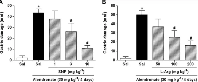
3.1. Effect of SNP orL-arginine on alendronate-induced gastric damage
In the present study, we observed that pretreatment with either
the NO donor, sodium nitroprusside (SNP;Fig. 1A) or the NOS
sub-strate,L-arginine (L-Arg;Fig. 1B) prevented alendronate-induced
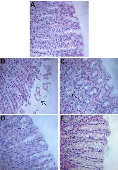
macroscopic gastric damage in a dose-dependent manner. Further-more, the results of microscopic analysis indicated that alendro-nate administration induced alterations in the gastric region characterized by epithelial cell loss, inflammatory cell, edema,
and intense hemorrhage (Fig. 2andTable 1). These changes were significantly prevented in rats pretreated with either SNP (Fig. 2D) orL-Arg (Fig. 2E).
3.2. Nitrite/nitrate production
As shown inFig. 3, administration of alendronate to gastric
tis-sue significantly reduced the generation of nitrite and nitrate metabolites of NO compared to controls. Pretreatment with SNP orL-Arg reversed this effect of alendronate; nitrite and nitrate lev-els were raised to values similar to those observed in the control group.
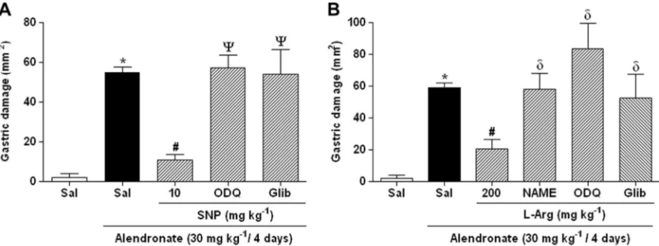
3.3. Role of soluble guanylate ciclase and ATP-sensitive K+channels
on gastroprotective effect of SNP orL-arginine
To assess the contribution of soluble guanylate cyclase and KATP
channels to the protective effects of SNP and L-Arg, rats were
pretreated with either ODQ, a guanylate cyclase inhibitor, or
gli-benclamide, a KATPchannel blocker.Fig. 4shows that pretreatment
with ODQ or glibenclamide significantly (P< 0.05) reversed the
positive effect that SNP (Fig. 4A) andL-Arg (Fig. 4B) had on alendr-onate-induced gastric macroscopic damage.
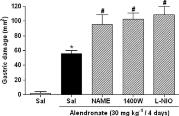
3.4. Role of nitric oxide synthase on alendronate-induced gastric damage
The role of NOS in alendronate-induced gastric damage was also
evaluated and the results are shown inFig. 5. Pretreatment with
L-NAME, 1400W, or L-NIO markedly aggravated the severity of
alendronate-induced gastric macroscopic damage.
The expression of iNOS (Fig. 6A) and eNOS (Fig. 6B) was evident from immunohistochemical analysis of the gastric mucosa of con-trol rats. However, lower staining levels of both iNOS and eNOS were observed in rats treated with alendronate. The results of protein expression experiments by western blotting were in
Fig. 2.Histopathological changes in the gastric mucosa (40magnification). (A) Control (saline) group showing gastric mucosal integrity. Alendronate (30 mg/kg, po) group showing lesions in the superficial gastric glandular region with (B) epithelial cell loss, (C) and bleeding. (D) Treatment with sodium nitroprusside (SNP; 10 mg/kg, po) + alendronate (30 mg/kg po), and with (E)L-Arginine (L-Arg; 200 mg/kg, ip) + alendronate (30 mg/kg po), showing reduction in alendronate-induced microscopic lesions.
agreement with immunohistochemical findings; protein expres-sion levels of iNOS (Fig. 7A) and eNOS (Fig. 7B) were significantly (P< 0.05) reduced after treatment with alendronate.
3.5. GSH, MDA, MPO and cytokine levels
Alendronate significantly reduced GSH levels, increased MDA concentrations, and increased MPO activity in gastric tissue when
compared to control, as shown inTable 2. Pretreatment with SNP
orL-Arg significantly increased GSH levels, reduced MDA
concen-trations, and reduced MPO levels (P< 0.05) in rats that received
alendronate. Furthermore, we observed that administration of either ODQ or glibenclamide reversed the gastroprotective effects
of SNP orL-Arg, as evidenced by changes in GSH, MDA and MPO.
Similarly, L-NAME combined with L-Arg treatment significantly
reduced GSH levels, increased MDA concentrations, and increased
MPO activity compared to rats treated withL-Arg alone. In contrast,
pretreatment withL-NAME, 1400W, andL-NIO had no effect on
GSH levels, MDA concentration, or MPO activity in the gastric
mucosa of rats treated with alendronate alone (Table 2).
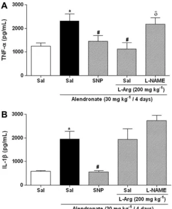
Treatment with either SNP or L-Arg significantly (P< 0.05)
reduced TNF-
a
levels in rats with alendronate-induced gastricdamage. In addition, SNP treatment led to a reduction in IL-1b
lev-els (Fig. 8). The observed changes in TNF-
a
were significantlyreversed withL-NAME pretreatment, but not withL-Arg
pretreat-ment.L-NAME pretreatment had no impact on IL-1blevels.
4. Discussion
In the present study, we show that SNP andL-Arg, acting via NO/
cGMP/KATP-dependent mechanisms, protect the gastric mucosa
against alendronate-induced damage. In addition, we demon-strated that alendronate reduced NO generation by modulating NOS expression.
Consistent with previous studies [4,20,21], we observed that
when given as a single injection for 4 days to fasting rats, alendro-nate damaged the gastric mucosa causing lesions to appear in the antrum and extend further to develop into ulcers with a white cap.
Table 1
Effect of sodium nitroprusside orL-arginine in alendronate-induced gastric microscopic damage.
Experimental group Hemorrhagic damage (score 0–4) Edema (score 0–4) Epithelial cell loss (score 0–3) Inflammatory cells (score 0–3) Total (score 0–14)
Saline 0 (0–1) 0 0 (0–1) 0 0
Saline + alendronate 3 (1–3)a 3 (2–3)a 3 (2–3)a 2 (2–3)a 11 (2–3)a
SNP + alendronate 2 (0–2) 0 (0–1)b 0 (0–1)b 0 (0–1)b 2 (0–2)b
L-Arg + Alendronate 1 (0–2)b 1 (0–1)b 0 (0–1)b 1 (0–1)b 3 (0–1)b
Data shown are medians with minimal and maximal scores shown in parentheses. Kruskal–Wallis nonparametric test, followed by Dunn’s test was used for multiple comparisons for histological assessment.
aP< 0.05, when compared with saline group. b P< 0.05, when compared with alendronate group.
Fig. 3.Nitric oxide levels (NO3/NO2) in rats pretreated with sodium nitroprusside (SNP) andL-arginine (L-Arg) in rats with alendronate-induced gastric damage. Rats were pretreated with either saline, SNP (10 mg/kg, po), orL-Arg (200 mg/kg, ip) 30 min before alendronate (30 mg/kg) administration. The control group received only saline. All drugs were administered once daily for 4 days. Data are expressed as the mean ± S.E.M. of 5–6 rats per group.⁄P< 0.05 vs. saline group,#P< 0.05 vs. alendronate group, one-way ANOVA and Newman–Keuls test.
Fig. 4.Role of soluble guanylate cyclase (sGC) and ATP-sensitive K+channels (K
In contrast, lesions that formed in the corpus healed. A previous study demonstrated that the white cap covering the damaged mucosa is composed mainly of inflammatory cells and fibrin-like
substances [22]. Histologically, we observed that gastric ulcers
caused by alendronate administration induced severe edema, hem-orrhagic damage, epithelial cell loss, and infiltration of inflamma-tory cells in the submucosa. Other studies have reported that when alendronate is applied to the gastric mucosa it causes a decrease in transmucosal potential difference of the stomach, which is suggestive of a disruption to surface epithelial cells due
to a direct action[12]. These toxic effects of alendronate in the
stomach have been linked to a direct effect of this agent on the
mucosal surface[22,23].
Several studies have shown that biphosphonates also interfere with cell migration at the site of inflammation, and this effect is
associated with increased production of proinflammatory
cyto-kines, IL-1b, TNF-
a
, and IL-6, and oxidative stress[24,25]. In thepresent study, increased MDA and MPO levels, increased
accumu-lation of TNF-
a
and IL-1b, and decreased GSH levels by alendronatein the gastric mucosa suggest that neutrophil infiltration and
pro-inflammatory cytokines (TNF-
a
and IL-1b) contribute to oxidativegastric damage. Activated neutrophils are also a potential source of oxygen metabolites that can contribute to gastric mucosal injury
[26]. It has been suggested that oxygen-derived free radicals may
contribute to alendronate-induced gastric mucosal lesions[20,21].
NO plays a critical role in mucosal integrity, either by direct
action, or by modulating the effects of other substances [27]
involved in important physiological functions, such as regulation
of mucosal blood flow and mucus generation[28]. In the present
study, we demonstrated that both the NO donor, SNP, and the
NOS substrate,L-Arg protected the gastric mucosa against
alendro-nate-induced macroscopic damage. Histological observations
fur-ther validated macroscopic findings that SNP and L-Arg prevent
gastric damage induced by subchronic treatment with oral
alendr-onate. Furthermore, SNP andL-Arg inhibited alendronate-induced
elevations in MDA, MPO, TNF-
a
, and IL-1b levels, andalendro-nate-induced reductions in GSH. Together, these data suggest that
the gastroprotective effects of SNP andL-Arg may be dependent on
their inhibitory effects on neutrophil infiltration and
neutrophil-associated TNF-
a
and IL-1b responses. Thus, the mechanismthrough which NO exerts its gastroprotective effect appears to involve a reduction of lipid peroxidation induced by alendronate in the gastric mucosa. Therefore, the effects of NO may result in a decreased redox state in alendronate-induced gastropathy.
It is well known that NO modulates the activity of mucosal immunocytes, as well as modulating leukocyte-endothelial inter-actions. NO inhibits recruitment of neutrophils into the gastroin-testinal tract mucosa, and inhibitors of NO synthesis enhance
leukocyte recruitment [23,29]. The anti-inflammatory properties
of NO include inhibiting the production of important
pro-inflam-matory molecules[29].
Fig. 5.Role of nitric oxide synthase (NOS) in alendronate-induced gastric damage. Rats were treated with either saline,L-NAME (3 mg/kg, ip), 1400 W (10 mg/kg, ip), orL-NIO (30 mg/kg, sc). Alendronate (30 mg/kg) was administered after 1 h. The control group received only saline. All drugs were administered once daily for 4 days. Data are expressed as the means ± S.E.M. of 5–6 rats per group.⁄P< 0.05 vs. saline group,#P< 0.05 vs. alendronate group, one-way ANOVA and Newman–Keuls test.
Fig. 6.Photomicrographs of gastric mucosa at 100magnification. (A) iNOS and (C) eNOS immunoreactivity detected in normal gastric tissue. Decreased (B) iNOS and (D) eNOS immunoreactivity in gastric mucosal tissue after alendronate was administered once daily for 4 days.
Studies have also documented that NO donors andL-Arg
pro-mote gastroprotection[6]and accelerate the healing of
experimen-tal gastric ulcers [30]. Therefore, it can be inferred that NO
synthesis plays an essential role in gastric protection against alendronate. To demonstrate this, we measured the nitrite and nitrate levels in the gastric mucosa as an estimation of NO
produc-tion. The inorganic anions, nitrate (NO3) and nitrite (NO2), are
products of endogenous NO metabolism. NO generated by NOS enzymes is oxidized in the blood and tissues to form nitrate and
nitrite[8,28]. We observed that alendronate significantly reduced
nitrite and nitrate levels in gastric tissue, and that pretreatment
with either SNP or L-Arg reversed these effects; raising nitrite
and nitrate levels to values similar to those observed in the controls.
Several actions of NO are mediated by activation of the intracel-lular second messenger, cyclic GMP (cGMP). Levels of cGMP are increased in response to activation of soluble guanylate cyclase by NO. In addition, it is well known that NO and cyclic GMP can
activate different types of KATPchannels[31,32]and that the
acti-vation of the NO/cGMP/KATPpathway has gastroprotective effects
[6]. We investigated whether the cGMP/KATPpathway participated
in the gastroprotective effects exhibited by NO in rats with alendr-onate-induced gastric damage. Using pharmacological approaches, we demonstrated that inhibition of soluble guanylate cyclase by
ODQ, and blockade of KATPchannels with glibenclamide, reversed
the protective effects of SNP and L-Arg against
alendronate-induced damage, and reversed the deleterious changes in GSH, MDA, and MPO levels in the gastric mucosa. Likewise,
pretreat-ment with the non-selective NOS inhibitor, L-NAME, abolished
the effects ofL-Arg. Thus, our results indicate that the NO/cGMP/
KATP pathway is of primary importance in the protection of the
gastric mucosa.
The three enzymatic sources of NO, nNOS, eNOS, and iNOS, have been characterized in the gastrointestinal tract. Several studies have demonstrated that NO plays a dual role in the ulcerogenic response of the gastrointestinal mucosa depending on the NOS iso-form involved; a protective effect of NO is derived from nNOS and eNOS, and the inhibition of these enzymes can result in distur-bances in GI motility, blood flow, secretion, and gastric ulcers [8,23]. In contrast, iNOS, which produces large amounts of NO under certain pathological conditions, is thought to contribute to
mucosal injury and dysfunction[23]. However, there is little data
to support this theory. Several other studies have suggested the possibility that excessive endogenous NO production, although having potentially detrimental hypotensive effects, serves an
essential beneficial role that has yet to be elucidated [33–38].
Nishio et al. (2006) also demonstrated that endogenous NO derived from both constitutive and inducible forms of NOS contributes to
Fig. 7.Protein expression of (A) iNOS and (B) eNOS determined by Western blot analysis of tissue from rats with alendronate-induced gastric damage. The results are reported as the relative density of iNOS/b-actin and eNOS/b-actin bands. ⁄P< 0.05 vs. saline group, one-way ANOVA and Newman–Keuls test.
Table 2
GSH, MDA, and MPO levels in alendronate-induced gastric damage.
Experimental group GSH (lg/g tissue) MDA (nmol/g tissue) MPO (U/mg tissue)
Saline 446.4 ± 21.4 26.6 ± 4.4 3.9 ± 0.9
Saline + alendronate 160.9 ± 8.7a 124.1 ± 4.2a 36.0 ± 5.9a
SNP + alendronate 471.6 ± 45.2b 85.6 ± 3.6b 15.1 ± 1.7b
ODQ + SNP + alendronate 220.2 ± 30.3c 98.6 ± 10.6 30.9 ± 1.6c
Glibenclamide + SNP + alendronate 239.4 ± 33.6c 95.4 ± 9.4 32.6 ± 6.6c
L-Arg + alendronate 318.7 ± 16.8b 56.4 ± 6.0b 16.3 ± 2.4b
L-NAME +L-Arg + alendronate 204.3 ± 16.9d 97.2 ± 10.6d 37.4 ± 4.5d
ODQ +L-Arg + alendronate 190.9 ± 32.0d 102.2 ± 19.9d 45.4 ± 3.8d
Glibenclamide +L-Arg + alendronate 223.7 ± 8.1d 113.3 ± 11.7d 28.8 ± 2.8d
L-NAME + alendronate 192.6 ± 28.4 99.9 ± 7.3 40.6 ± 4.2
1400W + alendronate 178.5 ± 32.5 125.8 ± 27.5 37.8 ± 5.1
L-NIO + alendronate 174.4 ± 23.0 107.1 ± 20.1 45.2 ± 4.4
Results are expressed as the means ± S.E.M. of 5–6 rats per group. aP< 0.05, when compared with saline group.
mucosal defense against iodoacetamide-induced gastric damage, partly by reducing acid secretion and maintaining mucosal integ-rity[39].
The roles played by different isoforms of NOS during the devel-opment of alendronate-induced ulcers are not well understood. In our study, we demonstrated that NO produced by eNOS and iNOS are essential for the promotion of gastroprotection against alendr-onate-induced damage. We showed that the administration of a nonselective inhibitor of NOS (L-NAME), a selective inhibitor of iNOS (1400W), or a selective inhibitor of eNOS (L-NIO), aggravated the severity of the lesions induced by alendronate, but did not alter the biochemical parameters analyzed (MPO, GSH and MDA). Since subchronic administration of alendronate diminished NO levels in the gastric mucosa, we believe that regardless of source, increased levels of NO would be beneficial. Indeed, eNOS and iNOS may pro-vide an endogenous mechanism to increase local NO levels, mini-mize gastric dysfunction, and increase gastric mucosal defense. We also demonstrated using immunohistochemistry that alendro-nate reduced the expression of iNOS and eNOS in the gastric mucosa. This finding was consistent with the observed reduction in protein expression levels of both iNOS and eNOS after treatment with alendronate. These findings strongly suggest that the increased gastric ulcerogenic response induced by alendronate is mediated by a decrease in NO derived from eNOS and iNOS.
Other possible protective mechanism of NO may be a decrease in gastric acid secretion and gastric acidity. In fact, it seems con-sensus that NO, either generated endogenously or administered exogenously, produced in low or large quantity, inhibits acid secre-tion under basal and stimulated condisecre-tions in rats[40], rabbits[41]
and isolated human gastric glands[42]. The literature also show
that alendronate increase the gastric acid secretion and gastric
acidity in rats[43]and this condition is essential to increase the
irritating effect of alendronate in gastric mucosa, since at very low pH conditions (pH < 2) alendronate is known to be more harm-ful. Thus, the absorption profile and consequent gastric damage of alendronate may differ when NO-donors or NO-inhibitors are used. Since NO produced by eNOS is generally considered to be
important in maintaining mucosal integrity[8,10], it is reasonable
to assume that alendronate-induced lesions are aggravated by
L-NAME and L-NIO. However, we show that the involvement of
iNOS/NO in gastric mucosal defense against irritation induced by subchronic administration of alendronate further suggests that iNOS is also responsible for the production of NO under such con-ditions. Moreover, it was recently reported that NO generated by iNOS contributes to mucosal protection during the late phase of adaptative cytoprotection, while increasing tissue injury in the
chronic phase [37]. Indeed, the suppression of NO synthesis by
cNOS and/or iNOS inhibition during the late phase may render
the gastric mucosa more susceptible to injury [44], whereas
administration of NO donors can protect the stomach from injury [45].
5. Conclusions
In summary, eNOS- and iNOS-derived NO prevents alendro-nate-induced gastric damage by activation of soluble guanylate
cyclase and KATPchannels. Furthermore, it decreases direct
oxida-tive damage, and causes inhibition of neutrophil infiltration. Although there are many mechanisms through which this effect can occur, our data support the hypothesis that activation of the
NO/cGMP/KATPpathway is of primary importance. These
observa-tions also raise the possibility that NO-releasing agents could be used to improve resistance to gastric mucosa injury.
Acknowledgments
The authors gratefully acknowledge the financial support from National Counsel of Technological and Scientific Development – CNPq (Brazil) and Research foundation for the state of Piauí – FAP-EPI. This work is part of the requirements to obtain a Master of Sci-ence degree in Biotechnology, Federal University of Piauí (R.O. Silva).
References
[1]C.M. Brandão, M.G. Lima, A.L. Silva, G.D. Silva, A.A. Guerra, F.A. Acúrcio, Treatment of postmenopausal osteoporosis in women: a systematic review, Cad. Saúde Pública 24 (2008) 592–606.
[2]P.D. Papapetrou, Biphosphonate-associated adverse events, Hormones 8 (2009) 96–110.
[3]K. Amagase, A. Inaba, T. Senta, Y. Ishikawa, K. Nukui, T. Murakami, K. Takeuchi, Gastric ulcerogenic and healing impairment effects of risedronate, a nitrogen-containing bisphosphonate in rats. Comparison with alendronate and minodronate, J. Physiol. Pharmacol. 62 (2011) 609–618.
[4]N.R. Costa, R.O. Silva, L.A. Nicolau, L.T. Lucetti, A.P. Santana, K.S. Aragão, P.M.G. Soares, R.A. Ribeiro, M.H.L.P. Souza, A.L. Barbosa, J.V. Medeiros, Role of soluble guanylate cyclase activation in the gastroprotective effect of the HO-1/CO pathway against alendronate-induced gastric damage in rats, Eur. J. Pharmacol. 700 (2013) 51–59.
[5]L.A.D. Nicolau, R.O. Silva, S.R.B. Damasceno, N.S. Carvalho, N.R.D. Costa, K.S. Aragão, A.L.R. Barbosa, P.M.G. Soares, M.H.L.P. Souza, J.V.R. Medeiros, The hydrogen sulfide donor, Lawesson’s reagent, prevents alendronate-induced gastric damage in rats, Braz. J. Med. Biol. Res. 00 (2013) 1–7.
[6]J.V. Medeiros, G.G. Gadelha, S.J. Lima, J.A. Garcia, P.M. Soares, A.A. Santos, G.A. Brito, R.A. Ribeiro, M.H. Souza, Role of the NO/cGMP/K(ATP) pathway in the protective effects of sildenafil against ethanol-induced gastric damage in rats, Br. J. Pharmacol. 153 (2008) 721–727.
[7]M.M. Khattab, M.Z. Gad, D. Abdallah, Protective role of nitric oxide in indomethacin-induced gastric ulceration by a mechanism independent of gastric acid secretion, Pharmacol. Res. 43 (2001) 463–467.
[8]S. Moncada, R.M.J. Palmer, E.A. Higgs, Nitric oxide: physiology, pathology and pharmacology, Pharmacol. Rev. 43 (1991) 109–142.
Fig. 8.Levels of cytokines TNF-aand IL-1bin rats with alendronate-induced gastric damage. Rats were pretreated with either sodium nitroprusside (SNP; 10 mg/kg, po) orL-arginine (L-Arg; 200 mg/kg, ip). A third group receivedL-NAME (10 mg/kg, ip) +L-Arg (200 mg/kg, ip). Alendronate (30 mg/kg) was administered after 1 h. The control group received only saline. All drugs were administered once daily for 4 days. Data are expressed as mean ± S.E.M. of 5–6 rats per group.⁄P< 0.05 vs. saline group, #P< 0.05 vs. alendronate group, dP< 0.05 vs. L-Arg + alendronate group, one-way ANOVA and Newman–Keuls test.
[9]C. Nathan, Nitric oxide as a secretory product of mammalian cells, FASEB J. 6 (1992) 3051–3064.
[10]J. Lopez-Belmonte, B.J.R. Whittle, S. Moncada, The action of nitric oxide donors in the prevention or induction of injury to the rat gastric mucosa, Br. J. Pharmacol. 108 (1993) 73–78.
[11]A.K. Nussler, T.R. Billiar, Inflammation, immunoregulation, and inducible nitric oxide synthase, J. Leukoc. Biol. 54 (1993) 171–178.
[12]K. Kanatsu, E. Aihara, M. Okayama, S. Kato, K. Takeuchi, Mucosal irritative and healing impairment action of risedronate in rat stomachs: comparison with alendronate, J. Gastroenterol. Hepatol. 19 (2004) 512–520.
[13]L.C. Green, D.A. Wagner, J. Glogowski, P.L. Skipper, J.S. Wishnok, S.R. Tannenbaum, Analysis of nitrate, nitrite, and [15N] nitrate in biological fluids, Anal. Biochem. 126 (1982) 131–138.
[14]J. Sedlak, R.H. Lindsay, Estimation of total, protein-bound, and nonprotein sulfhydryl groups in tissue with Ellman’s reagent, Anal. Biochem. 25 (1968) 192–205.
[15]M. Mihara, M. Uchiyama, Determination of malonaldehyde precursor in tissues by thiobarbituric acid test, Anal. Biochem. 86 (1978) 271–278. [16]P.P. Bradley, R.D. Christensen, G. Rothstein, Cellular and extracellular
myeloperoxidase in pyogenic inflammation, Blood 60 (1982) 618–622. [17]F.Q. Cunha, M.A. Boukili, J.I. Motta, B.B. Vargaftig, S.H. Ferreira, Blockade by
fenspiride of endotoxin-induced neutrophil migration in the rat, Eur. J. Pharmacol. 238 (1993) 47–52.
[18]L. Laine, W.M. Weinstein, Histology of alcoholic hemorrhagic ‘‘gastritis’’: a prospective evaluation, Gastroenterology 94 (1988) 1254–1262.
[19]S. Kato, F. Ohkawa, Y. Ito, K. Amagase, K. Takeuchi, Role of endothelial nitric oxide synthase in aggravation of indomethacin-induced gastric damage in adjuvant arthritic rats, J. Physiol. Pharmacol. 60 (2009) 147–155.
[20]G. Sener, K. Paskaloglu, C. Kapucu, S. Cetinel, G. Contuk, G. Ayanoglu-Dulger, Octreotide ameliorates alendronate-induced gastric injury, Peptides 25 (2004) 115–121.
[21]G. Sener, C. Kapucu, S. Cetinel, E. Cikler, R.G. Ayanoglu-Dulge, Gastroprotective effect of leukotriene receptor blocker montelukast in alendronate-induced lesions of the rat gastric mucosa, Prostaglandins Leukot. Essent. Fatty Acids 72 (2005) 1–11.
[22]Y. Ohashi, E. Aihara, K. Amagase, K. Takeuchi, Induction of antral ulcers by alendronate, a nitrogen-containing bisphosphonate, in rat stomachs, Gastroenterology 134 (2009). A–134.
[23]J.L. Wallace, L. Ma, Inflammatory mediators in gastrointestinal defense and injury, Exp. Biol. Med. 226 (2001) 1003–1015.
[24]K. Yamaguchi, K. Motegi, Y. Iwakura, Y. Endo, Involvement of interleukin-1 in the inflammatory actions of aminobisphosphonates in mice, Br. J. Pharmacol. 130 (2000) 1646–1654.
[25]D. Thieabaud, A. Sauty, P. Burckhard, P. Leuenberger, L. Sitzler, J.R. Green, A. Kandra, J. Zieschang, P. Ibarra de Palacios, An in vitro and in vivo study of cytokines in the acute-phase response associated with bisphosphonates, Calcif. Tissue Int. 61 (1997) 386–392.
[26]G.W. Sullivan, I.J. Sarembock, J. Linden, The role of inflammation in vascular diseases, J. Leukoc. Biol. 67 (2000) 591–602.
[27]S. Calatayud, D. Barrachina, J.V. Esplugues, Nitric oxide: relation to integrity, injury, and healing of the gastric mucosa, Microsc. Res. Tech. 53 (2001) 325– 335.
[28]J.O. Lundberg, E. Weitzberg, M.T. Gladwin, The nitrate–nitrite–nitric oxide pathway in physiology and therapeutics, Nat. Rev. Drug Discov. 7 (2008) 156– 167.
[29]D.M. McCafferty, E. Sihota, M. Muscara, J.L. Wallace, K.A. Sharkey, P. Kubes, Spontaneously developing chronic colitis in IL-10/iNOS double-deficient mice, Am. J. Physiol. Gastrointest. Liver Physiol. 279 (2000) 90–99.
[30]S.N. Elliott, W. McKnight, G. Cirino, J.L. Wallace, A nitric oxide-releasing nonsteroidal anti-inflammatory drug accelerates gastric ulcer healing in rats, Gastroenterology 109 (1995) 524–530.
[31]S.L. Archer, J.M. Huang, V. Hampl, D.P. Nelson, P.J. Shultz, E.K. Weir, Nitric oxide and cGMP cause vasorelaxation by activation of a charybdotoxin-sensitive K channel by cGMP-dependent protein kinase, Proc. Natl. Acad. Sci. 91 (1994) 7583–7587.
[32]V.M. Bolotina, S. Najibi, J.J. Palacino, P.J. Pagano, R.A. Cohen, Nitric oxide directly activates calcium-dependent potassium channels in vascular smooth muscle, Nature 368 (1994) 850–853.
[33]B.L. Tepperman, B.D. Soper, Nitric oxide synthase induction and cytoprotection of rat gastric mucosa from injury by ethanol, Can. J. Physiol. Pharmacol. 72 (1994) 1308–1312.
[34]S. Mariotto, M. Menegazzi, A. Carcereri de Prati, L. Cuzzolin, A. Adami, H. Suzuki, G. Benoni, Protective effect of NO on gastric lesions and inhibition of expression of gastric inducible NOS by flurbiprofen and its nitro-derivative, nitroflurbiprofen, Br. J. Pharmacol. 116 (1995) 1713–1714.
[35]H. Yu, E.F. Sato, Y. Minamiyama, T. Arakawa, K. Kobayashi, M. Inoue, Effect of nitric oxide on stress-induced gastric mucosal injury in the rat, Digestion 58 (1997) 11–318.
[36]Y. Li, C.H. Cho, The ulcer healing effect of protamine sulphate in rat stomach, Aliment. Pharmacol. Ther. 13 (1999) 1351–1362.
[37]H. Yamamoto, A. Tanaka, T. Kunikata, T. Hirata, S. Kato, K. Takeuchi, Inducible types of cyclooxygenase and nitric oxide synthase in adaptive cytoprotection in rat stomachs, J. Physiol. (Paris) 93 (1999) 405–412.
[38]S. Kato, A. Tanaka, A. Konaka, T. Kunikata, K. Takeuchi, Changes in gastric mucosal ulcerogenic responses in rats with adjuvant arthritis: role of nitric oxide, Aliment. Pharmacol. Ther. 13 (1999) 833–840.
[39]H. Nishio, Y. Hayashi, S. Terashima, K. Takeuchi, Role of endogenous nitric oxide in mucosal defense of inflamed rat stomach following iodoacetamide treatment, Life Sci. 79 (2006) 1523–1530.
[40]S. Kato, M. Kitamura, R.P. Korolkiewicz, K. Takeuchi, Role of nitric oxide in regulation of gastric acid secretion in rats: effects of NO donors and NO synthase inhibitor, Br. J. Pharmacol. 123 (1998) 839–846.
[41]H. Kim, K.H. Kim, Effects of a nitric oxide donor and nitric oxide synthase inhibitors on acid secretion of isolated rabbit gastric glands, Pharmacology 53 (1996) 331–339.
[42]A. Berg, S. Redeen, A.C. Ericson, S.E. Sjöstrand, Nitric oxide – an endogenous inhibitor of gastric acid secretion in isolated human gastric glands, BMC Gastroenterol. 4 (2004) 1–9.
[43]G. Sener, O. Sehirli, S. Cetinel, S. Midilliog˘lu, N. Gedik, G. Ayanog˘lu-Dülger, Protective effect of taurine against alendronate-induced gastric damage in rats, Fundam. Clin. Pharmacol. 19 (2004) 93–100.
[44]P. Kubes, S. Kanwar, X.F. Niu, J. Gaboury, Nitric oxide synthesis inhibition induces leukocyte adhesion via superoxide and mast cells, FASEB J. 7 (1993) 1293–1299.