The ro le o f nitric o xide
in re pro ductio n
1Pennington Biomedical Research Center, Louisiana State University,
Baton Rouge, LA, USA
2Centro de Estudios Farmacologicos y Botanicos,
Consejo Nacional de Investigaciones Cientificas y Tecnicas (CEFYBO -CO NICET), Buenos Aires, Argentina
S.M. McCann1,
C. Mastronardi1,
A. Walczewska1,
S. Karanth1, V. Rettori2
and W.H. Yu1
Abstract
Nitric oxide (NO) plays a crucial role in reproduction at every level in the organism. In the brain, it activates the release of luteinizing hormone-releasing hormone (LHRH). The axons of the LHRH neu-rons project to the mating centers in the brain stem and by afferent pathways evoke the lordosis reflex in female rats. In males, there is activation of NOergic terminals that release NO in the corpora cavernosa penis to induce erection by generation of cyclic guanosine monophos-phate (cGMP). NO also activates the release of LHRH which reaches the pituitary and activates the release of gonadotropins by activating neural NO synthase (nNOS) in the pituitary gland. In the gonad, NO plays an important role in inducing ovulation and in causing luteolysis, whereas in the reproductive tract, it relaxes uterine muscle via cGMP and constricts it via prostaglandins (PG).
Co rre spo nde nce
S.M. McCann
Pennington Biomedical Research Center
Louisiana State University 6400 Perkins Road Baton Rouge, LA 70808-4124 USA
Fax: + 1-225-763-3030 E-mail: mccannsm@ mhs.pbrc.edu
Presented at the Meeting “NO Brazil, Basic and Clinical Aspects of Nitric O xide”, Foz do Iguaçu, PR, Brazil, March 10-13, 1999.
Research supported by NIH grants (Nos. DK43900 and MH51853).
Received May 29, 1999 Accepted September 29, 1999
Ke y wo rds
·NO and synthase
·NG-monomethyl-L-arginine ·Nitroprusside
·NO ergic neurons ·FSH
·LH ·LHRH ·FSHRF
Intro ductio n
It is already apparent that nitric oxide (NO) plays a crucial role in reproduction at all levels from the brain to the gonads and to the accessory sex organs. In this review, we will concentrate on the role of NO in control of hypothalamic-pituitary function, and in sexual behavior, and only briefly describe its effects on the reproductive organs themselves.
Hypo thalamic co ntro l o f go nado tro -pin se cre tio n
The control of gonadotropin secretion is extremely complex as revealed by the re-search of the past 35 years since the
release of LH without altering FSH release (4). LH but not FSH pulses can be sup-pressed by alcohol (5), delta-9-tetrahydro-cannabinol and cytokines, such as interleu-kin-1 alpha (IL-1a) (6). In addition, a num-ber of peptides inhibit LH but not FSH re-lease and a few stimulate FSH without af-fecting LH (4,7).
The hypothalamic areas controlling LH and FSH are separable. Stimulation in the dorsal anterior hypothalamic area can cause selective FSH release, whereas lesions in this area selectively suppress the pulses of FSH and not LH (8). In contrast, stimula-tions or lesions in the medial preoptic region can augment or suppress LH release without affecting FSH release. Electrical stimulation in the preoptic region releases only LH, whereas lesions in this area inhibit LH re-lease without inhibiting FSH rere-lease. The medial preoptic area contains the perikarya of the LHRH neurons. The axons of these neurons project from the preoptic region to the anterior and mid-portions of the median eminence. Extracts of the anterior mid-me-dian eminence contain LH-releasing activity commensurate with the content of immu-noassayable LHRH, whereas extracts of the caudal median eminence and organum vasculosum lamina terminalis contain more FSH-releasing activity than can be accounted for by the content of LHRH (4).
Lesions confined to the rostral and mid-median eminence can selectively inhibit satile LH release without altering FSH pul-sations, whereas lesions which destroy the caudal and mid-median eminence can selec-tively block FSH pulses in castrated male rats (4,9,10). Therefore, it appears that the putative FSHRF is produced in neurons with perikarya in the dorsal anterior hypothalam-ic area with axons whhypothalam-ich project to the mid-and caudal median eminence to control FSH release selectively.
We, followed by several other groups, reported FSH-releasing activity in the stalk-median eminence. The activity was purified
and separated from the LH-releasing activity in 1965 as measured by in vivo bioassays
(11). Later, using radioimmunoassay for iden-tification of FSH and LH release, it was reported that these activities could not be separated; however, it has now been clearly shown that the FSH-releasing activity can be separated from bioactive and radioimmu-noassayable LHRH by gel filtration through Sephadex G-25 on the same type of column as used in the earlier research. FSH- and LH-releasing activity were assayed by the in-crease in plasma FSH and LH, respectively, in ovariectomized, estrogen-progesterone blocked rats. The separation was confirmed by radioimmunoassay of the LHRH in the fractions (11). The separation of the two activities was also demonstrable by assay of FSH and LH released from hemipituitaries incubated in vitro (12). In both assay
sys-tems, FSHRF emerged from the column just before elution of LHRH.
In the search for FSHRF, we first be-lieved that it might be an analogue of LHRH and we had many such analogues synthe-sized and tested the forms of GnRH that are known to exist in lower species. We had not tested lamprey (l) GnRH-III, but when we realized that antiserum which cross-reacted with l-GnRH-III and l-GnRH-I immuno-stained neural fibers in the arcuate nucleus that proceeded to the median eminence of human brain, it occurred to us that l-GnRH-III could be the FSHRF since l-GnRH-I had little activity to release either LH or FSH. Indeed, l-GnRH-III was a potent FSH-re-leasing factor with little or no LH-reFSH-re-leasing activity both in vitro when incubated with
hemipituitaries and in vivo when injected
There-fore, it is either FSHRF or a very closely related peptide (12).
FSH and LH themselves have intrahypo-thalamic actions to alter gonadotropin secre-tion which are probably of physiologic sig-nificance (13). Growth factors are also in-volved (14). Gonadal steroids play an impor-tant role in controlling LHRH release and pituitary responsiveness to the peptide (2-4). In the male, the influence is inhibitory by androgens at both the hypothalamic and pi-tuitary level, whereas in females there is a biphasic effect of estrogen to first suppress the release of LHRH and the pituitary re-sponsiveness to it, and then, after a delay to stimulate the release of LHRH and to aug-ment the responsiveness of the pituitary to the peptide. Furthermore, there is a self-priming action of LHRH to further augment the responsiveness of the gonadotropes to the peptide when the gland is under the influence of estrogen (2,3,7).
The pulse frequency and amplitude of pulses of LH are also altered by gonadal steroids and this plays an important role in the control of the menstrual cycle and in the induction of puberty (2-4). In turn, pulsatile release of LH and FSH is under the control of a host of classical transmitters and peptides as further discussed below.
Ro le o f NO in co ntro l o f LHRH re le ase
NO is formed in the body by NO synthase (NOS), an enzyme which converts arginine in the presence of oxygen and several cofac-tors into equimolar quantities of citrulline and NO. There are 3 isoforms of the enzyme. One of these, neural (n) NOS, is found in the cerebellum and various regions of the cere-bral cortex and also in various ganglion cells of the autonomic nervous system. Large num-bers of nNOS-containing neurons, termed NOergic neurons, were also found in the hypothalamus, particularly in the paraven-tricular and supraoptic nuclei with axons
projecting to the median eminence and neu-ral lobe which also contains large amounts of nNOS. These findings indicated that the enzyme is synthesized at all levels of the neuron from perikaryon to axon terminals (15).
Because of this distribution in the hypo-thalamus in regions which contain peptider-gic neurons that control pituitary hormone secretion, we decided to determine the role of this soluble gas in hypothalamic-pituitary function. The approach used was to use so-dium nitroprusside (NP) that spontaneously liberates NO to see if this altered the release of various hypothalamic transmitters. He-moglobin, which scavenges NO by a reac-tion with the heme group on the molecule and inhibitors of NOS, such as NG
-mono-methyl-L-arginine (NMMA), a competitive inhibitor of NOS, were used to determine the effects of decreased NO. Two types of stud-ies were performed. In the first set of experi-ments, medial basal hypothalamic (MBH) explants were preincubated in vitro and then
exposed to neurotransmitters which modify the release of the various hypothalamic pep-tides in the presence or absence of inhibitors of the release of NO. The response to NO itself, provided by sodium NP, was also evaluated. Anterior pituitaries were incu-bated similarly in vitro and the effect of
these compounds that increase or decrease the release of NO into the tissue on the release of pituitary hormones was examined. In order to determine if the results in vitro
also held in vivo, substances were
microin-jected into the third ventricle (3V) of the brain of conscious, freely moving animals to determine the effect on pituitary hormone release (6).
hypotha-lamic action. Our experiments showed that release of NO from sodium NP in vitro
pro-moted LHRH release and that the action was blocked by hemoglobin, a scavenger of NO. NP also caused an increased release of pros-taglandin E2 (PGE2) from the tissue, which
in previous experiments was shown to play an important role in release of LHRH. Fur-thermore, it caused the biosynthesis and re-lease of prostanoids from 14
C arachidonic acid. The effect was most pronounced for PGE2, but there also was release of
lipoxy-genase products that have been shown to play a role in LHRH release. Inhibitors of cyclooxygenase, the enzyme responsible for prostanoid synthesis such as indomethacin and salicylic acid blocked the release of LHRH induced by norepinephrine (NE), pro-viding further evidence for the role of NO in the control of LHRH release via the activa-tion of cyclooxygenase-1 (6,16,17). The ac-tion is probably mediated by the associaac-tion of NO with the heme group of cyclooxy-genase thus altering its conformation. The action on lipoxygenase is similar; although it contains ferrous iron, the actual presence of heme in lipoxygenase has yet to be demon-strated (6,16,17).
The previously accepted pathway for the physiologic action of NO is by activation of soluble guanylate cyclase by interaction of NO with the heme group of this enzyme, thereby causing conversion of guanosine tri-phosphate into cyclic guanosine monophos-phate (cGMP), which mediates the effects on smooth muscle by decreasing the intra-cellular [Ca2+
]. On the other hand, Muallems group (18) has shown in incubated pancre-atic acinar cells that cGMP has a biphasic effect on intracellular [Ca2+
], increasing it at low concentration and lowering it at higher concentrations. We postulate that the NO released from the NOergic neurons, near the LHRH neuronal terminals, increases the in-tracellular free calcium required to activate phospholypase A2 (PLA2). PLA2 causes the
conversion of membrane phospholipids in
the LHRH terminal to arachidonate, which then can be converted to PGE2 via the
acti-vated cyclooxygenase. The released PGE2
activates adenyl cyclase causing an increase in cAMP release, which activates protein kinase-A, leading to exocytosis of LHRH secretory granules into the hypophyseal por-tal capillaries for transmission to the anterior pituitary gland (19).
NE has been shown to be a powerful releaser of LHRH. In the present experi-ments, we showed it acted by activation of the NOergic neurons since the activation of these neurons and the release of LHRH could be blocked by a competitive inhibitor of NOS, NMMA. NE acts to stimulate the re-lease of NO from the NOergic neurons by a1
adrenergic receptors since its action can be blocked by phentolamine, an a receptor blocker, and prazosine, an a1 receptor
blocker. Activation of the a1 receptors is
postulated to increase intracellular [Ca2+
] that combines with calmodulin to activate NOS leading to generation of NO.
We measured the effect of NE on the content of NOS in the MBH explants at the end of the experiments by homogenizing the tissue and adding 14
citrul-line. The conversion was significantly in-creased by NE (19).
Glutamic acid (GA), acting at least in part through n-methyl-d-aspartate (NMDA) receptors, also plays a physiologically sig-nificant role in controlling the release of LHRH. Therefore, we determined where GA fit into the picture. It also acted via NO to stimulate LHRH release, but we showed that the effect of GA could be completely inhib-ited by the a-receptor blocker phentolamine. Consequently, we concluded that GA acted by stimulation of the noradrenergic termi-nals in the MBH to release NE which then initiated NO release and stimulation of LHRH release (21).
Oxytocin has actions within the brain to promote mating behavior in the female and penile erection in the male rat. Since LHRH mediates mating behavior, we hypothesized that oxytocin would stimulate LHRH release that, after secretion into the hypophyseal portal vessels, mediates LH release from the pituitary. Consequently, we incubated MBH explants and demonstrated that oxytocin (0.1 to 100 nM) induced LHRH release via NE stimulation of nNOS. Therefore, oxytocin may be very important as a stimulator of LHRH release. Furthermore, NO acted by negative feedback to block oxytocin release (22).
One of the few receptors to be identified on LHRH neurons is the gamma amino bu-tyric acid a (GABAa) receptor. Consequently,
we evaluated the role of GABA in LHRH release and the participation of NO in this. The data showed that GABA blocked the response of the LHRH neurons to NP that acts directly on the LHRH terminals. We concluded that GABA suppressed LHRH release by blocking the response of LHRH neuronal terminals to NO. Additional ex-periments showed that NO stimulated the release of GABA, thereby providing an in-hibitory feed-forward pathway to inhibit the pulsatile release of LHRH initiated by NE. As NE stimulated the release of NO, this
would stimulate the release of GABA, which would then block the response of the LHRH neuron to the NO released by NE (23).
Other studies indicated that NO would suppress the release of dopamine and NE. We have already described the ability of NE to stimulate LHRH release and the fact that dopamine also acts as a stimulatory trans-mitter in the pathway. Therefore, there is an ultra short-loop negative feedback mechan-ism to terminate the pulsatile release of LHRH since the NO released by NE would diffuse to the noradrenergic terminals and inhibit the release of NE, thereby terminat-ing the pulse of NE, LHRH and finally LH (24).
Effe ct o f cyto kine s (IL-1 and granulo cyte macro phage co lo ny-stim ulating facto r) o n the NO e rgic co ntro l o f LHRH re le ase
The cytokines so far tested, for example IL-1 (25) and granulocyte macrophage colony-stimulating factor (GMCSF) (26), act within the hypothalamus to suppress the re-lease of LHRH as revealed in both in vivo
and in vitro studies. We have examined the
mechanism of this effect and found that for IL-1, it occurs by inhibition of cyclooxy-genase as shown by the fact that there is blockage of the conversion of labeled ara-chidonate to prostanoids, particularly PGE2,
and the release of PGE2 induced by NE is
also blocked (25).
A principal mechanism of action is by suppression of the LHRH release induced by NO donors such as NP (25). We first believed that there were IL-1 and GMCSF receptors on the LHRH neuron which blocked the response of the neuron to NO. However, since we had also shown that GABA blocks the response to NP and earlier work had shown that GABA recep-tors are present on the LHRH neurons, we evaluated the possibility that the action of cytokines could be mediated by stimula-tion of GABAergic neurons in the MBH. Indeed, in the case of GMCSF, its inhibi-tory action on LHRH release can be par-tially reversed by the GABAa receptor
blocker, bicuculline, which also blocks the inhibitory action of GABA, itself, on the response of the LHRH terminals to NO. Furthermore, GMCSF reduced the NOS content measured by the citrulline method, suggesting that there were GMCSF receptors of the NOergic neurons that inhibited them leading to decreased synthesis of NOS. Therefore, we believe that the inhibitory action of cytokines on LHRH release not only is mediated by stimulation of GABA neurons but also by inhibition of NOergic neurons (26).
Ro le o f NO in mating be havio r
LHRH controls lordosis behavior in the female rat and is also involved in mediating male sex behavior. Studies in vivo have shown
that NO stimulates the release of LHRH that induces sex behavior. This behavior can be stimulated by 3V injection of NP and is blocked by inhibitors of NOS. Apparently, there are two LHRH neuronal systems: one, with axons terminating on the hypophyseal portal vessels, the other with axons terminat-ing on neurons which mediate sex behavior (27). NO is also involved in inducing penile erection by the release of NO from NOergic neurons innervating the corpora cavernosa of the penis. The role of NO in sex behavior in both sexes has led us to change the name of NO to the sexual gas (15).
Effe ct o f NO in the re le ase o f o the r hypo thalamic pe ptide s
NO appears to act similarly by stimulat-ing the release of corticotropin-releasstimulat-ing hormone (CRH) (28), growth hormone-re-leasing hormone (GHRH) (29), somatosta-tin (30), but not FSHRF (6) since this is not affected by inhibitors of NOS or by donors of NO. On the other hand, the release of vasopressin and oxytocin release is also sup-pressed by NO (22).
Actio n o f NO to co ntro l re le ase o f ante rio r pituitary ho rmo ne s
NOS is localized in certain pituitary cells, principally the folliculostellate cells, which are modified glial cells that bear a resem-blance to macrophages, and also in the LH gonadotropes as revealed by immunocy-tochemistry. When pituitaries are incubated
in vitro, most pituitary hormones are
dopa-mine (31). In the case of pituitary hormones that are secreted at low levels because of lost stimulatory hypothalamic input, NO donors increase hormone release, for example, in the case of LH and GH. In contrast, in the case of prolactin, which is released in large amounts, NO donors suppress the release of the hormone and inhibitors of NOS usually enhance the release, indicating that the gland can further increase the release of prolactin
in vitro.
Dopamine, the most important prolactin-inhibiting factor, acts on dopamine type 2 receptors (D2 receptors) in the gland. The
dramatic inhibitory action of dopamine can be prevented by D2 receptor blockers and
also is prevented by incubation in the pres-ence of inhibitors of NOS. Therefore, we conclude that the primary inhibitory action of dopamine is mediated by the stimulation of D2 receptors on the NOS-containing cells
in the pituitary gland, with the resultant re-lease of NO which diffuses to the lactotropes and activates guanylate cyclase, causing the release of cGMP that mediates the inhibition of prolactin secretion. Consistent with this hypothesis is the fact that NO donors sup-press prolactin release and the addition of cyclic GMP can also lower the release of the hormone from incubated pituitaries (31).
The LH-releasing action of LHRH has long been known to be caused by an increase in intracellular free calcium and this is also true for FSH (2,3,32). Since NOS has been localized to gonadotropes, it occurred to us that NO might play a role in control of gonadotropin secretion. Indeed, in early work, we found that cGMP, but not cAMP, would activate both FSH and LH release (33). We have now shown that blockade of NO forma-tion by the use of the inhibitor of NOS, NMMA, inhibits the release of FSH and LH induced by LHRH. Presumably, NO acti-vates guanylyl cyclase (GC) causing the re-lease of cGMP, which, as already indicated, can release both gonadotropins, presumably by acting on protein kinase-G (34).
Certain cytokines can also affect gonado-tropin secretion by the pituitary including GMCSF (Kimura M and McCann SM, un-published data), which increases LH release from the gland. It has not yet been deter-mined whether these effects are also medi-ated via NO.
Po te ntial ro le o f the adipo cyte ho rmo ne , le ptin, in re pro ductio n
The hypothesis that leptin may play an important role in reproduction stems from several findings. First, the Ob/Ob mouse, lacking the leptin gene, is infertile and has atrophic reproductive organs (35). Gonado-tropin secretion is impaired and very sensi-tive to negasensi-tive feedback by gonadal steroids as is the case for prepubertal animals (36). It has now been shown that treatment with leptin can recover the reproductive system in the Ob/Ob mouse by leading to growth and function of the reproductive organs and fer-tility (37) via secretion of gonadotropins (38).
The critical weight hypothesis for the development of puberty states that puberty occurs when body fat stores have reached a certain level (39). This hypothesis in its origi-nal form does not hold since if animals are underfed, puberty is delayed but with access to food, rapid weight gain leads to onset of puberty at weights well below the critical weight under normal nutritional conditions (40). We hypothesized that during this pe-riod of refeeding or at the time of the critical weight in the normally fed animal, there is increased release of leptin into the blood stream from the adipocytes and that this acts on the hypothalamus to stimulate the release of LHRH with the resultant induction of puberty. Indeed, leptin has recently been shown to induce puberty (41).
effect on the release of FSH and LH from hemipituitaries, and also its possible action to release LHRH from MBH explants in vitro. To determine if it was active in vivo,
we used a model that we employed to evalu-ate stimulatory effects of peptides on LH release, namely the ovariectomized, estro-gen-primed rat. Since our supply of leptin was limited, we began by microinjecting it into the 3V in conscious animals bearing implanted third ventricular cannulae, and also catheters in the external jugular vein extending to the right atrium, so that we could draw blood samples before and after the injection of leptin and measure the effect on plasma FSH and LH (42).
Effe ct o f le ptin o n LH re le ase
We found that, under our conditions, leptin had a bell-shaped dose-response curve to release LH from anterior pituitaries incu-bated in vitro. There was no consistent
stim-ulation of LH release with a high concentra-tion of 10 µM. The effects became signifi-cant with 100 nM and remained on a plateau through 10 pM with the reduced release at a concentration of 1.0 pM that was no longer statistically significant. The release was not significantly less than that achieved with 40 nM LHRH. Under these conditions, there was no additional release of LH when 100 nM leptin was incubated together with 40 nM LHRH. In some experiments, there was an additive effect when leptin was incubated with LHRH; however, this effect was not always observed. The data indicate that leptin was only slightly less effective than LHRH itself to release LH.
Effe ct o f le ptin o n FSH re le ase
In the incubates from these same glands, we also measured FSH release and found that it showed a pattern similar to that of LH, except that the sensitivity for FSH release was much less than that for LH. The minimal
effective dose for FSH was 1.0 nM, whereas it was 10 pM for LH. The responses were roughly of the same magnitude at the effec-tive concentrations as obtained with LH and the responses were clearly equivalent to those observed with 4 nM LHRH. The combina-tion of LHRH with leptin at a concentracombina-tion which was less than required to give an effect, gave a clear additive effect.
Effe ct o f le ptin o n pro lactin re le ase
The results with prolactin differed from those with FSH and LH in that the maximal response (a 4.5-fold increase) was obtained with 10 µM, the highest concentration tested and prolactin release declined with lower concentrations, such that there was no longer a significant effect with 10nM leptin.
Effe ct o f le ptin o n LHRH re le ase
There was no significant effect of leptin in a concentration range of 1 pM to 1 µM on LHRH release during the first 30 min of incubation; however, during the second 30 min, the highest concentration, 1 µM, pro-duced a borderline significant decrease in LHRH release followed by a tendency to increase with lower concentrations and a significant, plateaued increase with 1 to 100 pM, with the lowest concentrations tested. The overall statistical significance obtained by combining the results with both effective doses was P<0.01.
Effe ct o f intrave ntricularly inje cte d le ptin o n plasma go nado tro pin co nce ntratio n in o varie cto mize d, e stro ge n-prime d rats
min, so that the maximal increase in LH from the starting value was highly significant (P<0.01) and constituted a mean increase of 60% above the initial concentration. In con-trast, leptin inhibited FSH release when com-pared with the diluent, but the effect was delayed and occurred mostly during the sec-ond hour. Therefore, at this dose of estrogen, it appears that leptin stimulates the release of LHRH and inhibits the release of FSHRF (43).
Me chanism o f actio n o f le ptin o n the hypo thalamic pituitary axis
Recently, we have shown that leptin ex-erts its action at both the hypothalamic and pituitary level by activating NOS since its effect to release LHRH, FSH and LH in vitro
is blocked by NMMA (34).
Leptin in essence is a cytokine secreted by the adipocytes which, like the cytokines, appears to reach the brain via a transport mechanism mediated by the Ob/Oba
recep-tors (44) in the choroid plexus (45). These receptors have an extensive extracellular domain, but a greatly truncated intracellular domain (44) and mediate transport of the cytokine by a saturable mechanism (46). Following uptake into the cerebrospinal fluid (CSF) through the choroid plexus, leptin is carried by the flow of CSF to the 3V, where it either diffuses into the hypothalamus through the ependymal layer lining the ven-tricle or combines with Ob/Obb (44)
recep-tors on terminals of responsive neurons that extend to the ventricular wall.
The Ob/Obb receptor has a large
intracel-lular domain which presumably mediates the action of the protein (45). These recep-tors are spread widely throughout the brain (45), but localized particularly in the region of the paraventricular (PVN) and arcuate nuclei (AN). Leptin activates stat 3 within 30 min after its intraventricular injection (47). Stat 3 is a protein which is important for carrying information to the cell nucleus to
initiate DNA-directed mRNA synthesis. Fol-lowing injection of bacterial lipopolysac-charide (LPS), stat 3 is also activated, but in this case, the delay is 90 min presumably because LPS induces IL-1ß mRNA in the same areas, namely the PVN and AN (48). IL-1ß mRNA would then cause production of IL-1ß that would activate stat 3. On en-trance into the nucleus, stat 3 would activate or inhibit DNA-directed mRNA synthesis. In the case of leptin, it activates CRH mRNA in the PVN, whereas in the AN, it inhibits neuropeptide Y (NPY) mRNA resulting in increased CRH synthesis and presumably release in the PVN and decreased NPY syn-thesis and release in the AN (45). Presum-ably, combination of leptin with these trans-ducing receptors also either increases or de-creases the firing rate of that particular neu-ron. In the case of the AN-median eminence (ME) area, leptin may enter the median emi-nence by diffusion between the tanycytes or alternatively by combining with its receptors on terminals of neurons projecting to the tanycytes. Activation or inhibition of these neurons would induce LHRH release.
receptors on the NOergic neurons causing the release of NO which diffuses to the LHRH terminals and activates LHRH re-lease by activating guanylate cyclase and cyclooxygenase1 as shown in our prior
ex-periments reviewed above. Leptin acts to activate NOS as indicated since its release of LHRH is blocked by inhibition of NOS (42). LHRH enters the portal vessels and is car-ried to the anterior pituitary gland where it acts to stimulate FSH and particularly LH release by combining with its receptors on the gonadotropes. The release of LH and to a lesser extent FSH is further increased by the direct action of leptin on its receptors in the pituitary gland (50).
We hypothesize that leptin may be a criti-cal factor in induction of puberty as the animal nears the so-called critical weight. Either metabolic signals reaching the adipo-cytes, or signals related to their content of fat cause the release of leptin which increases LHRH and gonadotropin release, thereby initiating puberty and finally ovulation and onset of menstrual cycles. In the male, the system would work similarly; however, there is no preovulatory LH surge brought about by the positive feedback of estradiol. Sensi-tivity to leptin is undoubtedly under steroid control and we are actively working to eluci-date this problem.
During fasting, the leptin signal is re-moved and LH pulsatility and reproductive function decline quite rapidly. In women with anorexia nervosa, this causes a rever-sion to the prepubertal state which can be reversed by feeding. Thus, leptin would have a powerful influence on reproduction throughout the reproductive life-span of the individual. The consequences to gonadotro-pin secretion of overproduction of leptin, as has already been demonstrated in human obesity, are not clear. There are often repro-ductive abnormalities in these circumstances and whether they are due to excess leptin production or other factors, remains to be determined (51).
In conclusion, it is now clear that leptin plays an important role in control of repro-duction by acting on the hypothalamus and pituitary. It may also act in the gonads since its receptors have been demonstrated there.
Ro le o f NO in acce sso ry se x o rgans
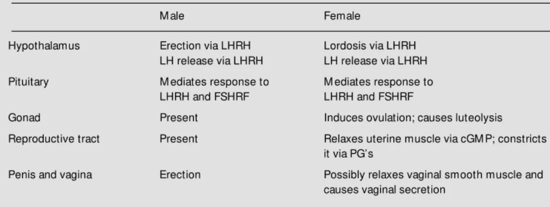
NO is also involved in the control of uterine smooth muscle via NOergic termi-nals on the uterine smooth muscle cells (53), and it has been shown to induce ovulation in rats (54), and luteal regression in rats (55) and cows (Hansel WF, unpublished data).
NO appears to be present in all parts of the male reproductive system as well and will undoubtedly be shown to play a role in testicular, epididymal and vas deferens func-tion (56) (Table 1). Lastly, NOS is present in
Table 1 - Role of NO in rat reproduction.
LHRH, Luteinizing hormone-releasing hormone; FSHRF, follicle stimulating hormone-releasing factor; cGM P, cyclic guanosine monophosphate; PG, prostaglandin. Present indicates that NOS has been found, but the function of NO has not been determined.
M ale Female
Hypothalamus Erection via LHRH Lordosis via LHRH LH release via LHRH LH release via LHRH
Pituitary M ediates response to M ediates response to LHRH and FSHRF LHRH and FSHRF
Gonad Present Induces ovulation; causes luteolysis
Reproductive tract Present Relaxes uterine muscle via cGM P; constricts it via PG’s
Penis and vagina Erection Possibly relaxes vaginal smooth muscle and causes vaginal secretion
spermatazoa and induces sperm motility (57). Indeed, in view of its multifaceted role to promote reproduction at all levels in the organism, it has been aptly named the sexual gas (15).
Ackno wle dgm e nts
We would like to thank Judy Scott and Jill Lucius for their excellent secretarial as-sistance.
Re fe re nce s
1. M cCann SM , Taleisnik S & Friedman HM (1960). LH-releasing activity in hypotha-lamic extracts. Proceedings of the Soci-ety for Experimental Biology and M edi-cine, 104: 432-434.
2. M cCann SM & Ojeda SR (1996). The an-terior pituitary and hypothalamus. In: Grif-fin J & Ojeda SR (Editors), Textbook of Endocrine Physiology. 3rd edn. Oxford University Press, New York, 101-133. 3. Reichlin S (1992). Neuroendocrinology. In:
Foster DW & Wilson JD (Editors), Text-book of Endocrinology. WB Saunders, Philadelphia, 135-219.
4. M cCann SM , M arubayashi U, Sun H-Q & Yu WH (1993). Control of follicle stimulat-ing hormone and luteinizstimulat-ing hormone re-lease by hypothalamic peptides. Intrao-varian regulators and polycystic oIntrao-varian syndrome: recent progress on clinical and therapeutic aspects. Annals of the New
York Academy of Sciences,687: 55-59. 5. Dees WL, Rettori V, Kozlow ski JG &
M cCann SM (1985). Ethanol and the pul-satile release of luteinizing hormone, fol-licle stimulating hormone and prolactin in ovariectomized rats. Alcohol, 2: 641-646. 6. Rettori V, Belova N, Dees WL, Nyberg CL,
Gimeno M & M cCann SM (1993). Role of nitric oxide in the control of luteinizing hormone-releasing hormone release in vivo and in vitro. Proceedings of the Na-tional Academy of Sciences, USA, 90: 10130-10134.
7. M cCann SM & Krulich L (1989). Role of neurotransmitters in control of anterior pituitary hormone release. In: deGroot LJ (Editor), Endocrinology. 2nd edn. W B Saunders, Philadelphia, 117-130. 8. Lumpkin M D, M cDonald JK, Samson WK
& M cCann SM (1989). Destruction of the dorsal anterior hypothalamic region
sup-presses pulsatile release of follicle stimu-lating hormone but not luteinizing hor-mone. Neuroendocrinology, 50: 229-235. 9. M arubayashi U, M cCann SM & Antunes-Rodrigues J (1989). Altered gonadotropin and prolactin release induced by median eminence (M E) lesions and pharmacolo-gical manipulation of prolactin release: Further evidence for separate hypotha-lamic control of FSH and LH release. Brain Research Bulletin, 23: 193-200. 10. M arubayashi U, Yu WH & M cCann SM
(1999). M edian eminence lesions reveal separate hypothalamic control of pulsatile follicle-stimulating hormone and luteiniz-ing hormone release. Proceedings of the Society for Experim ental Biology and M edicine, 220: 139-146.
LHRH of mammalian, avian, and piscian origin. Brain Research Bulletin, 18: 175-178.
12. Yu WH, Karanth S, Walczew ska A, Sow er SA & M cCann SM (1997). A hypothalamic f ollicle-st im ulat ing horm one-releasing decapeptide in the rat. Proceedings of the National Academy of Sciences, USA, 94: 9499-9503.
13. Yu WH & M cCann SM (1991). Feedback of follicle-stimulating hormone to inhibit luteinizing hormone and stimulate follicle-stimulating hormone release in ovariecto-mized rats. Neuroendocrinology, 53: 453-459.
14. Rage F, Hill DF, Sena-Est eves M , Breakefield XO, Coffey RJ, Costa M E, M cCann SM & Ojeda SR (1997). Target-ing transformTarget-ing grow th factor a expres-sion to discrete loci of the neuroendo-crine brain induces female sexual precoc-ity. Proceedings of the National Academy of Sciences, USA, 94: 2735-2740. 15. M cCann SM & Rettori V (1996). The role
of nitric oxide in reproduction. Proceed-ings of the Society for Experimental Biol-ogy and M edicine, 211: 7-15.
16. Canteros G, Rettori V, Franchi A, Genaro A, Cebral E, Saletti A, Gim eno M & M cCann SM (1995). Et hanol inhibit s luteinizing hormone-releasing hormone (LHRH) secretion by blocking the re-sponse of LHRH neuronal terminals to nitric oxide. Proceedings of the National Academy of Sciences, USA, 92: 3416-3420.
17. Rettori V, Gimeno M , Lyson K & M cCann SM (1992). Nitric oxide mediates norepi-nephrine-induced prostaglandin E2
re-lease from the hypothalamus. Proceed-ings of the National Academy of Sciences, USA, 89: 11543-11546.
18. Xu X, Star RA, Tortorici G & M uallem S (1994). Depletion of intracellular Ca2+
stores activates nitric-oxide synthase to generate cGM P and regulate Ca2+ influx.
Journal of Biological Chem istry, 269: 12645-12653.
19. Canteros G, Rettori V, Genaro A, Suburo A, Gimeno M & M cCann SM (1996). Nitric oxide synthase (NOS) content of hypotha-lamic explants: Increased by norepineph-rine and inactivated by NO and cyclic GM P. Proceedings of the National Acade-my of Sciences, USA,93: 4246-4250. 20. Bredt DS & Snyder SH (1989). Nitric oxide
mediates glutamate-linked enhancement of cGM P levels in the cerebellum. Pro-ceedings of the National Academy of Sci-ences, USA, 86: 9030-9033.
21. Kamat A, Yu WH, Rettori V & M cCann SM
(1995). Glutamic acid stimulated luteiniz-ing hormone-releasluteiniz-ing hormone release is mediated by alpha adrenergic stimula-tion of nitric oxide release. Brain Research Bulletin, 37: 233-235.
22. Rettori V, Canteros G, Renoso R, Gimeno M & M cCann SM (1997). Oxytocin stimu-lates the release of luteinizing hormone-releasing hormone from medial basal hy-pothalamic explants by releasing nitric ox-ide. Proceedings of the National Acade-my of Sciences, USA, 94: 2741-2744. 23. Seilicovich A, Duvilanski BH, Pisera D,
Thies S, Gimeno M , Rettori V & M cCann SM (1995). Nitric oxide inhibits hypotha-lamic luteinizing hormone-releasing hor-mone release by releasing g-aminobutyric acid. Proceedings of the National Acade-my of Sciences, USA, 92: 3421-3424. 24. Seilicovich A, Lasaga M , Befum o M ,
Duvilanski BH, Dias M del C, Rettori V & M cCann SM (1995). Nitric oxide inhibits the release of norepinephrine and dopa-mine from the medial basal hypothala-mus of the rat. Proceedings of the Na-tional Academy of Sciences, USA, 92: 11299-11302.
25. Rettori V, Belova N, Kamat A, Lyson K & M cCann SM (1994). Blockade by interleu-kin-1-alpha of the nitricoxidergic control of luteinizing hormone-releasing hormone release in vivo and in vitro. Neuroimmu-nomodulation, 1: 86-91.
26. Kimura M , Yu WH, Rettori V, Walczew ska A & M cCann SM (1997). Granulocyte-macrophage colony stimulating factor sup-presses LHRH release mediated through nitric oxide and g-aminobutyric acid. Neu-roimmunomodulation, 4: 237-243. 27. M ani SK, Allen JM C, Rettori V, O’M alley
BW, Clark JH & M cCann SM (1994). Nitric oxide mediates sexual behavior in female rats by stimulating LHRH release. Pro-ceedings of the National Academy of Sci-ences, USA, 91: 6468-6472.
28. Karanth S, Lyson K & M cCann SM (1993). Role of nitric oxide in interleukin 2-in-duced corticotropin-releasing factor re-lease from incubated hypothalami. Pro-ceedings of the National Academy of Sci-ences, USA, 90: 3383-3387.
29. Rettori V, Belova N, Yu WH, Gimeno M & M cCann SM (1994). Role of nitric oxide in control of grow th hormone release in the rat. Neuroimmunomodulation, 1: 195-200. 30. Aguila M C (1994). Grow th hormone-leasing factor increases somatostatin re-lease and mRNA levels in the rat periven-tricular nucleus via nitric oxide by activa-tion of guanylate cyclase. Proceedings of the National Academy of Sciences, USA,
91: 782-786.
31. Duvilanski BH, Zambruno C, Seilicovich A, Pisera D, Lasaga M , Diaz M del C, Belova N, Rettori V & M cCann SM (1995). Role of nitric oxide in control of prolactin release by the adenohypophysis. Proceedings of the National Academy of Sciences, USA, 92: 170-174.
32. Wakabayashi K, Kamberi IA & M cCann SM (1969). In vitro responses of the rat pituitary to gonadotrophin-releasing fac-tors and to ions. Endocrinology,85: 1046-1056.
33. Nakano H, Faw cet t CP, Kim ura F & M cCann SM (1978). Evidence for the in-volvement of guanosine 3' ,5' -cyclic mon-ophosphate in the regulation of gonado-tropin release. Endocrinology, 103: 1527-1533.
34. Yu WH, Walczew ska A, Karanth S & M cCann SM (1997). Nitric oxide mediates leptin-induced luteinizing hormone-releas-ing hormone (LHRH) and LHRH and leptin-induced LH release from the pituitary gland. Endocrinology, 138: 5055-5058. 35. Sw erdloff R, Batt R & Bray G (1976).
Re-productive hormonal function in the ge-netically obese (ob/ob) mouse. Endocri-nology, 98: 1359-1364.
36. Sw erdloff R, Peterson M , Vera A, Batt R, Heber D & Bray G (1978). The hypotha-lamic-pituitary axis in genetically obese (ob/ob) mice: response to luteinizing hor-mone-releasing hormone. Endocrinology, 103: 542-547.
37. Chehab FF, Lim M E & Ronghua L (1996). Correction of the sterility defect in ho-mozygous obese female mice by treat-ment w ith the human recombinant leptin.
Nature Genetics, 12: 318-320.
38. Barash IA, Cheung CC, Weigle DS, Ren H, Kabigting EB, Kuijper JL, Clifton DK & Steiner RA (1996). Leptin is a metabolic signal to the reproductive system. Endo-crinology, 137: 3144-3147.
39. Frisch RE & M cArthur JW (1974). M en-strual cycles: Fatness as a determinant of minimum w eight for height necessary for their maintenance or onset. Science, 185: 949-951.
40. Ronnekleiv OK, Ojeda SR & M cCann SM (1978). Undernutrition, puberty and the development of estrogen positive feed-back in the female rat. Biology and Repro-duction, 19: 414-424.
41. Chehab FF, M ounzih K, Lu R & Lim M E (1997). Early onset of reproductive func-tion in normal female mice treated w ith leptin. Science, 275: 88-90.
leptin in hypothalamic-pituitary function.
Proceedings of the National Academy of Sciences, USA, 94: 1023-1028.
43. Walczew ska A, Yu WH, Karanth S & M cCann SM (1999). Estrogen and leptin have differential effects on FSH and LH release in female rats. Proceedings of the Society for Experim ental Biology and M edicine, 222: (in press).
44. Cioffi J, Shafer A, Zupancic T, Smith-Gbur J, M ikhail A, Platika D & Snodgrass H (1996). Novel B219/ob receptor isoforms: possible role of leptin in hematopoiesis and reproduction. Nature M edicine, 2: 585-588.
45. Schw artz M W, Seeley RJ, Campfield LA, Burn P & Baskin DG (1996). Identification of targets of leptin action in rat hypothala-mus. Journal of Clinical Investigations, 98: 1101-1106.
46. Banks WA, Kastin AJ, Huang W, Jaspan JB & M aness LM (1996). Leptin enters the brain by a saturable system independ-ent of insulin. Peptides, 17: 305-311. 47. Vaisse C, Halaas JL, Horvath CM , Darnell
Jr JE, Stoffel M & Friedman JM (1996). Leptin activation of stat3 in the hypothala-mus of w ildtype and Ob/Ob mice but not Db/Db mice. Nature Genetics, 14: 95-97. 48. W ong M -L, Bongiorno PB, Rettori V,
M cCann SM & Licinio J (1997). Interleu-kin 1ß, interleuInterleu-kin 1 receptor antagonist, interleukin 10, and interleukin 13 gene expression in the central nervous system and anterior pituitary during systemic in-flammation: Pathophysiological implica-tions. Proceedings of the National Acade-my of Sciences, USA, 94: 227-232. 49. Reznikov AG & M cCann SM (1993).
Ef-fects of neuropeptide Y on gonadotropin and prolactin release in normal, castrated or flutamide-treated male rats. Neuroen-docrinology, 57: 1148-1154.
50. Naivar JS, Dyer CJ, M atteri RL & Keisler DH (1996). Expression of leptin and its receptor in sheep tissues. Proceedings of the Society for the Study of Reproduc-tion, Seattle, WA, Abstract No. 391, 154. 51. Licinio J, Negrão AB, M ant zoros C, Kaklamani V, Wong M -L, Bongiorno PB, M ulla A, Cearnal L, Veldhuis JD, Flier JS, M cCann SM & Gold PW (1998). Synchro-nicity of frequently-sampled 24-hour con-centrations of circulating leptin, luteiniz-ing hormone, and estradiol in healthy w omen. Proceedings of the National A-cademy of Sciences, USA, 95: 2541-2546. 52. Burnett AL, Low enstein CJ, Bredt DS, Chang TSK & Snyder SH (1992). NO: A physiologic mediator of penile erection.
Science, 257: 401-403.
53. Franchi AM , Chaud M , Rettori V, Suburo A, M cCann SM & Gimeno M (1994). Role of nitric oxide in eicosanoid synthesis and uterine motility in estrogenized rat uteri.
Proceedings of the National Academy of Sciences, USA, 91: 539-543.
54. Shukovski L & Tsafiri A (1994). The in-volvement of NO in the ovulatory process in the rat. Endocrinology, 135: 2287-2290. 55. M otto AB & Gimeno M AF (1997). Nitric oxide participates in the corpus luteum regression in ovaries isolated from pseu-dopregnant rats. Canadian Journal of Physiology and Pharmacology, 75: 1335-1339.
56. Burnett AL, Ricker DD, Chamness SL, M aguire M P, Crone JK, Bredt DS, Snyder SH & Chang TS (1995). Localization of NO synthase in the reproductive organs of the male rats. Biology and Reproduction, 52: 1-7.