vol. 42, n. 3, jul./set., 2006
Study of activity transcription factors C/EBP
α
α
α
α
α
in region - 53 to - 33 of
promoter apolipoprotein B gene
Kátia Cristina Dantas1, Sérgio Paulo Bydlowski 1,2, Estela Maria Novak1,2*
1Research and Molecular Biology Division, Fundação Pró-Sangue Hemocentro de São Paulo, 2Department of Hematology, University of São Paulo Medical School, Brazil
Apolipoprotein B (ApoB) plays a major role in the regulation of cellular cholesterol homeostasis and pathogenesis of atherosclerosis. This protein acts as a ligand for the cellular recognition and catabolism of low density lipoprotein (LDL) by the LDL receptor. Previous studies have shown that the expression of apoB in hepatic cells is regulated by the interaction of factors binding to enhancer elements in intron 2 and three elements designated III, IV and V. These elements lie within regions respectively –86 to –62, -72 to –53 and –53 to –33 from the ApoB promoter. In this study, we have suggested that transcription factor C/EBPα, which binds to the –53 to –33 region of the apoB, interacts with the HNF-4 synergistic complex and C/EBPα factors within -86 to -53 and may contribute to increase transcription of the ApoB gene.
INTRODUCTION
Apolipoproteins are important constituents of lipoprotein particles and play a central role in the transport and metabolism of lipids (Goldstein et al.,
1983). Different lipoproteins and apo have been identified. Recent epidemiological studies have shown the importance of the apolipoprotein B, and its direct correlation with the incidence of coronary heart disease (Sattaret al., 2004; Olofsson, Borén, 2005; Barter et al.,
2006; Thompson, Danesh, 2006). Apolipoprotein B (apo B) is a constituent of several lipoproteins and acts as a ligand for cell recognition and catabolism of low density lipoprotein (LDL) by the LDL receptor (Goldstein, Brown 1977; Brown, Goldstein, 1986). It is the structural protein moiety of plasma low density lipoprotein (LDL), comprising 23.8% of the LDL particle (Goldstein,
Brown, 1983). Thus, it is reasonable to expect that changes in the regulation of apolipoprotein B synthesis will affect the plasma lipids or lipoprotein profiles and contribute to the pathogenesis of coronary heart disease (Grundy, 2005; Barter et al., 2006). Apo B gene is
localized in chromosome 2pter-2p24 and comprises 29 exons and 28 introns (Huang, Manson, 1986), and it is expressed in the liver, intestine and placenta, showing that ApoB gene transcription is regulated in a tissue-specific manner (Zannis et al., 2001). In humans, there
are two forms of apolipoprotein B, namely apoB-100 and apoB-48. Apolipoprotein B-100 is expressed in the liver and is the main protein part of low density lipoprotein. In contrast, Apo B-48 is synthesized in the small intestine and is a constituent of chylomicrons and their remnants (Breslow, 1988; Lilja et al., 1999; Zannis et al., 2001).
Hepatic apoB gene transcription is regulated by several
*Correspondence:
E. M. Novak Research and Molecular Biology Division Fundação Pró-Sangue Hemocentro de São Paulo Av. Dr. Enéas de Carvalho Aguiar, 155-1o Andar.
05403-000 - São Paulo, SP - Brazil E-mail:enovak@usp.br
Uniterms
regions, including the proximal 150 bases pairs of the promoter and enhancer elements in intron 2 (Brooks et al., 1992). Hepatic apoB gene transcription is regulated
by regions localized in the -150 to +124 proximal promoter region of apoB (Kardassis et al., 1992).
Specifically, the expression of ApoB in hepatic cells is controlled mainly by interaction of regulator proteins that bind to three elements designated III, IV and V, located respectively within the regions –86 to –62, -72 to –53 and –53 to –33, respectively (Kardassis et al., 1992;
Zanniset al., 2001). These regions interact specifically
with proteins regulatory present in the liver as HNF-4 and C/EBPα (Kardassis et al.,1992; Metzger et al.,
1993). HNF-4 (Hepatic nuclear factor-4) was classified as an orphan nuclear receptor and belongs to the superfamily of steroid-thyroid hormone receptors (Sladeket al., 1990), while C/EBPα (CAAT enhancer-binding protein alpha) is a positive acting transcription factor of the bZip family of proteins (Landschultz et al.,
1988; Metzger et al., 1993). Both transcription factor
have an overlapping binding site within the region -86 to -53 from the ApoB promoter, and act synergically to activate transcription (Kardassis et al., 1992). We have
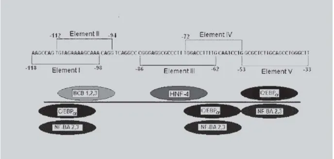
shown previously that mechanisms involving the interaction between HNF-4 and C/EBPα factors in Apo B promoter require a perfect 5´-CCTTTGGA-3’ motif to facilitate the interaction between these two factors (Novaket al., 1998). As shown in figure 1, C/EBPα
factor binds in two locations, element IV (-72 to -53) and element V (-53 to -33). Element IV contains the core recognition sequence GCAAT of the transcription factor C/EBP± and is localized within -72 to- 53 Apo B region. However, C/EBPα also recognizes the GCAAG sequence located within - 53 to -33 region from element V of the Apo B promoter (Kardassis et al., 1992). In this
study, we have examined murine melanoma cells (B16/ F10), whether C/EBP a bound to -53 to -33 may also interact synergically with HNF-4 and C/EBP a proteins localized in the -86 to -33 region of the ApoB promoter and to stimulate transcription of the apoB gene.
MATERIAL AND METHODS
Material
B16-F10 murine melanoma cells were purchased from American Type Culture Collection (Manassas, VA), while [g 32P] ATP (5000 Ci/mmol) and [3H]
chloramphenicol (30 Ci/mmol) were purchased from Amersham (Amersham Pharmacia Biotech Inc., Piscataway, NJ, USA). Transformation competent bacterial HB101 cells, polynucleotide kinase, O-Nitrophenyl-β-D-galactopyranoside, RSV-β -galacto-sidase plasmids and double-stranded poly (dI-dC) were all acquired from Invitrogen (Life Technology Inc.,Rockville, MD, USA).
FIGURE 1 -Schematic representation of transcription factors binding to regulatory region I to V on the promoter region -118 to -33 of the human ApoB gene.The five regulatory elements and the binding activities that interact with them are
shown as described by Kardassis et al. (1992). The circles represent transcription factors BCB 1, 2, 3; HNF-4 (NF- BA1);
Methods
Nuclear extract preparation and protein purification
Male Sprague-Dawley rats (200-250g) were housed in a climate controlled (21°C) room with a 12-hour light-dark cycle and were given tap water. The Animal Ethics Committee of the University of Sao Paulo Medical School approved all experimental protocols. Rat liver nuclear extracts were prepared as previously described (Kardassis et al., 1990; Ogami et al., 1990;
Kardassiset al., 1992). Two hundred mg of crude rat
liver nuclear extracts in NDB buffer (25 mM Hepes, pH 7, 40mM KCl, 0.1mM EDTA, 10% glycerol, 5mM MgCl2,1mM dithiotreitol were heated at 85 °C for 5 minutes. The extracts were placed on ice for 5 min and centrifuged for 5 min at 4°C. The supernatants were transferred and protein concentrations were determined spectrophotometrically using Bradford assay (Bradford, 1976). HNF-4 protein was purified from rat liver nucle-ar extract as described by Sladek et al., (1990). C/EBPα
was purified from rat liver nuclear extract as described by Metzger et al., (1993). Crude rat liver nuclear extract
was passed over a heparin - agarose column followed by a double-stranded DNA-cellulose column (Amersham Pharmacia Biotech Inc., Piscataway, NJ, USA). Both columns were washed with 2ml salt gradients (0.1-1M KCl). The fractions were assayed for activity by a gel retardation assay as described by Metzger et al., (1993),
using double-stranded synthetic oligonucleotides C/EBPα with a sequence corresponding to -86 to -33 of the human apoB promoter (Figure 2).The active fractions from the DNA-cellulose column were then mixed, diluted to reduce salt. Next, they were heated for 6 min at 85 °C in a boiling water bath. The sample was cooled on ice immediately and then centrifuged at 9,500 rpm for 15 min to precipitate the insoluble material (Mertzger et al., 1993). After centrifugation, the heat–soluble material
was passed over an affinity column made with the C/EBPα oligo, as described by Kadonaga and Tijan (1986).The C/EBPα site specific DNA binding activity was eluted in 0.8 M NaCl. This C/EBPα and HNF-4 transcription factors were used in the eletrophoretic mobility shift assays (EMSA).
Gel eletrophoretic mobility shift assays
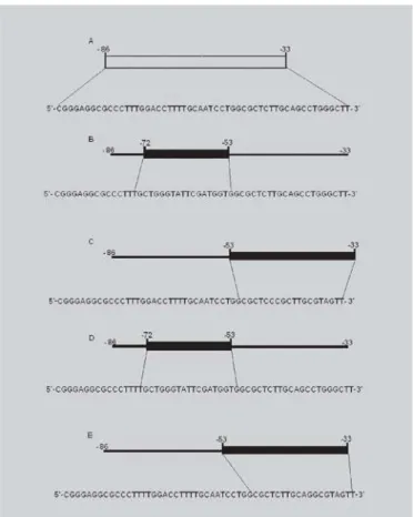
Synthetic double stranded oligonucleotides containing wild type -86 to -33 region apoB region (WT) and mutations that abolished binding of the C/EBPα to -53 to -33 region (M4), C/EBPα -72 to - 53 region (M3),
mutation M3 with a thymidine inserted at -71 position (M5) and mutation M4 with a thymidine inserted at -71 position (M6) were used in EMSA (Figure 2). EMSA were performed using 2 µl C/EBPα and/or 2 µl HNF-4 of
purified nuclear protein prepared as above. DNA binding reactions were in 20 µl reaction volume containing 25nM
Hepes pH 7.6, 8% Ficoll 400, 40 mM KCl, 1 mM dithiothreitol, 5 mM MgCl2, 3 µg of poly (dI-dC).
Following a 15 min incubation on ice, 3 fmol g P32[ATP
(30,000 cpm) of labeled double stranded oligonucleotides WT, M3, M4, M5 and M6 were added, followed by incubation for 30 min on ice. The reaction mixture was then loaded directly onto a 4% polyacrylamide gel 1 X TAE (6.7 mM Tris, 3.3 mM sodium acetate, 1 mM EDTA, pH 7.9) and electrophoreses at 10 Volts/cm for 2-3 h at 4 °C with recirculization of the buffer (miniVE Vertical Electrophoresis system, Amersham Pharmacia Biotech Inc., Piscataway, NJ, USA). After the run, the gel was then dried and analyzed by autoradiography. For quantification, autoradiograms were scanned in a densitometer (LKB UltroScan XI Laser Densitometer, Amersham Pharmacia Biotech Inc., Piscataway,NJ, USA).
Plasmid constructions
To create the chloramphenicol acetyltransferase (CAT) constructions, double –stranded synthetic oligonucleotide containing promoter fragments with sequence to bind the -86 to -33 (WT), -86 to -53 (72BCAT) and -53 to -33 (33BCAT) of the ApoB promoter plus GATC at the 5’ ends were generated by the polymerase chain reaction (PCR) amplification and fused to the CAT gene plasmids (pUSHCAT) as described by Kardassiset al. (1992). To CAT construct 86/33 BCAT,
two ApoB promoter fragments extending from - 86 to -72 and -53 to -33 were obtained by PCR amplification and further ligated. Double–stranded oligonucleotide was cloned into SmaI and Asp -718 sites of the pUCSH-CAT vector as described previously (Ogami et al. 1990).
Transactivation assay in murine melanoma cells (B16/ F10) was performed with pMT2 expression vector containing the HNF-4 cDNA as described by Sladek et al.,
(1990) and pMT2 expression vector containing the C/EBPα cDNA as described by Metzger et al. (1993).
Cell cotransfection and chloramphenicol acetyltransferase assays
streptomycin, and 2 mM L-glutamine for DNA transfection experiments. Cells were placed in 60 mm culture dishes at approximately 60 confluences and cultured for 18 h prior to transfection. Transfection experiments were performed with 5 µg of the constructs: WT, or 72BCAT, or 86/33BCAT, or 33BCAT and cotransfected with 5µg of pMT2-HNF-4, 5 µg of pMT2-C/EBPα, and 3 µg of RSV-β-galactosidase plasmids.
Cells were harvested 42 h post-transfection and lysed by freeze-thawing. Plasmid constructs were transfected into cells by the calcium phosphate DNA co-precipitation method (Graham and Van de Ebb, 1973). The
β-glycosidase activity of the cell lysates was determined by spectrophotometrically by monitoring the hydrolysis of the synthetic substrate O-nitrophenyl galactoside, at a wavelength of 410 nm using a microplate reader (Diagnostic Pasteur-LP 400, BioRad, Lexington, USA), as described by Miller et al. (1976). The protein
concentrations were determined using the Bradford assay (Bradford, 1976). The CAT activity of the cell lysate was determined in triplicate, as described by Neumann et al.
(1987). The b-galactosidase activity of the cell extracts was used to normalize the efficiency of transfection (Gormanet al. 1982).
RESULTS AND DISCUSSION
As seen in Figure 3, the direct binding of the C/EBPα with oligonucleotides which contains wild type (- 86 to - 33) region interacts with both region -53 to33 (M4) (Figure 3, lane 1, Panel A), as with 72 to -53 region (M3) (Figure 3, lane 2, Panel A). Kardassis et al. (1992) suggested that C/EBPα protein bind to different affinities within these regions. Our results have confirmed this observation. In our case, we observed that C/EBPα protein alone binds more strongly to the M3 region than to the M4 region (Figu-re 3, lane 1 and 2, Panel A). HNF-4 protein also recognized the wild type region with a higher affinity (Figure 3, lane 3, Panel A). In addition, C/EBPα bound to element V and IV formed a protein-DNA complex that migrated slower than the HNF-4 and C/EBPα
protein alone (Figure 3, lane 4 and 6, Panel A). Our results using mutations in element V showed that C/EBPα bound to element V (Figure 3, lane 6, Panel A) formed ternary complex with HNF-4 protein. These results are similar to observed with C/EBPα bound to element IV (Figure 3, lane 4, Panel A). We hypothesized that despite C/EBPα bound to element V produces a weak interaction with HNF-4, this heat-stable factor also may be forming a complex with HNF-4 and C/EBPα factors
bound to element III and IV, as reported by Kardassis et al.
(1992) and Metzger et al. (1993). HNF-4 factor binds to
regulatory region -86 to –62, inducing a DNA helix bend, facilitating communication with C/EBPα proteins bound to element IV and located one helix turn from this HNF-4 (Novak et al. 1998).
To define a probable physical interaction of the complex HNF4 factor bound -86 to -53 with C/EBPα
proteins bound to -53 to -33 apoB region, we abolished by mutation At –71 position of apoB promoter, the interaction between HNF-4 and C/EBPα proteins bound to element
V( Figure 3, lane 5 and 7).
FIGURE 2 -Schematic representation of the oligonucleotide sequence of the promoter region of the ApoB gene used in EMSA. (A) Oligonucleotide sequence in the promoter region
(-86 to -33) (WT). (B) Oligonucleotide sequence with
mutation in element IV (-72 to -53) (M3). (C) Oligonucleotide
sequence with mutation in element V (- 53 to -33), (M4). (D)
Oligonucleotide sequence with mutation M3 and insertion of a thymidine nucleotide at the -71 position of the apoB promoter (M5). (E) Oligonucleotide sequence with mutation
Our findings showed that, under these conditions, the interactions between HNF-4 and both C/EBPα proteins bound to element IV or V were affected (Figure 3, lane 5 and 7, respectively). These results suggest that flexibility of the HNF-4 proteins is important factors to facilitate the interaction with C/EBPα factor bound to element V. Probably, C/EBPα proteins bound to -53 to -33 region may be acting synergistically with complex formed between HNF-4 protein and C/EBPα proteins localized in -86 to -53 region of the apoB promoter. Murine melanoma cell were used due to the lack of expression of apoB or HNF-4 or C/EBPα transcription factor (Figure 3, lane 1 and 2, Panel B). To further examine the effect of activity of the C/EBPα
protein bound to the element V on apoB transcription, we studied the activation of CAT reporter constructs by C/EBPα
in murine melanoma cells, B16/F10 (Figure 4).These findings
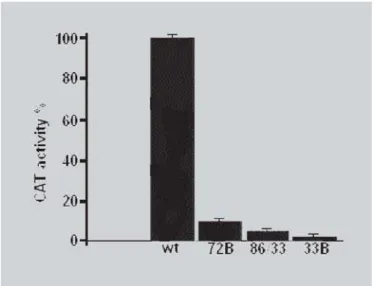
have shown that transient transfections in B16/F10 cells with plasmid -33BCAT reduced transcription of 2% control (wt), while the transfections with plasmid -86/ -33BCAT reduced the transcription of the apoB gene to 10%. These results showed that the binding of C/EBPα in the -53 to -33 regions is also important for the transcription of the apoB gene. Moreover, transients transfection by plasmid -86/-53BCAT in melanoma cells (B16/F10) reduced to 6% of the wild type. Similar results were found for HepG2, where mutations that prevent bonds of HNF-4 in the -86 to -62 region and C/EBPα in the -72 to -53
region to their cognate sites have reduced the promoter activity to 2-13% of control (wt) (Kardassis et al., 1992). Transcription
from the apoB promoter is strongly dependent on the specific binding of C/EBPα proteins to element IV and V and their
interaction with HNF-4. Finally, these findings suggest that C/EBPα bound in the -53 to -33 regions may contribute to a
higher level of expression of the ApoB gene. Thus, CAT transfection can only provide suggestive evidence of the physiological role of the C/EBPα in the -53 to -33 regions. However, the precise role of this transcription factor in apoB expression still remains unclear. In this reporter, we do argue that C/EBPα bound -53 to -33 region could be physically
interacting with HNF-4 and C/EBPα (-86 to -53) affecting apoB gene transcription.
FIGURE 3 -Gel electrophoretic mobility shift analysis. (A) The
transcription factors HNF-4 and C/EBPα from liver nuclear
extracts incubated with radiolabelled double-stranded oligonucleotides WT, M3, M4, M5 and M6 were performed as described in material and methods. Lane 1- 2 µg of C/EBPα
protein incubated with M4. Lane 2- 2 µg of C/EBPα protein
incubated M3. Lane 3- 2 µg of HNF-4 protein incubated with WT. Lane 4- 2µg of HNF-4 and 2 µg of C/EBPα protein
incubated with M3. Lane 5- 2 µg of HNF-4 and 2 µg of C/EBPα protein incubated with M5. Lane 6- 2µg of HNF-4 and 2 µg of C/EBPα proteins binding to M4. Lane 7- 2 µg of HNF-4 and 2 µg of C/EBPα proteins binding to M6; (B) The
transcription factors HNF-4 and C/EBPα from melanoma cells B16/F10 extracts were incubated with WT and M4 as described in materials and methods. Lane 1 - 2 µg of C/EBPα protein incubated with probe M3. Lane 2- 2 µg of HNF-4 protein incubated with probe WT. The arrow shows the low mobility band derived from HNF-4 and C/EBPα specific complexes. High mobility bands correspond to unbound probes.
FIGURE 4 - Transactivation of the region of apoB promoter. CAT assays were analyzed as described in materials and methods. B16/F10 cells were transfected with 5 mg of the four constructs: WT, 72BCAT, 86/-33BCAT, 86/-33BCAT, and co-transfected with 5 µg of
RESUMO
Estudo da atividade do fator de transcrição C/EBPααααα
na região -53 a -33 do promoter do gene da apolipoproteina B
A apolipoproteina B (apoB) tem um importante papel na regulação na homeostasia celular, do colesterol e na patogênese da aterosclerose. Esta proteína age como ligante para o reconhecimento e catabolismo lipoproteínas de baixa densidade (LBD) através do re-ceptor de LDL. Estudos anteriores mostraram que a ex-pressão do gene da apolipoproteína B (APOB) em célu-las hepáticas é regulada pela interação de fatores liga-dos ao elemento enhancer no intron 2, e em 3 elementos denominados de III, IV e V localizados nas regiões -86 a -62, -72 a -53 e -53 a -33 , respectivamente, do promo-tor do gene da APOB. Neste trabalho, nós sugerimos que o fator de transcrição C/EBPα ligado a região -53 a -33 da APOB interage com o complexo HNF-4 e C/EBPα
localizado dentro da região -86 a -53 do APO B e con-tribui para aumentar a transcrição do gene APOB.
UNITERMOS: Apolipoproteína B. Fator de transcrição C/EBPa. Fator de transcrição HNF-4. Células de melanoma -B16/F10.
REFERENCES
BARTER, P. J.; BALLANTYNE, C. M.; CARMENA, R.; CASTRO CABEZAS, M., Apo B versus cholesterol in estimating cardiovascular risk and in guiding therapy: report of the thirty-person/ten-country panel. J. Intern. Med., v.259, p.247-258, 2006.
BRADFORD, M. M. A rapid and sensitive method for the quantification of microgram quantities of protein utilizing the principle of protein-dye binding. Anal Biochem., v.72, p.248-254, 1976.
BRESLOW, J. L. Human apolipoprotein genetic variation.
Physiol. Rev., v. 68, p. 85-132, 1988.
BROOK, A. R.; Levy-Wilson, B. Hepatocyte nuclear factor 1 and C/EBP are essential for the activity of the human apolipoprotein B gene second intron enhancer. Mol. Cell. Biol., v.12, p.1134-1148, 1992.
BROWN, M.S.; GOLDSTEIN, J.L. receptor – mediated pathway for cholesterol homeostasis. Science, v. 232,
p. 34-47, 1986.
GOLDSTEIN, J.L.; Brown, M.S. Atherosclerosis: the low – density lipoprotein receptor hypothesis. Metabolism,
v. 26, p. 1257-75, 1977.
GOLDSTEIN, J.L.; KITO, T.; BROWN, M.S. Defective lipoprotein receptors and atherosclerosis. New Engl. J. Med., v. 309, p. 288-296, 1983.
GORMAN, C.M.; MOFFAT, L.F.; HOWARD, B.H. Recombinant genomes which express chloramphenicol acetyltransferase in mammalian cells.Mol. Cell Biol., v.2,
p.1044-1051, 1982.
GRAHAM,F.L.; VAN der EB. A new technique for the assay of infectivity of human adenovirus 5 DNA. Virology,
v. 52, p. 456, 1973.
GRUNDY, S.M. Metabolic syndrome: therapeutic considerations.Hand. Exp. Pharmacol., v. 170, p.
107-133. 2005.
HUANG, C.; MASON, J.T. Structure and properties of mixed – chain phospholipids assemblies. Biochim. Biophys. Acta, v. 864, p. 423-70, 1986.
KADONAGA, J.T.; TJIAN, R. Affinity purification of sequence-specific DNA binding proteins. Nucleic Acids Res., v. 15, p. 8125-8143, 1986.
KARDASSIS, D.; HADZOPOULOU-CLARADAS, M.; RAMJI, D.P.; CORTESE, R.; ZANNIS, V.I.; CLARADAS, C. Characterization of the promoter elements required for hepatic and intestinal transcription of the human apoB: gene definition of the DNA-Binding site of a tissue specific transcriptional factor. Mol. Cell. Biol., v.10, p. 2653-2659, 1990.
KARDASSIS, D.; ZANNIS, V.I.; CLARADAS C. Organi-zation of the regulatory Elements and Nuclear Activities Participating in the Transcription of the Human Apolipo-protein B Gene. J. Biol. Chem., v. 267, p.2622-2632, 1992.
LANDSCHULTZ, W. H.; JOHNSON, P. F.; McKnight, S. I. The leucine zipper: A hypothetical structure common to a new cass of DNA binding proteins. Science, v. 240, p.
1759-1764, 1988.
LILJA, H.; KAMOHARA, Y.; NEUMANN, T.; DEMETRIOU, A.A.; ROZGA , J. Transformation growth factor b 1 helps maintain differentiated functions in mitogen-treated primary rat hepatocyte cultures.
MERTZGER, S.; HALA, J.; BRESLOW, J. L.; SLADEK, F. M. Orphan receptor HNF-4 and bZip protein C/EBPα
bind to overlapping regions of the apolipoprotein B gene promoter and synergically active transcription. J. Biol. Chem., v.268, p. 16831-1638, 1993.
MILLER, J. H. Experiments in molecular genetics. J. Biol. Chem., v. 251, p. 3774-3779,1976.
NEUMANN, J. R.; MORENCY, A.C.; RUSSIAN, K.O. A novel rapid assay for chloramphenicol acetyltransferase gene expression. Biotechniques, v. 5, p.444,1987.
NOVAK, E.M.; DANTAS, K.C.; CHARBEL, C. E.; BYDLOSWKI, S. P. Association of hepatic nuclear factor-4 in the Apolipoprotein B promoter: a preliminary report.Braz. J. Med. Biol. Res., v. 31, p. 140-3, 1998.
OGAMI, K.; HADZOPOULOU-CLARADAS, M.; CLARADAS, C.; ZANNIS, V. I. Promoter elements and factors require for hepatic and intestinal transcription of the human Apo CIII gene. J. Biol. Chem., v. 265, p.
9908-9815, 1990.
OLOFSSON S, BORÉN J. Apolipoprotein B: a clinically important apolipoprotein which assembles atherogenic lipoproteins and promotes the development of atherosclerosis.J. Intern. Med., v. 258, p.359-410, 2005.
SATTAR, N.; WILLIAMS, K.; SNIDERMAN,A.L.; D´ÁGOSTINO, R.; HAFFNER, S. Comparison of the associations of apolipoprotein B and Non-High-density lipoprotein cholesterol with other cardiovascular risk factor in patients with the metabolic syndrome in the insulin resistance atherosclerosis study. Circulation,
v.110, p.2687-2693, 2004.
SLADEK, F. M.; ZHONG, W.; LAI, E.; DARNELL, Jr. J. E. Liver, enriched transcription factor HNF-4 is a novel member of the steroid hormone receptor super family.
Genes Dev., v. 4, p. 2353-2365, 1990.
THOMPSON A.; DANESH J. Associations between apolipoprotein B, apolipoprotein AI, the apolipoprotein B/AI ratio and coronary heart disease: a literature-based meta-analysis of prospective studies. J. Intern. Med.,
v.259, p.481-492, 2006.
ZANNIS, V. I.; KAN, H. Y.; KRITIS, A. Transcriptional regulation of the human Apolipoprotein genes in vitro and in vivo. Curr. Opin. Lipidol., v.12, p. 181-207, 2001.