Immunohistochemistry expression of tumor markers CD34 and
Immunohistochemistry expression of tumor markers CD34 and
Immunohistochemistry expression of tumor markers CD34 and
Immunohistochemistry expression of tumor markers CD34 and
Immunohistochemistry expression of tumor markers CD34 and
P27 as a prognostic factor of clinically localized prostate
P27 as a prognostic factor of clinically localized prostate
P27 as a prognostic factor of clinically localized prostate
P27 as a prognostic factor of clinically localized prostate
P27 as a prognostic factor of clinically localized prostate
adenocarcinoma after radical prostatectomy
adenocarcinoma after radical prostatectomy
adenocarcinoma after radical prostatectomy
adenocarcinoma after radical prostatectomy
adenocarcinoma after radical prostatectomy
Expressão imunohistoquímica do marcador tumoral CD34 e
Expressão imunohistoquímica do marcador tumoral CD34 e
Expressão imunohistoquímica do marcador tumoral CD34 e
Expressão imunohistoquímica do marcador tumoral CD34 e
Expressão imunohistoquímica do marcador tumoral CD34 e
P27 como fator prognóstico em adenocarcinoma de próstata clinicamente
P27 como fator prognóstico em adenocarcinoma de próstata clinicamente
P27 como fator prognóstico em adenocarcinoma de próstata clinicamente
P27 como fator prognóstico em adenocarcinoma de próstata clinicamente
P27 como fator prognóstico em adenocarcinoma de próstata clinicamente
localizado após prostatectomia radical
localizado após prostatectomia radical
localizado após prostatectomia radical
localizado após prostatectomia radical
localizado após prostatectomia radical
AISSAR EDUARDO NASSIF, ACBC-PR1; RENATO TÂMBARA FILHO2
A B S T R A C T A B S T R A C T A B S T R A C T A B S T R A C T A B S T R A C T
O O O O
Objectivebjectivebjectivebjectivebjective: to analyze the immunohistochemical expression of P27 and CD34 markers as prognostic factors in patients with localized prostate cancer. MethodsMethodsMethodsMethods: analysis of 100 patients with localized prostate cancer submitted to curative surgery. We carried out theMethods usual histological preparation, followed by immunohistochemistry to detect the accumulation of P27 and CD34 protein followed by statistical analysis. ResultsResultsResultsResultsResults: in the evaluation of P27 marker and on the correlation with the variables we found significant difference in Gleason score with positive expression (positive P27) related to lower mean PSA (p = 0.091), lower Gleason score (p < 0.0001) and smaller tumor area in CD34 (p = 0.036). Regarding the CD34 marker at the tumor area, it was observed that the smaller the positive CD34, the lower the PSA value (p < 0.0001) and lower the Gleason score (r = 0.5726, p < 0.0001), and the higher the positive CD34, the higher the staging (r = 0.3305, p <0.0001) and the chance of recurrence (p = 0.002). Patients with higher stage also displayed larger positive CD34 areas (p < 0.0001). ConclusionConclusionConclusionConclusionConclusion: the markers CD34 and P27 are associated with events specific to prostate cancer, however, only CD34 was able to determine the possibility of biochemical recurrence.
Key words Key words Key words Key words
Key words: Prostatectomy. Neoplasms of the prostate. Rex gene products. CD34 antigens.
INTRODUCTION
INTRODUCTION
INTRODUCTION
INTRODUCTION
INTRODUCTION
P
rostate cancer is the most common visceral malignancy in men and its incidence tends to increase in the coming decades with the increased life expectation1,2.3.Approximately 543,000 new cases are diagnosed each year worldwide. In Brazil, the National Cancer Institute predicted 49,530 new cases in 2008, a number that corresponds to the estimated risk of 52 new cases per 100,000 men; it is the most common non-skin tumor diagnosed in all regions of the country4.
Despite the gain in epidemiology and biomolecular knowledge of prostate cancer, one cannot predict which patients will develop clinically significant disease and which will remain with restricted tumor5,6.7. Early
detection of prostate cancer with prostate-specific antigen (PSA) has allowed many patients the possibility of radical treatment with curative intent. However, up to 30% of patients undergoing radical prostatectomy for clinically localized disease will experience biochemical recurrence.
In some cases, biochemical recurrence represents micro-metastatic disease not detected before the operation and almost always not yet detectable at the time of PSA recurrence6.
In prostate cancer, histopathological analysis has great clinical relevance8,9. Histological and serological exams
describe a number of important changes, allowing monitoring of the disease progression10,11. However,
qualitative immuno-histochemistry has shown great disparity and variability of results among different observers1,12,13. In
order to provide a numerical scale and reproducible standards for tissue marking, thus increasing sensitivity analysis and quality control, technological refinements using automated morphometric methods have been increasingly sought14.
The search for an ideal prognostic factor in prostate cancer, including the patient’s decision about the type of treatment, has been one of the great challenges of medicine. When the biological variables that help to indicate adjuvant therapy can be identified, it is likely that tumoral recurrence rates will be reduced15,16,17,18. Many studies have
This study was done at the Graduate Program in Principles of Surgery, Hospital de Clínicas / UFPR - Curitiba-PR-Brasil.
demonstrated the importance of new immunohistochemical markers that may in the future be used as predictors of prognosis and tumoral development11,12,15,16,17,18.
This paper aims to analyze the immunohisto-chemical expression of P27 and CD34 markers as prognostic factors in patients with localized prostate cancer.
METHODS
METHODS
METHODS
METHODS
METHODS
This study was approved by the Ethics Committee on Human Research of the Clinical Hospital of the Federal University of Paraná and it is in accordance with national legislation and the International Committee on Harmonization of rules of good clinical practice. (Res. CNS 196/96 MS and ICH-GCP).
Sample characterization Sample characterizationSample characterization Sample characterizationSample characterization
Patients were identified using the database of more than 500 samples from men with clinically localized prostate cancer undergoing radical prostatectomy and pelvic lymphadenectomy between January 2000 and December 2006. After assuring anonymousness and exclusion of patients who received preoperative 5-alpha reductase inhibitors or neoadjuvant antiandrogen therapy, 100 patients were enrolled. Mean follow-up was 36 months.
All were diagnosed with prostate adenocarcinoma, usually acinar type, with preoperative clinical stages T1c and T2c. Clinical history, physical examination, and urological and general laboratory tests (general and specific hematologic evaluation with PSA) were systematically analyzed by the same observer.
The evaluation of transrectal ultrasound and biopsy were done by the same professional. Transrectal biopsies were performed on a broader scale, with samples of the lateral and transition zones, 12 or 14 samples. Patients were tested for the preoperative staging by bone scintigraphy, chest radiography and computed tomography or magnetic resonance imaging of the pelvis, as needed. They were classified according to the latest update proposed by the International Union Against Cancer, TNM. After confirming the diagnosis by biopsy, a period of four to six weeks elapsed prior to the operation. Surgical specimens were collected and analyzed in the same laboratory, and these blocks and reports were reviewed by an uropathologist.
Histological processing Histological processingHistological processing Histological processingHistological processing
The surgical specimens were submitted to serial sections in slices 0.5 cm thick in the entire gland. The material was embedded in Indian ink for better identification and assessment of surgical margins. The slices representing macroscopic cuts with rotation microtome (American Optical Instruments®) were subjected to paraffin embedding for
histological cuts 3 µm thick and stained with hematoxylin-eosin. Once the diagnosis was confirmed by the pathologist
tumor areas were separated from the paraffin blocks in order to be studied.
The glass slide with the best tumor representation was selected for immunohistochemical study. The microscope used was a Nikon® Eclipse E-400 model. All
slides were evaluated by the same Uropathologist.
Immunohistochemical staining and Immunohistochemical staining and Immunohistochemical staining and Immunohistochemical staining and Immunohistochemical staining and microscopic evaluation
microscopic evaluation microscopic evaluation microscopic evaluation microscopic evaluation
Histological grading was established according to the Gleason criteria. Immunohistochemistry was prepared to detect the accumulation of P27 and CD34. Sections were rewashed and hydrated in decreasing concentrations of ethanol and washed with distilled water. They were then placed in a citrate solution (pH 6.0) and led to the microwave oven on medium-temperature for a maximum of 15 minutes after being removed and left to stand at room temperature for 15 minutes. After adding the material in PBS buffer solution (pH = 7.6), endogenous peroxidase blockers were used; the sections were incubated in a solution of 3% hydrogen peroxide in methanol for 30 minutes at room temperature.
We then proceeded to a wash cycle with distilled water followed by incubation for 30 minutes in a solution containing 4% normal serum in PBS. The production of specific antibodies (DO-7, Dako A / S, Denmark) were prepared with 1:50 dilution in PBS and incubated with the sections for 12 hours in a moist chamber. After a new cycle of washes the sections were again placed in a moist chamber for 30-minute incubation with secondary IgG biotin anti-mouse antibody (Vectastain, Vector Lab, CA, USA) diluted 1:600 in PBS. After another round of washes, the material was incubated in a moist chamber for 60 minutes with the avidin / biotin complex (Streptococcus ABC, Vectastain, Vector Lab CA, USA) at dilution of 1:800 in PBS. The color of immunoperoxidase reaction was achieved by immersion for eight minutes in a solution containing the DAB chromogen (3,5-diamino-benzidine tetra-hydrochloride) and hydrogen peroxide. After washing in tap water a counter-staining with Harris hematoxylin (Merck, Darmstadt, Germany) was carried out for two minutes.
Each group of sections was analyzed and positive and negative controls were also included. Reading of the reaction was performed under a light microscope and the positivity criteria for P27 used by other authors were observed, the minimum percentage of tumors being 40% or more of tumor cells stained per high power field (400X). The pathological study of identical circles in the slides stained with CD34+ ensured that the same areas were counted in each slide. The counting method was a modified form of the protocol described by Weidner et al.19 The same area
microvessel. If two or more positive foci appeared to belong to the same, single and continuous vessel, they were counted as one microvessel. Counted MVD (microvascular density) was defined as the sum of the three highest counts.
Statistical analysis Statistical analysis Statistical analysis Statistical analysis Statistical analysis
The correlation between CD34 and P27 expression and clinicopathologic characteristics were evaluated by parametric Student t, and nonparametric Mann-Whitney, tests; for comparison of two proportions we used the software Primer of Biostatistics, Chi-square and Fisher exact tests (Epi-Info). The level of significance was less than 5% (p<0.05).
RESULTS
RESULTS
RESULTS
RESULTS
RESULTS
Demographic Evaluation Demographic Evaluation Demographic Evaluation Demographic Evaluation Demographic Evaluation
We evaluated 100 patients with prostate cancer, mean age 63.7 ± 6.8 years, ranging from 44.0 to 75.0 years with a high concentration for the 60 to 69 years age group (58.0%). PSA in these patients was 7.8 ± 4.5 ng/dl (median 6.5) ranging from 2.3 to 24.0 ng/dl, and most patients (70.0%) had PSA ranging between 4.0 and 9.9 ng/ dl. There was a greater proportion of Gleason score up to six (72.0%); the predominant pathological stage was pT2c (65.0%).
Expression of the P27 marker Expression of the P27 marker Expression of the P27 marker Expression of the P27 marker Expression of the P27 marker
With the marker P27 it was observed, in general, higher proportion of positive expression (60.0%) versus negative (40%) (p<0.0007).
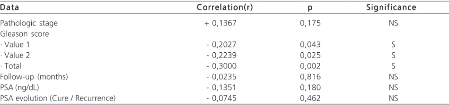
In the evaluation of P27 and the correlation with the variables it was observed a significant difference in Gleason score, independent of the values, where patients with positive expression (P27 Positive) had a higher proportion of scores less than or equal to 6 (p=0.015). When comparing the P27 marker variables we observed that patients with positive expression (P27 Positive) also had lower mean PSA (p=0.091), lower Gleason score (p<0.0001) and smaller tumor area in CD34 (p=0.036). (Table 1)
Expression of CD34 marker Expression of CD34 marker Expression of CD34 marker Expression of CD34 marker Expression of CD34 marker
The correlation of the CD34 at the tumor site showed that the lower the CD34, the lower the PSA value (p<0.0001); the lower CD34, the lower the Gleason score (r=0.5726, p<0, 0001), indicating that the greater the CD34, the higher the stage (r=0.3305, p<0.0001) and the chance of recurrence (p=0.002). Patients with higher stage also had the largest CD34 area (p<0.0001) (Table 2).
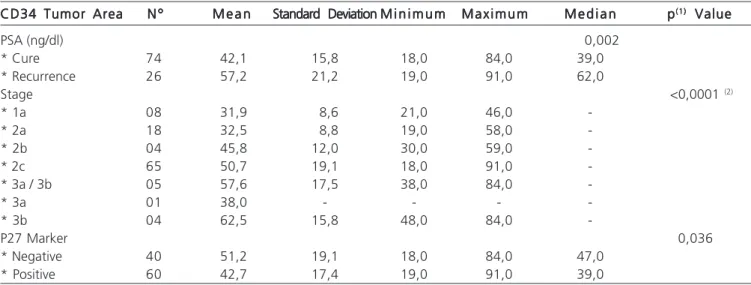
Correlation between CD34 and P27 Correlation between CD34 and P27 Correlation between CD34 and P27 Correlation between CD34 and P27 Correlation between CD34 and P27 Correlation analysis showed that patients with positive P27 expression had lower CD34 area (p = 0.036) (Table 3).
DISCUSSION
DISCUSSION
DISCUSSION
DISCUSSION
DISCUSSION
Cell proliferation and the gradual acquisition of specialized phenotype show that development processes can be influenced by several variables, including physical, extracellular matrix components, cell adhesion molecules and junction complexes between the apposed cell membranes. The exponential growth of tumor cells also requires support from nurturing blood vessels20.At the cellular
level the onset of neovascularization increases tumor growth through perfusion and paracrine effects. It is shown that there is a higher density of microvessels in the center of the prostate tumor than in the periphery, suggesting that the angiogenic promoters are more active there20, 21.
From a clinical perspective, neovascularization allows the tumor to grow and spread. Hence, angiogenesis is an important factor in the progression and increase in solid tumors. Recent studies of bladder, uterus, cervix, breast cancers and melanoma have shown that the tumor vasculature in invasive cancers is a very significant predictor of overall prognosis and recurrence-free time20 22.23.
The microvessel density (MVD) in prostate cancer was described in 199320. The first observation was the
significant difference in MVD when comparing benign areas with neoplastic ones. Other authors have shown that MVD in tumors from patients who developed metastatic disease
Table 1 – Table 1 – Table 1 – Table 1 –
Table 1 – Correlation of data relating to P27 in 100 patients with prostate adenocarcinoma. D a t a
D a t a D a t a D a t a
D a t a Correlation(r)Correlation(r)Correlation(r)Correlation(r)Correlation(r) ppppp S i g n i f i c a n c eS i g n i f i c a n c eS i g n i f i c a n c eS i g n i f i c a n c eS i g n i f i c a n c e
Pathologic stage + 0,1367 0,175 NS
Gleason score
· Value 1 - 0,2027 0,043 S
· Value 2 - 0,2239 0,025 S
· Total - 0,3000 0,002 S
Follow-up (months) - 0,0235 0,816 NS
PSA (ng/dL) - 0,1351 0,180 NS
PSA evolution (Cure / Recurrence) - 0,0745 0,462 NS
was significantly higher than in those without metastases24,25. In multivariate analysis, MVD was an
important predictor of metastatic disease and an independent predictor of disease progression after radical prostatectomy20,22.26. It is known that MVD is associated with
recurrence, but there is a cutoff point set for this avaliation24, 26. The CD34 biomarker can be used to quantify MVD in
prostate cancer and stratify patients at greatest risk of recurrence after radical prostatectomy20.
The importance of angiogenesis in prostate cancer is well established. Many studies have now demonstrated a direct correlation with Gleason score, tumor stage, progression, metastasis and survival27,28,29,30,31.In
addition to MVD, other biomarkers such as VEGF, MMP-2, MMP-9 and HIF-1 are associated with CD34, tumor stage, grade of disease and specific survival in patients with prostate cancer20,22,24,32,33.
Gleason score, PSA and clinical and pathological stages were used individually and combined to improve prediction of relapse after radical prostatectomy30. MVD can
be used to more accurately predict recurrence, especially in those patients who are classified as intermediate-risk by preoperative Gleason score and pathologic stage20,30.33. In
this study the CD34 biomarker was significantly associated with the nuclear count and degree, with the sum of the Gleason score and with the pathological stage; MVD remained significant in predicting recurrence.
The P27 gene, whose protein product is a negative regulator of cell cycle and a potential tumor suppressor, belongs to the Cip/Kip inhibitors of cyclin-dependent protein kinase family, which promote a decrease in cell proliferation. Low levels of P27 are associated with worse prognosis in patients with breast, colon and lung cancers, brain astrocytoma, oral squamous cell carcinoma, lymphoma and ovarian cancer; P27 is considered one of the most promising markers in prostate cancer34, 35,36,37,38.
The low expression of the P27 marker is considered an independent predictor of poor prognosis in prostate cancer. Its evaluation in biopsies and samples of
Table 2 Table 2Table 2 Table 2
Table 2 - Correlation of data relating to CD34 and tumor area. D a t a
D a t aD a t a D a t a
D a t a Correlation(r)Correlation(r)Correlation(r)Correlation(r)Correlation(r) ppppp S i g n i f i c a n c eS i g n i f i c a n c eS i g n i f i c a n c eS i g n i f i c a n c eS i g n i f i c a n c e
Age (years) - 0,0724 0,474 NS
PSA (ng/dL) + 0,6307 <0,0001 S
Gleason score
* Value 1 + 0,2153 0,031 S
* Value 2 + 0,5115 <0,0001 S
* Total + 0,5726 <0,0001 S
Patologic stage + 0,3305 <0,0001 S
Follow-up (months) + 0,1179 0,243 NS
CD34 - normal area + 0,1936 0,054 NS
Note: Correlation analysis.
Tabela 3 Tabela 3 Tabela 3 Tabela 3
Tabela 3 – Descriptive statistics of the evolution of PSA, pathologic stage and P27 in relation to CD34 and tumor area. CD34 Tumor Area
CD34 Tumor Area CD34 Tumor Area CD34 Tumor Area
CD34 Tumor Area N ºN ºN ºN ºN º M e a nM e a nM e a nM e a nM e a n Standard DeviationStandard Deviation M i n i m u mStandard DeviationStandard DeviationStandard DeviationM i n i m u mM i n i m u mM i n i m u mM i n i m u m M a x i m u mM a x i m u mM a x i m u mM a x i m u mM a x i m u m M e d i a nM e d i a nM e d i a nM e d i a nM e d i a n ppppp(1)(1)(1)(1)(1) Value Value Value Value Value
PSA (ng/dl) 0,002
* Cure 74 42,1 15,8 18,0 84,0 39,0
* Recurrence 26 57,2 21,2 19,0 91,0 62,0
Stage <0,0001 (2)
* 1a 08 31,9 8,6 21,0 46,0
-* 2a 18 32,5 8,8 19,0 58,0
-* 2b 04 45,8 12,0 30,0 59,0
-* 2c 65 50,7 19,1 18,0 91,0
-* 3a / 3b 05 57,6 17,5 38,0 84,0
-* 3a 01 38,0 - - -
-* 3b 04 62,5 15,8 48,0 84,0
-P27 Marker 0,036
* Negative 40 51,2 19,1 18,0 84,0 47,0
* Positive 60 42,7 17,4 19,0 91,0 39,0
radical prostatectomy specimens may help to distinguish between potentially aggressive and potentially non-aggressive disease in prostate cancer screening39. It is
associated with changes in apoptosis and expression of different markers as: Cadherins, Ki-67, BCL-2, p53 protein expression in the bladder and prostate cancer, Akt/protein kinase B, Skp2 (S phase protein kinase), changes in histones37,39,40.Several authors have described the correlation
between P27 and pre and postoperative parameters, such as Gleason score, extra-capsular extension, seminal vesicle involvement, pelvic lymph node metastasis, positive surgical margins, the coexistence of high-grade prostatic intraepithelial neoplasia, tumor size, prostate volume and PSA levels41-44.
Moreover, P27 is related to prediction of higher risk of recurrence and disease-specific survival and is useful as a potential molecular target for new systemic agents in recurrent prostate cancer37,39,40,41,43. This study showed a
correlation between the P27 marker, Gleason score and PSA values.
Roy et al.43 analyzed the role of inhibitors of
cyclin-dependent kinase and found that the lower expression of P27 protein in prostate cancer tissues is often associated with poor prognosis, and the markers P21 and P27 were associated with higher MVD and indicated that they have compensatory roles in advanced prostate cancer cells, and ablation or down-modulation of both these molecules essentially enhances the aggressive prostate carcinoma
phenotype. These results are similar to those found in this work, where the main areas of MVD/CD34 are associated with reduced expression of P27 protein.
We investigated the predictive clinical value of altered expression of P27 and CD34 in patients treated for localized prostate cancer. These data indicate that altered expression (negative) of P27 is a common biological event, which suggests they may play a role in the pathogenesis of prostate cancer, and clinical predictive value seems limited compared with PSA and Gleason score. The observation that the decreased expression of P27 is altered is comparable with published reports on P27 by other groups40,42,44. In this analysis, however, there
was a strong correlation between the fall of P27 expression and clinical outcome.
The P27 and CD34 tumor markers are common biological events in prostate cancer, but the P27 seems limited in comparison with the standard prognostic factors. On the other hand, angiogenesis may be clinically useful as a prognostic factor in prostate carcinoma and micro-vascular density measurement using CD34 immunohistochemistry is a prognostic factor associated with recurrence-free survival in radical prostatectomy.
The CD34 and P27 markers are associated with events specific to prostate cancer. However, only CD34 was able to determine the possibility of biochemical recurrence.
R E S U M O R E S U M O R E S U M O R E S U M O R E S U M O
Objetivo: Objetivo: Objetivo: Objetivo:
Objetivo: Analisar a expressão imunoistoquímica do marcador CD34 e p27, como fator prognóstico em pacientes com neoplasia de próstata localizada. Métodos: Métodos: Métodos: Métodos: Análise de 100 casos de pacientes portadores de neoplasia prostática localizada submetida à cirurgiaMétodos: curativa. Realizou-se o preparo histológico habitual, seguido da reação imunoistoquímica para a detecção do acúmulo da proteína CD34 e p27 seguida de análise estatística. Resultados:Resultados:Resultados:Resultados:Resultados: Na avaliação do marcador P27 e na correlação com as variáveis, observou-se diferença significativa no escore de Gleason com expressão positiva (P27 positivo) relacionada com PSA médio mais baixo (p=0,091), escore de Gleason mais baixo (p<0,0001) e menor área de tumor no CD34 (p=0,036). Correlacionando-se o marcador CD34 na área tumoral observou-se quanto menor o CD34 positivo menor é o valor do PSA (p<0,0001), e menor é o escore de Gleason (r=0,5726 ; p<0,0001) e quanto maior o CD34 positivo maior é o estadiamento (r=0,3305 ; p<0,0001) e a chance de recidiva (p=0,002). Os pacientes com estadiamento mais alto, também tinham maior área CD34 positivo (p<0,0001). Conclusão: Conclusão: Conclusão: Conclusão: OsConclusão: marcadores P27 e CD34 estão associados com os eventos próprios ao câncer de próstata; contudo, apenas o CD34 foi capaz de determinar a possibilidade de recidiva bioquímica.
Descritores: Descritores: Descritores: Descritores:
Descritores: Prostatectomia. Neoplasias da próstata. Produtos do gene rex. Antígenos CD34.
REFERENCES
REFERENCES
REFERENCES
REFERENCES
REFERENCES
1. Ercole B, Marietti SR, Fine J, Albertsen PC. Outcome following active surveillance of men with localized prostate cancer diagnosed in the prostate specific antigen era. J Urol. 2008;180(4):1336-9; discussion 1340-1. Epub 2008 Aug 15.
2. Kuo NW, Lin HC, Lee HC. Physician clinical experience and inappropriate prostate specific antigen screening: evidence from an Asian country. J Urol. 2008;180(5):1954-8; discussion 1958. Epub 2008 Sep 17.
3. Wilt TJ, MacDonald R, Rutks I, Shamliyan TA, Taylor BC, Kane RL. Systematic review: comparative effectiveness and harms of treatments for clinically localized prostate cancer. Ann Intern Med. 2008;148(6):435-48. Epub 2008 Feb 4. Erratum in: Ann Intern Med. 2008;148(1):888.
4. INSTITUTO NACIONAL DE CÂNCER. Estimativa 2008 - Incidência de câncer no Brasil. Rio de Janeiro: INCA; 2008.
preoperative risk categories. J Urol. 2008;179(6):2212-7.Epub 2008 Apr 18.
6. Neal DE. Can we accurately identify men with low risk prostate cancer? J Urol. 2008;180(4):1217-18. Epub 2008 Aug 15. 7. Simone NL, Singh AK, Cowan JE, Soule BP, Carolli PR, Litwin MS.
Pretreatment predictors of death from other causes in men with prostate cancer. J Urol. 2008; 180(6):2447-51; discussion 2451-2. Epub 2008 Oct 19.
8. Billis A, Guimarães MS, Freitas LLL, Meirelles L, Magna LA, Ferreira U. The impact of the 2005 international society of urological pathology consensus conference on standard Gleason grading of prostatic carcinoma in needle biopsies. J Urol. 2008; 180(2):548-52; discussion 552-3. Epub 2008 Jun 11.
9. Liska J, Repiska V, Polak S, Varga I, Blasko M, Macejova D, et al. Prostate tumors-histological classification and molecular aspects of prostate tumorigenesis. Endocr Regul. 2007;41(1):45-57.
10. Arlen PM, Bianco F, Dahut WL, D´amico A, Figg WD, Freedland SJ, et al. Prostate specific antigen working group guidelines on prostate specific antigen doubling time. J Urol. 2008;179(6):2181-5; discussion 2185-6. Epub 2008 Apr 18.
11. Freedland SJ, Moul JW. Prostate specific antigen recurrence after definitive therapy. J Urol. 2007;177(6):1985-91.
12. Doganavsargil B, Simsir A, Boyaciogluc H, Cal C, Hekimgli M. A comparison of p21 and p27 immunoexpression in benign glands, prostatic intraepithelial neoplasia and prostate adenocarcinoma. BJU Int. 2006;97(3):644-8.
13. Rhodes DR, Sanda MG, Otte AP, Chinnaiyan AM, Rubin MA. Multiplex biomarkers approach for determining risk of prostate-specific antigen-defined recurrence of prostate cancer. J Nat Cancer Inst. 2003;95(9):661-8.
14. Dhir R. Prostate cancer biobanking. Curr Opin Urol. 2008;18(3):309-14.
15. Concato J, Jaind D, Li WW, Risch HA, Uchio EM, Wells CK. Molecular markers and mortality in prostate cancer. BJU Int. 2007;100(6):1259-63. Epub 2007 Sep 10.
16. Grignon DJ, Caplan R, Sarkar FH, Lawton CA, Hammond EH, Pilepich MV, et al. p53 status and prognosis of locally advanced prostatic adenocarcinoma: a study based on RTOG 8610. J Nat Cancer Inst. 1997;89(2):158-65.
17. Jin S. p53. Autophagy and tumor suppression. Autophagy. 2005;1(3):171-3.Epub 2005 Oct 21.
18. Mohaptra S, Chu B, Zhao X, Pledger WJ. Accumulation of p53 and reductions in XIAP abundance promote the apoptosis of prostate cancer cells. Cancer Res. 2005;65(17):7717-23.
19. Weidner N, Carroll PR, Flax J, Blumenfeld W, Folkman J. Tumor angiogenesis correlates with metastasis in invasive prostate carcinoma. Am J Pathol. 1993; 143(2):401-9.
20. Bettencourt MC, Bauer JJ, Sesterhenn IA, Connelly RR, Moul JW. CD34 immunohistochemical assessment of angiogenesis as a prognostic marker for prostate cancer recurrence after radical prostatectomy. J Urol. 1998;160(2):459-65.
21. Bono AV, Celato N, Cova V, Salvadore M, Chinetti S, Novario R. Microvessel density in prostate carcinoma. Prostate Cancer Prostatic Dis. 2002;5(2):123-7.
22. Izawa JI, Dinney CP. The role of angiogenesis in prostate and other urologic cancer: a review. CMAJ. 2001;164(5):663-70. 23. C h a r l e s w o r t h P J , H a r r i s A L . M e c h a n i s m s o f d i s e a s e :
angiogenesis in urologic malignancies. Nat Clin Prac Urol. 2006;3(3):157-69.
24. Taille A, Katz AE, Bagiella E, Buttyan R, Sharir S, Olsson CA, et al. Microvessel density as a predictor of PSA recurrence after radical prostatectomy. Am J Clin Pathol. 2000;113(4):555-62.
25. Arakawa A, Soh S, Chakraborty S, Scardino PT, Wheeler TM. Prognostic significance of angiogenesis in clinically localized prostate cancer (staining for factor VIII-related antigen and CD34 antigen). Prostate Cancer Prostatic Dis. 1997;1(1):32-8.
26. Pang RW, Poon RT. Clinical implication of angiogenesis in cancers. Vasc Heal Risk Manag. 2006;2(2):97-108.
27. Hall MC, Troncoso P, Pollack A, Zhau HY, Zagars GK, Chung LW, et al. Significance of tumor angiogenesis in clinically localized prostate carcinoma treated with external beam radiotherapy. Urology. 1994;44(6):869-75.
28. Borre M, Offersen BV, Nerstrøm B, Overgaard J. Microvessel density predicts survival in prostate cancer patients subjected to watchful waiting. Br J Cancer. 1998, 78(7):940-4.
29. Halvorsen O, Haukaas S, Høisaeter P, Akslen LA. Independent prognostic importance of microvessel density in clinically localized prostate cancer. Anticancer Res. 2000;20(5C):3791-9.
30. Rubin MA, Buyyounouski M, Bagiella E, Sharir S, Neugut A, Benson M, et al. Microvessel density in prostate cancer: lack of correlation with tumor grade, pathologic stage, and clinical outcome. Urology. 1999;53(3):542-7.
31. Strohmeyer D, Rössing C, Strauss F, Bauerfeind A, Kaufmann O, Loening S. Tumor angiogenesis is associated with progression after radical prostatectomy in pT2/pT3 prostate cancer. Prostate. 2000;42(1):26-33.
32. Gettman MT, Pacelli A, Slezak J, Bergstralh EJ, Blute M, Zincke H, Bostwick DG. Role of microvessel density in predicting recurrence in pathologic Stage T3 prostatic adenocarcinoma. Urology. 1999;54(3):479-85.
33. Krupski T, Petroni GR, Frierson HF Jr, Theodorescu JU. Microvessel density, p53, retinoblastoma, and chromogranin A immunohistochemistry as predictors of disease-specific survival following radical prostatectomy for carcinoma of the prostate. Urology. 2000;55(5):743-9.
34. Barbareschi M. p27 expression, a cyclin dependent kinase inhibitor in breast carcinoma. Adv Clin Path. 1999;3(4):119-27.
35. Tsuchiya A, Zhang GJ, Kanno M. Prognostic impact of cyclin-dependent kinase inhibitor p27kip1 in node-positive breast cancer. J Surg Oncol. 1999;70(4):230-4.
36. Catzavelos C, Tsao MS, DeBoer G, Bhattacharya N, Shepherd FA, Slingerland JM. Reduced expression of the cell cycle inhibitor p27Kip1 in non-small cell lung carcinoma: a prognostic factor independent of Ras. Cancer Res. 1999;59(3):684-8.
37. Vis AN, Noordzij MA, Fitoz K, Wildhagen MF, Schröder FH, van der Kwast TH. Prognostic value of cell cycle proteins p27 (kip1) and MIB-1, and the cell adhesion protein CD44s in surgically treated patients with prostate cancer. J Urol. 2000;164(6):2156-61. 38. Yang RM, Naitoh J, Murphy M, Wang HJ, Phillipson J, DeKernion
JB, et al. Low p27 expression predicts poor disease-free survival in patients with prostate cancer. J Urol. 1998;159(3):941-5. 39. Yu DS. Apoptosis-related markers for predicting progression of
prostate cancer. J Chin Med Assoc. 2007;70(1):3.
40. Nguyen PL, Lin DI, Lei J, Fiorentino M, Mueller E, Weinstein MH, et al. The impact of Skp2 overexpression on recurrence-free survival following radical prostatectomy. Urol Oncol. 2009 May 16. [Epub ahead of print]
41. Revelos K, Petraki C, Gregorakis A, Scorilas A, Papanastasiou P, Tenta R, Koutsilieris M. p27(kip1) and Ki-67 (MIB1) immunohistochemical expression in radical prostatectomy specimens of patients with clinically localized prostate cancer. In Vivo. 2005;19(5):911-20.
42. Zheng XY, Ding W, Xie LP, Chen ZD. [Correlation of Skp2 and P27kip1 protein expression and clinicopathological features of prostate cancer]. Ai Zheng. 2004; 23(2):215-8.
43. Roy S, Singh RP, Agarwal C, Siriwardana S, Sclafani R, Agarwal R. Downregulation of both p21/Cip1 and p27/Kip1 produces a more aggressive prostate cancer phenotype. Cell Cycle. 2008;7(12):1828-35. Epub 2008 Jun 30.
44. Drobnjak M, Melamed J, Taneja S, Melzer K, Wieczorek R, Levinson B, et al. Altered expression of p27 and Skp2 proteins in prostate cancer of African-American patients. Clin Can Res. 2003;9(7):2613-9.
Received in 29/07/2009
Accepted for publication in 30/09/2009 Conflict of interest: none
How to cite this article: How to cite this article: How to cite this article: How to cite this article: How to cite this article:
Nassif AE,Tâmbara Filho R. Immunohistochemistry expression of tumor markers cd34 and p27 as a prognostic factor of clinically localized prostatic adenocarcinoma after radical prostatectomy. Rev Col Bras Cir. [periódico na Internet] 2010; 37(5). Disponível em URL: http:// www.scielo.br/rcbc