Evaluation of the capillaroscopy using endothelin-1 as a marker
Evaluation of the capillaroscopy using endothelin-1 as a marker
Evaluation of the capillaroscopy using endothelin-1 as a marker
Evaluation of the capillaroscopy using endothelin-1 as a marker
Evaluation of the capillaroscopy using endothelin-1 as a marker
of endothelial activation in microvascular injury and cutaneous
of endothelial activation in microvascular injury and cutaneous
of endothelial activation in microvascular injury and cutaneous
of endothelial activation in microvascular injury and cutaneous
of endothelial activation in microvascular injury and cutaneous
ulcerations
ulcerations
ulcerations
ulcerations
ulcerations
Avaliação da capilaroscopia usando Endotelina-1 como um marcador de
Avaliação da capilaroscopia usando Endotelina-1 como um marcador de
Avaliação da capilaroscopia usando Endotelina-1 como um marcador de
Avaliação da capilaroscopia usando Endotelina-1 como um marcador de
Avaliação da capilaroscopia usando Endotelina-1 como um marcador de
ativação endotelial na lesão microvascular e úlceras cutâneas
ativação endotelial na lesão microvascular e úlceras cutâneas
ativação endotelial na lesão microvascular e úlceras cutâneas
ativação endotelial na lesão microvascular e úlceras cutâneas
ativação endotelial na lesão microvascular e úlceras cutâneas
THIAGO MICHAELIS1; MARIANNE ANDRETTA2; CAROLINA ALBERS2; THELMA LAROCCA SKARE3; CARMEN AUSTRALIA PAREDES MARCONDES RIBAS3;
LUCIANA BUGMANN MOREIRA3
A B S T R A C T A B S T R A C T A B S T R A C T A B S T R A C T A B S T R A C T
Objective Objective Objective Objective
Objective: To evaluate the presence of ET-1 in patients with scleroderma and its correlation with the level of disease activity; to verify if the levels of endothelin are associated with the clinical profile and autoantibodies of scleroderma, and even if there is an association with microvascular injury detected by nailfold capillaroscopy. MethodsMethodsMethodsMethodsMethods: A total of 74 patients, 37 patients with scleroderma, the remaining being controls, were subjected to measurement of ET-1 by ELISA. Patients with scleroderma were evaluated through a questionnaire about characteristics of the disease and determination of autoantibodies. Disease severity was defined by the criteria of Medsger and microvascular disease was accessed through nailfold capillaroscopy. Results
Results Results Results
Results: Of the 37 patients with scleroderma, three (8.1%) were men and 34 (91.89%) women, with a mean age of 48.97 ± 13.36 years and mean disease duration of 42.54 ± 13, 35. The amounts of ET-1 in the controls was 0.41 to 5.65 pg / ml (median of 2.26 pg / ml) and, in the scleroderma group, from 0.41 to 8.82 pg / ml (median, 0.41 pg / ml), with p = 0.0007. There was no correlation with disease duration, patient age and the degree of skin involvement. No correlation was found between serum levels of ET-1 and disease severity (p = 0.13). Higher levels of ET-1 were observed in the form of overlap (1.49 to 6.82 pg / ml). ConclusionConclusionConclusionConclusionConclusion: The levels of ET-1 in scleroderma were inferior to controls. There was no association of ET-1 levels with the variables studied.
Key words: Key words: Key words: Key words:
Key words: Scleroderma, diffuse. Biological markers. Endothelin-1. Microscopic angioscopy. Skin ulcer.
Work done at the Post-Graduation Program in Principles of Surgery, Medical Research Institute of the Evangelical School of Paraná / Evangelical University Hospital of Curitiba, Curitiba, Paraná State, Brazil.
1. Master’s Degree, Post-Graduation Program in Principles of Surgery, Medical Research Institute of the Evangelical School of Paraná / Evangelical University Hospital of Curitiba; 2. Medical School Graduate, Evangelical School of Paraná / Evangelical University Hospital of Curitiba; 3. PhD, Permanent Professor, Post-Graduation Program in Principles of Surgery, Medical Research Institute of the Evangelical School of Paraná / Evangelical University Hospital of Curitiba.
INTRODUCTION
INTRODUCTION
INTRODUCTION
INTRODUCTION
INTRODUCTION
V
ascular lesions are important causes of morbidity and mortality in scleroderma (SSc)1,2. They foster the appearance of ischemic lesions that alter the function of kidneys, heart, lungs and the musculoskeletal system. In the latter case, Raynaud’s phenomenon is considered one of the most striking findings, leading to ischemia and finger amputation and ulcerated lesions on members that not only impair their functional performance but also patient’s quality of life. Ischemic ulcers are extremely painful and difficult to heal2,3.The pathogenesis of scleroderma is still debated. The reason for the vascular injury appears to be endothelial and fibrous intimal thickening2. This process is seen initially in small and medium-sized arteries, resulting in hardening and thickening of the blood vessel wall2. As a
result, blood vessels lose their normal ability to relax, becoming prone to episodes of vasospasm triggered by cold temperatures and other stimuli such as emotional stress. Hypoxia caused by decreased blood flow leads to the production of substances such as oxygen radicals and cytokines that may cause tissue damage4. Patients prognosis and disease evolution are dependent on the extent and severity of the vascular lesions1.
vasospasm, causing repeated episodes of tissue ischemia and reperfusion4.
On the other hand, it is also postulated that the cellular injury due to vascular damage triggers ischemia and, consequently, inflammatory response. The pro-inflammatory cytokines, growth factors and the ET-1 generated by this process would be ultimately responsible for the attraction and activation of fibroblasts5.
ET-1 is a natural peptide, with multiple effects on the vasculature. It has essential functions in the development and regulation of normal physiology, including cardiovascular homeostasis, electrolyte balance and respiratory function6. However, ET-1 also mediates multiple pathogenic harmful effects, including vasoconstriction, uncontrolled proliferation of fibroblasts and collagen, causing fibrosis, vascular hypertrophy and inflammation6. There is a wide range of physiological and pathological effects showing the role of systemic endothelin6.
So, it is understood that ET-1 is a mediator of the deleterious effects observed in patients with SSc and that the degree of vascular involvement predicts the prognosis in this disease.
In this context, we carried out the present study that aims : a) to compare the levels of ET-1 in patients with SSc with the normal population and to assess its correlation with the level of disease severity; b) to verify if the levels of ET-1 are associated with the clinical profile and autoantibodies in SSc; c) to check whether the levels of ET-1 are associated with microvascular injury detected by nailfold capillaroscopy.
METHODS
METHODS
METHODS
METHODS
METHODS
The present study is transversal, observational and case-control, and was approved by the Ethics Committee in Research from the local institution (SEB - FEPAR). All participants signed an informed consent.
We included 74 patients in the period between March 2010 and August 2011. Thirty seven patients had a diagnosis of SSc and the other 37 belonged to the control group. The inclusion of SSc patients was made by order of arrival for consultation at the Rheumatology Unit at the Evangelical University Hospital of Curitiba and willingness to participate in the study. In order to be included, patients should complete the Preliminary Qualifying Criteria of the American College of Rheumatology7 for classification of this disease, sign the consent form and be older than 18 years. We excluded pregnant patients, those under 18 or with cognitive incapacity to understand and sign the consent form.
The control sample was obtained from the Vascular Surgery Outpatient Clinic and was composed of patients who sought the treatment of varicose veins and who had no known rheumatic inflammatory disease.
SSc patients’ and controls’ data were collected in individual records containing name, gender, age and risk factors for atherosclerosis (diabetes mellitus, hypertension, smoking, obesity, family history of cardiovascular disease, dyslipidemia) and inflammatory activity (ESR and C-reactive protein).
From patients with SSc we collected demographic data, data on the scleroderma form, on skin and visceral involvement and data on the presence of autoantibodies, such as antinuclear factor (ANA), anticardiolipin (aCl) IgG and IgM, LAC (lupus anticoagulant), anti-RNP (or ribonucleoprotein), anti Scl 70 (or anti-topoisomerase) and anti-centromere. The measure of disease severity was made by the Medsger index8.
The Medsger8 severity index has a scale from zero to 36 and was scored according to impairment in the locomotor system, skin, muscle, general symptoms, such as weight loss, gastrointestinal involvement, presence of Raynaud’s and cardiac, pulmonary and renal involvement. In this scale zero means absence of involvement and 36 the maximum severity.
Patients and controls underwent venous puncture to obtain 5 ml of venous blood, which was centrifuged to separate serum and aliquoted and stored at -16oC to the preparation of tests. The dose of ET-1 was carried out with Human kit Endothelin-1 (EIA-3111 - DRG Internation Inc., USA) according to manufacturer’s instructions.
Periungual capillary was performed by a single observer using a Colemam stereomicroscope apparatus with low heat radiation source, using a 10-fold increase9. After dipping the area to be studied in drying enamel oil, we analyzed the periungual bed from the 2nd to 5th digits of both hands, proceeding to count the number of loops per millimeter (mm) and observing the presence of deletions and changes in capillary morphology. We considered the absence of at least two successive capillary as a deletion or avascular focus 9.
For statistical purposes, the count value used for each subject was the mean of all the analyzed nails. Data were collected in frequency and contingency tables. Mean and standard deviation were used as measures of central tendency for the parametric variables and the median for non-parametric ones. For studies of associations of nominal data we used Fisher’s exact and chi-square tests; for numeric data, the unpaired t and Mann-Whitney tests, according to the parametric distribution of the sample or not. For correlation studies we used Spearman tests. Calculations were made with the help of Graph Pad Prism software - version 5.0, adopting a 5% significance level.
RESULTS
RESULTS
RESULTS
RESULTS
RESULTS
Sample characteristics Sample characteristics Sample characteristics Sample characteristics Sample characteristics
18 to 79 years (mean 48.97 ± 13.36) and the duration of disease, from 10 to 69 years (mean 42.54 ± 13.35). There was exposure to tabacco in 11/37, or 29.72%. The most prevalent form of SSc was the limited (54.05% of the sample), followed by the diffuse (35.13%) and overlap (10.8%).
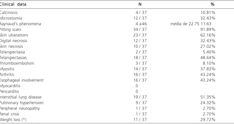
The prevalence of clinical findings in the studied population of scleroderma can be seen in Table 1, which shows that the most common findings were those of Raynaud’s phenomenon, pitting scars, interstitial lung disease and telangiectasia.
The Medsger severity index ranged from 2 to 13, with a mean 6.47±2.89.The profile of autoantibodies showed that ANA was present in 75.6% of patients. In 13.5% (5/34) anti topoisomerase or anti Scl-70 was present; in 34.3% (11/32) there was positivity of anticentromere; in 9.1% (3/33), anti ribonucleroproteine (or anti RNP) was present; in 8.3% (3/36) there was a positive anticardiolipin IgG; 11.7% (4/34) had anticardiolipin IgM and 5.5 % (1/ 18) had lupus anticoagulant.
Table 2- presents the data from the findings of nailfold capillaroscopy in the study sample.
Data from pairing SSc sample with Data from pairing SSc sample with Data from pairing SSc sample with Data from pairing SSc sample with Data from pairing SSc sample with controls
controlscontrols controls controls
The data regarding the pairing of the sample for risk factors of atherosclerosis are described in Table 3. Only the HDL displayed a significant difference.
Comparison of serum endothelin (ET-1) of controls
versus SSc patients
Values †of endothelin in the controls’ serum were from 0.41 to 5.65 pg/ml (median 2.26 pg/ml) and in patients with scleroderma from 0.41 to 8.82 pg/ml (median 0.41 pg / ml), with p = 0.0007 .
Values †of endothelin in the population of SSc according to demographic, clinical and serological data
Endothelin levels in male patients with scleroderma had a median value of 0.88 (0.41 to 2.73) and female of 0.41 to 6.82, p = 0.61. Serum endothelin levels in 37 patients with scleroderma according to the form of disease were (range †in pg/ml, median values in pg/ ml): 1) limited form – 0.41 to 2.73 and 0.4250; 2); diffuse form – 0.41 to 4.23 and 0.4100; 3); overlap form – 1.49 to 6.82 and 2.315 (p = 0.0424, Kruskall Wallis)
The analysis according to the scleroderma form showed that patients with overlap had higher levels of endothelin.
The variable age did not affect the levels of endothelin (p = 0.23, R Spearmnn -0.19, 95% CI of -0.49 to 0.14). The same was observed for disease duration (p = 0.67, Spearman R of -0.072, 95% CI -0.39 to 0.26)
The analysis of the variability of serum levels of endothelin is in accordance with the main clinical study, as can be seen in table 4.
The analysis of correlation between serum endothelin level and disease severity measured by the Medsger showed that p = 0.13, Spearman R 0.25, 95% CI -0091 to 0.54. Correlation analysis between the values of
Table 1 Table 1 Table 1 Table 1
Table 1 - Prevalence of clinical findings in the studied population (37 patients with scleroderma). Clinical data
Clinical dataClinical data Clinical data
Clinical data NNNNN %%%%%
Calcinosis 4 / 37 10.81%
Microstomia 12 / 37 32.43%
Raynaud’s phenomena 4 a 46 média de 22.75 11.63
Pitting scars 34 / 37 91.89%
Skin ulcerations 23 / 37 62.16%
Digital necrosis 12 / 37 32.43%
Skin necrosis 10 / 37 27.02%
Telangiectasia 2 / 37 5.40%
Telangiectasias 18 / 37 48.64%
Thromboembolism 3 / 37 8.10%
Myositis 14 / 37 37.83%
Arthritis 16 / 37 43.24%
Esophageal involvement 16 / 37 43.24%
Myocarditis 0
Pericarditis 0
Interstitial lung disease 19 / 37 51.35%
Pulmonary hypertension 9 / 37 24.32%
Peripheral neuropathy 1 / 37 2.70%
Renal crisis 1 / 37 2.70%
Weight loss (*) 11 / 37 29.72%
capillary periungual endothelin and serum can be assessed in table 5.
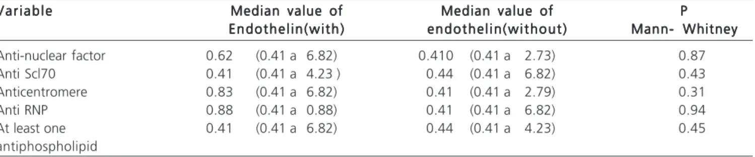
The analysis of endothelin serum levels according to the presence of autoantibodies is found in table 6.
DISCUSSION
DISCUSSION
DISCUSSION
DISCUSSION
DISCUSSION
There is large evidence that ET-1 modulates the tonus of peripheral blood vessels in humans and that the vasoconstrictor effect of ET-1 can be blocked by receptor antagonists (ETa)10,11. However, despite ET-1 is considered the most potent endogenous vasoconstrictor, its role in scleroderma – a disease where the vascular injury is the key to ethiopatogenesis of the majority of clinical manifestations – is still being investigated.
The idea that ET-1 may have elevated levels in patients with scleroderma has been suggested in several studies, especially those associated with pulmonary hypertension12. In this context, different antagonists of endothelin receptors have been developed and have shown promising treatment results13. However, the results obtained in clinical trials are difficult to be interpreted due to the great variety of types and density of receptors for ET-1 to the tissue studied. Furthermore, since endothelin is an agent with autocrine and paracrine activity, these performances are burdensome to be measured in vivo14.
In this study, an increased ET-1 in the sample of 37 scleroderma patients was not observed. In our sample, the patients had a mean disease duration of 42 years, were
predominantly female (92% of the sample) and the most prevalent form was the limited one – which affected 54% of all patients. The demographic pattern of the sample reflects a pattern that is typically found in this disease according to the literature15,16. The overlap form displayed higher levels of ET-1. In this sample we could not find an association between increased levels of ET-1 and degree of skin involvement, presence of Raynaud’s phenomenon and pulmonary hypertension. Raynaud’s phenomenon affected most of the studied individuals (92%). Few did not present with this finding, which may have hampered the comparative analysis. Moreover, the opposite case happens with pulmonary hypertension, one manifestation that appeared in only nine of 34 subjects in the sample. In patients with Raynaud there was a tendency to increased ET-1.
These results are similar to the study by Smyth et al.11 that evaluated a sample of 18 patients with primary Raynaud’s phenomena and 14 associated with scleroderma. Using plethysmography, These authors identified the vasospasm induced by cooling. They measured the ET-1 in that moment and compared to patients not exposed to vasospasm. In this study no significant changes in the levels of ET-1 were observed in cases compared to controls.These results and those of Smyth et al.11 are opposed to those of Biondi et al.17. The latter evaluated the serum levels of ET in 14 patients: 7 of them had primary Raynaud’s phenomenon and the others had Raynaud secondary to systemic sclerosis. They found higher levels of ET-1 in individuals with Raynaud than in controls.
Table 2 Table 2 Table 2 Table 2
-Table 2 - Findings of nailfold capillaroscopy in 37 scleroderma patients. Capillaroscopy findings
Capillaroscopy findings Capillaroscopy findings Capillaroscopy findings
Capillaroscopy findings R a n g eR a n g eR a n g eR a n g eR a n g e M e a nM e a nM e a nM e a nM e a n Capillary density ( number of capillary loops / mm) 3.3 a 7 5.08 /1.094
Deletions / finger 0 a 3 1.50 / 0.97
Dilated capillaries / finger 0 a 10 2.06 / 2.58
Table 3 Table 3 Table 3 Table 3
-Table 3 - Pairing scleroderma sample with controls
Patients with scleroderma (n=37) Patients with scleroderma (n=37) Patients with scleroderma (n=37) Patients with scleroderma (n=37)
Patients with scleroderma (n=37) Controls (n=37)Controls (n=37)Controls (n=37)Controls (n=37)Controls (n=37) PPPPP
Gender 3 men / 34 women 3 men / 34 women 1.00
Mean age (years) 48.97 / 3.36 49.57 / 11.29 0.83
Diabetics 1 / 33 3 / 37 0.61
Obesity 3 / 36 9 / 37 0.11
Smoking 11 / 37 10 / 37 0.58
Hypertension 7 / 32 11 / 37 0.58
Cholesterol (mg/dl) 169.1 / 47.66 182.2 / 36.83 0.22
Triglycerides (mg/dl) 114.8 / 62.52 108.6 / 55.14 0.68
HDL cholesterol (mg/dl) 45.89 / 12.18 57.09 / 9.850 0.0002
Fasting glucose (mg/dl) 90.53 / 15.22 91.86 / 16.62 0.77
In another study13 where the plasma level of ET-1 was also analyzed, 3ET-1 SSc patients had higher levels when compared to controls. In contrast to our study, diffuse sclerosis patients had higher levels than the ones with the limited form. It is also evident the variation of ET-1 with pulmonary disorders, with levels of ET-1 that showed inverse correlation with the ability to diffusion of carbon monoxide. Due to those data the authors raised the possibility of ET-1 levels be considered predictors of disease severity. The present study could not confirm this finding, since there was no association of serum endothelin findings with interstitial lung disease.
Furthermore, Hettema et al.10 studied 15 patients with limited form subjected to treatment with bosentan for 16 weeks and observed that the use of antagonists of
ET-1 showed no changes in either the vasodilator response or structural change or function of the microvasculature after treatment.
The microvasculature was examined in this study by nailfold capillaroscopy. Although this is an analysis based solely on morphological data, it reflects the evolutionary microvascular events in scleroderma18. In this study, neither deletion nor the degree of ectasia of the capillary nailfold capillaroscopy could be associated with serum levels of endothelin, showing no association between impaired microvascular morphology with ET-1 levels.
In this study, we found lower values of ET-1 in patients with limited scleroderma when compared to other groups, which could corroborate the minor effect of bosentan in these patients, as identified by Hettema et al.10 in their
Table 4 -Table 4 -Table 4 Table 4
-Table 4 - Comparison of serum median levels of endothelin (in pg/ml) according to clinical findings (n = 37). Clinical Data
Clinical DataClinical Data Clinical Data
Clinical Data Value of endothelin(with)Value of endothelin(with)Value of endothelin(with)Value of endothelin(with)Value of endothelin(with) Value endothelin (without)Value endothelin (without)Value endothelin (without)Value endothelin (without)Value endothelin (without) PPPPP
Calcinosis 0.41 (0.41 a 2.47) 0.44 (0.41 a6.820) 0.50
Microstomia 1.69 (0.41 a 6.82) 0.41 (0.41 a 2.47) 0.01
Raynaud’s Phenomena 0.63 (0.41 a 6.82 ) 0.41 (0.41 a 0.41) 0.053
Scars 0.41 (0.41 a2.730) 0.64 (0.41 a 6.82) 0.77
Cutaneous ulcers 0.41 (0.41 a 4.23) 0.44 (0.41 a 6.82) 0.69
Digital necrosis 0.41 (0.41 a 4.23) 0.83 (0.41 a 6.82) 0.74
Telangiectasias 0.62 (0.41 a 2.79) 0.41 (0.41 a 5.82) 0.74
Myositis 0.41 (0.41 a 6.82) 0.41 (0.41 a 2.79) 0.17
Arthritis 1.98 (0.41 a 6.82) 0.41 (0.41 a 1.48) 0.14
Esophageal Involvement 0.64 (0.41 a 6.82) 0.41 (0.41 a 2.79) 0.70
Interstitial pneumonitis 0.41 (0.41 a 4.23) 0.62 (0.41 a 2.31) 0.70
Pulmonary hypertension 0.98 (0.41 a 2.73) 0.41 (0.41 a 5.82) 0.57
Loss of weight 0.41 (0.41 a 2.63) 0.88 (0.41 a 6.82) 0.34
Table 5 Table 5Table 5 Table 5
Table 5 - Correlation between nailfold capillaroscopy and serum endothelin (in pg/ml) in 30 patients with scleroderma. R Spearman
R Spearman R Spearman R Spearman
R Spearman 95% I C95% I C95% I C95% I C95% I C PPPPP
Mean capillary density (capillary loops / mm) -0.14 -0.49 to 0.24 0.46
Mean deletion / finger 0.049 -0.30 to 0.39 0.78
Mean dilated capillaries / finger 0.20 -0.18 to 0.53 0.28
Table 6 Table 6 Table 6 Table 6
Table 6 - Comparison of serum median levels of endothelin (in pg / ml) in relation to autoantibodies profile (n = 37). V a r i a b l e
V a r i a b l eV a r i a b l e V a r i a b l e
V a r i a b l e Median value ofMedian value ofMedian value ofMedian value ofMedian value of Median value ofMedian value ofMedian value ofMedian value ofMedian value of PPPPP E n d o t h e l i n ( w i t h )
E n d o t h e l i n ( w i t h ) E n d o t h e l i n ( w i t h ) E n d o t h e l i n ( w i t h )
E n d o t h e l i n ( w i t h ) e n d o t h e l i n ( w i t h o u t )e n d o t h e l i n ( w i t h o u t )e n d o t h e l i n ( w i t h o u t )e n d o t h e l i n ( w i t h o u t )e n d o t h e l i n ( w i t h o u t ) Mann- WhitneyMann- WhitneyMann- WhitneyMann- WhitneyMann- Whitney
Anti-nuclear factor 0.62 (0.41 a 6.82) 0.410 (0.41 a 2.73) 0.87
Anti Scl70 0.41 (0.41 a 4.23 ) 0.44 (0.41 a 6.82) 0.43
Anticentromere 0.83 (0.41 a 6.82) 0.41 (0.41 a 2.79) 0.31
Anti RNP 0.88 (0.41 a 0.88) 0.41 (0.41 a 6.82) 0.94
At least one 0.41 (0.41 a 6.82) 0.44 (0.41 a 4.23) 0.45
15 patients with limited disease.
The role of ET-1 depends not only on its serum levels but also on the type and degree of healthiness of the tissue upon which it acts, as this can determine both the number of receptors and changes in their sensitivity19. For example, in normal tissue, the effect of type ET-B endothelin receptor is vasodilation, but under pathological conditions it is predominantly vasoconstriction. The reversal effect can result from increased levels of endothelin, as well as the location and adjustment of the ET-B receptors at endothelial level as there is concomitant up-regulation of ET-B receptors in some vascular smooth muscle cells7. Furthermore, a change in the proportions of receptors appears in pathological conditions, such as situations with myocardial ischemia and reperfusion with and without hypertension, which shows that ET-B blocks vasoconstriction in normotensive animals and causes vasodilatation in hypertensive ones20. This was also seen in scleroderma lung fibrosis, in tissues where the levels of the ET-A receptors were significantly reduced, while the levels of ET-B receptors were modestly elevated when compared to healthy tissue7. There are intriguing data about the complex role of 1 in the pathogenesis of SSc. If on one hand the ET-1 antagonist, bosentan, resulted in significant improvement in the prevention and formation of new digital ulcers, Korn et al.21, studying 122 patients from 17 centers in Europe and North America, have shown that it cannot improve the healing of pre-existing digital ulcers. It is unclear why a drug that supposedly promotes peripheral vasodilatation as an antagonist of ET-1 could not also promote healing of ulcers
more effectively. The ET-1 receptors were also found in keratinocytes, which suggests that ET-1 modulation can be a function of that cell, and therefore the receptor blockade could affect the re-epithetialisation process21.Thus, not only the serum levels of ET should be considered in the pathogenesis of the clinical findings of scleroderma, but also the levels and types of active receptors. This may explain the lack of association between serum levels of ET-1 and the main clinical findings of scleroderma in this study.
The present study has a small number of patients, which is expected in relatively rare diseases such as scleroderma. This may have disturbed the ability to demonstrate certain associations. Researches with larger samples are needed in order to strengthen the present findings. In addition, more detailed analysis involving the connection of this vascular mediator receptors are also interesting in order to help clarify the pathophysiology of vascular processes involved in scleroderma.
In conclusion, it can be stated that plasma levels of ET-1 are lower in patients with scleroderma when compared to controls. There was no association of these levels with disease severity. There was no significant association between the levels of ET-1 antibody and clinical profile. There was no correlation between the levels of endothelin and microvascular lesions detected by nailfold capillaroscopy.
Acknowledgements Acknowledgements Acknowledgements Acknowledgements Acknowledgements
To the Alvaro Laboratory from Curitiba-PR for helping in the determination of serum endothelin-1.
R E S U M O R E S U M O R E S U M O R E S U M O R E S U M O
Objetivo: Objetivo: Objetivo: Objetivo:
Objetivo: Avaliar a presença da ET-1 em pacientes portadores de esclerodermia e a sua correlação com o nível de atividade da doença; verificar se os níveis de endotelina estão associados com o perfil clínico e de autoanticorpos da esclerodermia e, ainda, se há associação com lesão microvascular detectada pela capilaroscopia periungueal. Métodos:Métodos:Métodos:Métodos:Métodos: Um total de 74 pacientes, sendo 37 portadores de esclerodermia e o restante controle, foram submetidos à dosagem de ET-1 por meio de teste de ELISA. Pacientes com esclerodermia foram analisados através de um questionário sobre características da doença e pesquisa de autoanticorpos. A gravidade da doença foi definida pelos critérios de Medsger e a doença microvascular foi acessada através de capilaroscopia periungueal. Resultados: Resultados: Resultados: Resultados: Resultados: Dos 37 pacientes com esclerodermia três (8,1%) eram homens e 34 (91,89%) mulheres, com idade média de 48,97 ? 13,36 anos e tempo médio de doença de 42,54 ? 13,35 anos. Os valores da ET-1 nos controles foram de 0,41 a 5,65 pg/ml (mediana de 2,26 pg/ml) e nos com esclerodermia de 0,41 a 8.82 pg/ml (mediana de 0,41 pg/ml) com p de 0,0007. Não houve correlação com o tempo de doença, idade do paciente e com o nível de acometimento cutâneo. Não encontrou-se correlação entre nível de ET-1 sérica e gravidade da doença (p=0,13). Níveis maiores de ET-1 foram observados na forma de superposição (1,49 a 6,82 pg/ml). Conclusão:Conclusão:Conclusão:Conclusão:Conclusão: Os níveis de ET-1 em esclerodérmicos mostraram-se inferiores aos controles. Não houve associação dos níveis de ET-1 com as variáveis estudadas.
Descritores: Descritores: Descritores: Descritores:
Descritores: Esclerodermia difusa. Marcadores biológicos. Endotelina-1. Angioscopia microscópica. Úlcera cutânea.
REFERENCES
REFERENCES
REFERENCES
REFERENCES
REFERENCES
1. Andersen GN, Caidahl K, Kazzam E, Petersson AS, Waldenström A, Mincheva-Nilsson L, et al. Correlation between increased nitric oxide production and markers of endothelial activation in systemic sclerosis: findings with the soluble adhesion molecules E-selectin, intercellular adhesion molecule 1, and vascular cell adhesion molecule 1. Arthritis Rheum. 2000;43(5):1085-93.
2. Silva LSM, Lima ARAG, Pucinelli MLC, Atra E, Andrade LEC. Capilaroscopia panorâmica periungueal e sua aplicação em doenças reumáticas. Rev Ass Med Bras. 1997;43(1):69-73. 3. Yamane K, Miyauchi T, Suzuki N, Yuhara T, Akama T, Suzuki H, et
al. Significance of plasma endothelin-1 levels in patients with systemic sclerosis. J Rheumatol. 1992;19(10):1566-71.
5. Dedola M, Godoi E, Coppé G, Cambou JP, Cantet C, Mas JL, et al. Risk factors management in 5708 ambulatory patients suffering from peripheral vascular disease followed in urban practice. Arch Mal Coeur Vaiss. 2005;98(12):1179-86.
6. Mayes MD. Endothelin and endothelin receptor antagonists in systemic rheumatic disease. Arthritis Rheum. 2003;48(5):1190-9. 7. Mayes MD. Systemic sclerosis: clinical aspects. In: Klippel JH, Stone JH, Crofford LJ, White PH, editors. Primer on rheumatic diseases. 13th ed. Atlanta: Springer; 2008. p.343-50.
8. Medsger TA Jr, Silman AJ, Steen VD, Black CM, Akesson A, Bacon PA, et al. A disease severity scale for systemic sclerosis: development and testing. J Rheumatol. 1999;26(10):2159-67.
9. Kayser C, Andrade LEC. Capilaroscopia periungueal: importância para a investigação do fenômerneo de Raynaud e doenças do espectro da esclerose sistêmica. Rev Bras Reumatol. 2004; 44(1):46-52.
10. Hettema ME, Zhang D, Stienstra Y, Smit AJ, Bootsma H, Kallenberg CG. No effects of bosentan on microvasculature in patients with limited cutaneous systemic sclerosis. Clin Rheumatol. 2009;28(7):825-33.
11. Smyth AE, Bell AL, Bruce IN, McGrann S, Allen JA. Digital vascular responses and serum endothelin-1 concentrations in primary and secondary Raynaud’s phenomenon. Ann Rheum Dis. 2000;59(11):870-4.
12. Oravec RM. NT-ProBNT e hieprtensão arterial pulmonar em esclerose sistemcia [dissertação]. Porto Alegre: Universidade Federal do Rio Grande do Sul, Faculdade de Medicina; 2007. 13. Yanagisawa M, Kurihara H, Kimura S, Tomobe Y, Kobayashi M,
Mitsui Y, et al. A novel potent vasoconstrictor peptide produced by vascular endothelial cells. Nature. 1988;332(6163):411-5. 14. Karkoulias K, Lykouras D, Sampsonas F, Drakatos P, Canova S,
Tsoukalas G, et al. The role of Endothelin-1 in obstructive sleep apnea syndrome and pulmonary arterial hypertension: pathogenesis and Endothelin-1 antagonists. Curr Med Chem. 2010;17(11):1059-66.
15. Assassi S, Del Junco D, Sutter K, McNearney TA, Reveille JD, Karnavas A, et al. Clinical and genetic factors predictive of mortality in early systemic sclerosis. Arthritis Rheum. 2009;61(10):1403-11.
16. Gabrielli A, Avvedimento EV, Krieg T. Scleroderma. N Engl J Med. 2009;360(19):1989-2003.
17. Biondi ML, Marasini B, Bassani C, Agastoni A. Increased plasma endothelin levels in patients with Raynaud’s phenomenon. N Engl J Med. 1991;324(16):1139-40.
18. Vane JR, Anggård EE, Botting RM. Regulatory functions of the vascular endothelium. N Engl J Med. 1990;323(1):27-36. 19. Saleh D, Furukawa K, Tsao MS, Maghazachi A, Corrin B,
Yanagisawa M, et al. Elevated expression of endothelin-1 and endothelin-converting enzyme-1 in idiopathic pulmonary fibrosis: possible involvement of proinflammatory cytokines. Am J Respir Cell Mol Biol. 1997;16(2):187-93.
20. Cowburn PJ, Cleland JG, McDonagh TA, McArthur JD, Dargie HJ, Morton JJ. Comparison of selective ET(A) and ET(B) receptor antagonists in patients with chronic heart failure. Eur J Heart Fail. 2005;7(1):37-42.
21. Korn JH, Mayes M, Matucci Cerinic M, Rainisio M, Pope J, Hachulla E, et al. Digital ulcers in systemic sclerosis: prevention by treatment with bosentan, an oral endothelin receptor antagonist. Arthritis Rheum. 2004;50(12):3985-93.
Received on 15/07/2011
Accepted for publication 23/09/2011 Conflict of interest: none
Source of funding: none
How to cite this article: How to cite this article:How to cite this article: How to cite this article:How to cite this article:
Michaelis T, Andretta M, Albers C, Skare TL, Ribas CAPM, Moreira LB. Evaluation of capillaroscopy using endothelin-1 as a marker of endothelial activation in microvascular injury and cutaneous ulcerations. Rev Col Bras Cir. [periódico na Internet] 2012; 39(2). Disponível em URL: http://www.scielo.br/rcbc
Correspondence to: Correspondence to:Correspondence to: Correspondence to:Correspondence to: Thiago Michaelis