CLINICAL SCIENCE
The effects of angiotensin-converting enzyme
inhi-bitors on peritoneal protein loss and solute transport
in peritoneal dialysis patients
Taner Basturk,IAbdulkadir Unsal,IYener Koc,IEren Nezaket,IIElbis Ahbap,ITamer Sakaci,IMustafa SevincI IDepartment of Nephrology, Sisli Etfal Research and Education Hospital, Istanbul, Turkey.IIDepartment of Biochemistry, Sisli Etfal Research and Education
Hospital, Istanbul, Turkey.
OBJECTIVE:The objective of this study was to examine the effects of angiotensin-converting enzyme inhibitors on peritoneal membrane transport, peritoneal protein loss, and proteinuria in peritoneal dialysis patients.
METHODS:Fifty-four peritoneal dialysis patients were included in the study. The patients were divided into two groups. Group 1 (n = 34) was treated with angiotensin-converting enzyme inhibitors. Group 2 (n = 20) did not receive any antihypertensive drugs during the entire follow-up. Eleven patients were excluded from the study thereafter. Thus, a total of 30 patients in Group 1 and 13 patients in Group 2 completed the study. We observed the patients for six months. Group 1 patients received maximal doses of angiotensin-converting enzyme inhibitors for six months. Parameters at the beginning of study and at the end of six months were evaluated. ClinicalTrial.gov: NCT01575652.
RESULTS: At the end of six months, total peritoneal protein loss in 24-hour dialysate effluent was significantly decreased in Group 1, whereas it was increased in Group 2. Compared to the baseline level, peritoneal albumin loss in 24-hour dialysate effluent and 4-hour D/P creatinine were significantly increased in Group 2 but were not significantly changed in Group 1. A covariance analysis between the groups revealed a significant difference only in the decreased amount of total protein loss in 24-hour dialysate. Proteinuria was decreased significantly in Group 1.
CONCLUSION:This study suggests that angiotensin-converting enzyme inhibitors reduce peritoneal protein loss and small-solute transport and effectively protect peritoneal membrane transport in peritoneal dialysis patients.
KEYWORDS: Angiotensin-converting enzyme inhibitors, peritoneal protein loss, peritoneal transport.
Basturk T, Unsal A, Koc Y, Nezaket E, Ahbap E, Sakaci T, et al. The effects of angiotensin-converting enzyme inhibitors on peritoneal protein loss and solute transport in peritoneal dialysis patients. Clinics. 2012;67(8):877-883.
Received for publication onNovember 28, 2011;First review completedMarch 5, 2012;Accepted for publication onApril 2, 2012
E-mail: [email protected]
Tel.: 90 5059271171
INTRODUCTION
Ever since peritoneal dialysis (PD) has been used in the treatment of chronic kidney disease (CKD), high peritoneal protein loss has been observed after each PD exchange. In adult patients, the loss has been estimated at 6 to 13 g daily (1). PD patients lose significant quantities of protein and albumin during the dialysis procedure (2). Low serum albumin has been associated with high peritoneal membrane transport status and also correlates with mortality (3). Loss of peritoneal function is a major complication associated with long-term peritoneal dialysis. Changes observed include loss and degeneration of the mesothelium, submesothelial thickening, alterations in the structure and number of
blood vessels, and reduplication of the vascular basement membrane (4).
The use of angiotensin-converting enzyme inhibitors (ACE-Is) in kidney disease has been demonstrated to be effective in reducing proteinuria and slowing the progression of kidney disease. The main mechanism of the antiprotei-nuric action of ACEIs is the reduction of the negative effects of angiotensin II on kidney hemodynamics (5). ACE-Is can preserve peritoneal histology, peritoneal function, and mesothelial cell remodeling (6).
Based on these beneficial effects of ACE-Is on proteinuria, we aimed to investigate the effects of ACE-Is on peritoneal membrane transport, peritoneal protein loss, and protei-nuria in patients being treated with PD.
PATIENTS AND METHODS
Study design and patients
This prospective cohort study was conducted at the Unit of Nephrology of Sisli Etfal Education and Research Hospital, Istanbul, Turkey. Prior to subject recruitment, the study protocol was reviewed and approved by the local Copyrightß2012CLINICS– This is an Open Access article distributed under
the terms of the Creative Commons Attribution Non-Commercial License (http:// creativecommons.org/licenses/by-nc/3.0/) which permits unrestricted non-commercial use, distribution, and reproduction in any medium, provided the original work is properly cited.
ethics committee in accordance with the ethical principles for human investigations, and written informed consents were obtained from all patients. Between June 2008 and January 2009, 54 age- and gender-matched continuous ambulatory peritoneal dialysis (CAPD) patients were included in the study consecutively.
Patients were divided into two groups according to decision of the physician: Group 1 (n = 34) consisted of patients treated with ACE-Is, and Group 2 (n = 20) consisted of patients not treated with ACE-Is. The inclusion criteria were chronic PD patients between 18 and 85 years old who had not received any antihypertensive drugs within the previous 12 months. All patients were on a standard CAPD program (2-2.5 L; 4 exchanges/day) without the use of icodextrin. The exclusion criteria were as follows: patients who had a history of antihypertensive treatment with ACE-Is or angiotensin-receptor blockers or aldosterone antagonists for the 12 months prior to the study time, intolerance to ACE-Is, CAPD-related peritonitis within six months prior to or during the study period, history of malignant hypertension or hypertensive encephalopathy, or cerebrovascular accident within the six months prior to the study, chronic liver disease, and recent acute illness and/or history of any overt chronic inflammatory disease. We excluded four patients from Group 1. One patient developed peritonitis during the study; two patients could not tolerate the medication because of hypotension and cough; one patient underwent renal transplantation. In Group 2, seven patients were excluded. Two patients developed peritonitis; two patients could not tolerate the medication due to hypotension and cough; two patients had uncontrolled hypertension; one patient did not attend follow-up visits. Thus, 30 patients in Group 1 and 13 patients in Group 2 completed the six-month study. The type of ACE-I prescribed to the patients was chosen randomly. Group 1 patients received the maximum tolerated doses of ACE-Is (such as lisinopril and perindopril) for six months.
Baseline definitions, measurements, and biochemical analysis
Demographic variables, including the etiology of CKD, age, and gender, were obtained from patients’ clinical charts. All blood samples were taken after 10 hours of overnight fasting. Serum urea, creatinine, and albumin levels were analyzed. Creatinine clearance [(CCr) dialysate, urine, and total] and Kt/V (dialysate, urine, and total) were calculated weekly. Daily volumes (UF), 24-hour protein, and albumin losses (dialysate, urine) were also recorded. Parameters at the beginning of study and at the end of the sixth month were evaluated. During the study, the dialysis regime remained the same for all patients. In both groups, we analyzed blood, 24-hour urine (in patients with residual diuresis.100 mL daily), and peritoneal effluent fluid at 4-and hour dwell times. Peritoneal effluent fluid at the 24-hour dwell time was used to determine total protein, albumin, urea and creatinine. The urea kinetic test in closest proximity to the time of the peritoneal equilibration test (PET) was used in the analysis.
After the subject had rested in the supine position for at least 15 minutes, blood pressure was measured with a standard mercury sphygmomanometer three times with the cuff around the right arm. Patients’ blood pressure measurements were taken on a regular basis every month. The mean values were calculated. All patients were on a standard 35-cal/kg/day carbohydrate, 1-2 g/kg/day
protein, and salt-restricted diet. Patients did not use essen-tial amino acids and peritoneal dialysis solutions containing amino acids. Serum urea, creatinine, and albumin levels were assessed by enzymatic colorimetric assay. Dialysate adequacy (Kt/V urea: dialysis and residual) and peritoneal transport (4-hour D/PCr) were measured using standard procedures (PD Adequest 1.4, 1994: Baxter Healthcare Corporation, Deerfield, IL, USA). Dialysate albumin loss was measured with the Bromo CresolGreen (BCG) method. Dialysate total protein loss was measured with the Biuret method. Urine protein concentration was determined with an immunoturbidimetric method.
Statistical analysis
Statistical analysis was performed using the SPSS 13.0 software package (SPSS Inc., Chicago, IL, USA). Kolmogorov-Smirnov tests were used to test the normality of data distribution. The data were expressed as arithmetic means and standard deviations. The chi-squared test was used to compare the categorical variables between groups. Inde-pendent sample T-testsandMann-Whitney U tests were used between groups for normally and abnormally distributed continuous variables, respectively. Paired t-tests and Wilco-xon signed-rank testswere used to analyze changes within each group. A two-sided p-value ,0.05 was considered to be statistically significant.
RESULTS
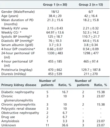
The baseline clinical, laboratory, and demographic char-acteristics of patients are presented in Table 1. There were
Table 1 -Patient Characteristics.
Group 1 (n = 30) Group 2 (n = 13)
Gender (Male/Female) 18/12 6/7
Age (years) 38.4¡20 42¡16.4
Mean duration of PD (months)
21.3¡15.6 18.2¡15.8
Weekly Kt/V urea 2.15¡0.43 2.21¡0.32
Weekly CCr * 64.97¡13.6 52.3¡14.2
Systolic BP (mmHg)* 125¡18.7 110.7¡21.7 Diastolic BP (mmHg)* 76¡10.3 64.6¡15.6 Serum albumin (g/dl) 3.7¡0.3 3.8¡0.34 4-hour D/P creatinine* 0.66¡0.07 0.54¡0.09 24-hour peritoneal UF
(ml)
1330¡483 1298¡477
4-hour peritoneal UF (ml)
455¡185 465¡97.4
Proteinuria (mg/day) 470¡662 129.7¡187.8
Diuresis (ml/day) 453¡539 211¡270
Primary kidney disease
Number of
patients Ratio, %
Number of
patients Ratio, %
Diabetic nephropathy 5 16.7 2 15.38
Chronic
glomerulonephritis
3 10 3 23.07
Chronic pyelonephritis 3 10 2 15.38
Polycystic renal disease 3 10 -
-Obstructive nephropathy 2 6.7 -
-Drug use 2 6.7 -
-Amyloidosis 1 3.3 3 23.07
Unknown 11 36.6 3 23.07
All values are given as the means¡standard deviations. NS: non-significant, CCr: creatinine clearance, BP: blood pressure, D/P: dialysate/ plasma, UF: daily volumes.
*
no significant differences in gender, age, or mean duration of PD between groups (p.0.05 for all) (Table 1). After six months, the decreases in systolic and diastolic blood pressures were statistically significant in Group 1 but not
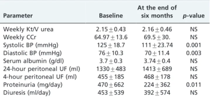
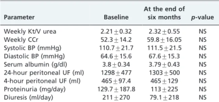
in Group 2. Only the decrease in peritoneal total protein loss at 24 hours of dwell time was significant following ACE-Is treatment (p,0.001). Statistically significant differ-ences were not identified in comparisons of the other studied parameters (p.0.05) (Table 2, Figures 1A, 1B, 3A). In Group 2, at the end of six months, 4-hour D/P creatinine and peritoneal albumin losses at 24 hours of dwell time were increased significantly. Other parameters did not change significantly in Group 2 (p.0.05) (Table 3, Figures 2A, 2B, 3B).
After ACE-Is were added to the treatment of the PD patients in Group 1, proteinuria levels were reduced significantly (p =0.011). Three patients became anuric; thus, the total number of anuric patients increased to twelve at the end of study. Residual renal function was surprisingly increased in eight patients at the end of six months and was decreased in ten patients. In total, the residual renal function decreased in Group 1, but this difference was not statistically significant (p.0.05) (Table 2). In Group 2, proteinuria levels did not change significantly during the study period (p.0.05). Three patients became anuric,
Table 2 -The effects of ACE-I treatment on the measured parameters in Group 1.
Parameter Baseline
At the end of six months p-value
Weekly Kt/V urea 2.15¡0.43 2.16¡0.46 NS
Weekly CCr 64.97¡13.6 69.5¡30. NS
Systolic BP (mmHg) 125¡18.7 111¡23.74 0.001 Diastolic BP (mmHg) 76¡10.3 70¡11.4 0.003 Serum albumin (g/dl) 3.7¡0.3 3.74¡0.4 NS 24-hour peritoneal UF (ml) 1330¡483 1413¡689 NS 4-hour peritoneal UF (ml) 455¡185 468¡178 NS Proteinuria (mg/day) 470¡662 224¡362 0.011
Diuresis (ml/day) 453¡539 392¡574 NS
All values are given as the means¡standard deviations. NS: non-significant, CCr: creatinine clearance, BP: blood pressure, UF: daily volumes.
increasing the number of anuric patients to ten in Group 2 at the end of six months. In addition, residual renal function was decreased in two patients, whereas it was increased in one patient. Overall, the residual renal function was also
decreased, but this difference was not statistically signifi-cant (p.0.05) (Table 3).
Covariance analysis between groups revealed a signifi-cant difference only in the amount of total protein loss in 24-hour dialysate in favor of reduction (p =0.048) in Group 1. No adverse effects, including hyperkalemia, were observed.
DISCUSSION
This study demonstrates that treatment with ACE-Is may preserve peritoneal membrane transport and may reduce peritoneal total protein loss and proteinuria in patients with PD.
ACE-Is affect the peritoneal membrane by increasing convective transport and decreasing diffusive transport— although in a limited way—and significantly reducing peritoneal protein losses at both 4-hour and 24-hour dwell times. The mechanisms of action of these effects have not been clearly described; however, they may be related to an effect on the permeability of the peritoneal membrane capillaries, where ACE-Is may act directly or indirectly by blocking the renin–angiotensin–aldosterone system (7).
Figure 2 -A. Total Loss of Protein in Group 2. B. Total Loss of Albumin in Group 2.
Table 3 -The parameters of untreated patients in Group 2.
Parameter Baseline
At the end of six months p-value
Weekly Kt/V urea 2.21¡0.32 2.32¡0.55 NS
Weekly CCr 52.3¡14.2 59.8¡16.05 NS
Systolic BP (mmHg) 110.7¡21.7 111.5¡21.5 NS Diastolic BP (mmHg) 64.6¡15.6 67.6¡15.3 NS Serum albumin (g/dl) 3.8¡0.34 3.79¡0.43 NS 24-hour peritoneal UF (ml) 1298¡477 1303¡500 NS 4-hour peritoneal UF (ml) 465¡97.4 465¡129 NS Proteinuria (mg/day) 129.7¡187.8 113¡225 NS
Diuresis (ml/day) 211¡270 79.1¡218 NS
Coronel et al. (8) demonstrated that captopril reduced peritoneal albumin loss without a significant change in systemic blood pressure. This effect likely decreases capillary permeability, either by a direct action of the drug or indirectly, mediated by AII, prostaglandins, or kinins. Jearnsujitwimol et al. (9) demonstrated that candesartan could provide a nutritional benefit by attenuating peritoneal loss of albumin and mediates an effective antihypertensive action. In addition, Agraharkar et al. (10) investigated the effect of RAS blockage on peritoneal protein loss in PD patients. These authors concluded that RAS blockage did not reduce protein loss into the peritoneal fluid during dialysis. In our study, peritoneal total protein loss was significantly reduced in patients treated with ACE-Is. However, peritoneal albumin loss did not change signifi-cantly. Additionally, peritoneal albumin loss increased significantly in Group 2.
Serum albumin is correlated with dietary protein intake in patients with renal disease. Peritoneal dialysis is associated with albumin and amino acid losses in the spent dialysate, which can reach 5–15 g/day. These losses may represent
,15% of the net daily protein intake (11). Kaysen GA et al.
(12) reported a strong correlation between serum albumin level and peritoneal protein loss in 18 patients. Pollock CA et al. (13) reported a weak correlation in 134 patients. This
correlation has not been confirmed in a number of other studies (14,15). Plasma volume expansion should cause the dilution of plasma proteins and a reduction in serum levels. Volume expansion commonly occurs in chronic renal failure, and the degree of fluid overload is associated with the serum albumin concentration (16). In this study, serum albumin concentrations were not significantly changed at the end of six months of treatment with ACE-Is. Although we did not evaluate the volume statuses of our patients, the reason for the stable plasma albumin levels may have been the increased plasma volume with no change in total albumin mass in PD patients.
PD solution. In addition, these authors found that ACEIs or AR blockers (or both) may preserve the viability of the peritoneum in continuous ambulatory PD patients over long periods.
The oral administration of enalapril ameliorates changes in peritoneal function and morphology (18), and the oral administration of lisinopril (an ACEI) and valsartan (an AR blocker) has similar beneficial effects on peritoneal function and morphology (19). Multiple studies have shown that ACE-Is prevent peritoneal protein loss (8,9). As a result, ACE-Is have a potential beneficial effect on the prevention of peritoneal fibrosis. Based on these findings, ACE-Is may preserve the viability of the peritoneum in PD patients over the long term.
In PD, both in vivo and in vitro studies involving the effects of ACE-Is on peritoneal membrane transporters have yielded different results. Kolesnyk et al. (20) have shown that treatment with ACEI/ARB in PD patients may prevent or retard the increase in D/P creatinine that occurs during long-term PD. In contrast, Jearnsujitwimol et al. (9) demonstrated that candesartan at a dose of 8-16 mg/day could effectively control blood pressure but caused no changes in peritoneal transport characteristics. We showed that peritoneal transport was not significantly decreased in Group 1 and was significantly increased in Group 2. The results of this study indicate that treatment with ACE-I in PD patients is likely to have a membrane-protective effect by preventing the increase in small-solute transport. Long-term treatment with ACE-Is attenuates the peritoneal alterations that can develop in long-term PD patients.
Ultrafiltration failure in patients undergoing PD is a condition with an incidence that increases over time. This complication is related to increased cardiovascular morbidity and mortality and is a major cause of the abandonment of the treatment technique (21). Duman et al. (17) demonstrated that an ACE-I, enalapril, improved ultrafiltration capacity when administered intraperitoneally. This ACE-I appeared to have a slower rate of decline in ultrafiltration, effectively protect-ing against peritoneal fibrosis in long-term peritoneal dialysis (17). ACE-Is may have an important role in PD by affecting the rate of decline of residual renal function in PD patients. Li et al. (22) investigated the effect of ACE-Is on residual renal function. Those authors concluded that the ACE-I ramipril might reduce the rate of decline of residual renal function in PD patients. At 12 months, 14 patients in the ramipril group (n = 30) and 22 in the control group (n = 30) became anuric. Two randomized controlled trials showed positive effects of A-II inhibitors on residual glomerular filtration rate (rGFR) in peritoneal dialysis patients (22,23). In another study, Kolesnyk et al. (24) found no difference with respect to the rate of decline of rGFR and time of the development of anuria. In our study, there were no significant differences in ultrafiltration volume and residual diuresis between groups, although proteinuria was significantly decreased in Group 1 (p =0.011). Therefore, the effect of ACE-Is may be important in preserving peritoneal function and in preventing protein losses, such as those experienced by PD patients, in whom considerable quantities of proteins are lost through the peritoneum and diuresis.
There are limitations in our study that must be consid-ered. The study groups were not large or well matched due to a limited number of patients. The follow-up period was
also short. The types and dosages of ACE-Is given to the patients were not strictly standardized.
Our findings suggest that ACE inhibitors support a decline in peritoneal protein losses and small-solute trans-port and effectively protect peritoneal membrane transtrans-port in long-term peritoneal dialysis.
AUTHOR CONTRIBUTIONS
Basturk T was responsible for the project and data collection. Unsal A was responsible for the project and editing the manuscript. Koc Y, Ahbap E, and Sakaci T collected the data and followed up with the patients. Nezaket E collected the data and performed the biochemical analyses. Sevinc M collected the data, followed up with the patients and edited the manuscript.
REFERENCES
1. Sandoz P, Walls J. Protein Losses in Continuous Ambulatory and Continuous Cyclic Peritoneal-Dialysis (Capd and Ccpd). Periton Dialysis B. 1984;4(2):109-10.
2. Krediet RT, Zuyderhoudt FMJ, Boeschoten EW, Arisz L. Peritoneal Permeability to Proteins in Diabetic and Nondiabetic Continuous Ambulatory Peritoneal-Dialysis Patients. Nephron. 1986;42(2):133-40, http://dx.doi.org/10.1159/000183652.
3. Davies SJ, Bryan J, Phillips L, Russell GI. Longitudinal changes in peritoneal kinetics: The effects of peritoneal dialysis and peritonitis. Nephrol Dial Transpl. 1996;11(3):498-506, http://dx.doi.org/10.1093/ oxfordjournals.ndt.a027318.
4. Go M, Kumano K, Sakai T. [Effect of angiotensin II(AII) on perito-neal transport during peritoperito-neal dialysis in rat]. Nihon Jinzo Gakkai shi. 1992;34(8):921-9, http://www.ncbi.nlm.nih.gov/entrez/query.fcgi?db = pubmed&cmd = Retrieve&dopt = AbstractPlus&list_uids = 1484411&query_ hl = 21&itool = pubmed_docsum.
5. Jafar TH, Schmid CH, Landa M, Giatras I, Toto R, Remuzzi G, et al. Angiotensin-converting enzyme inhibitors and progression of nondia-betic renal disease. A meta-analysis of patient-level data. Ann Intern Med. 2001;135(2):73-87.
6. Lewis EJ, Hunsicker LG, Bain RP, Rohde RD. The Effect of Angiotensin-Converting Enzyme-Inhibition on Diabetic Nephropathy. New Engl J Med. 1993;329(20):1456-62, http://dx.doi.org/10.1056/NEJM1993 11113292004.
7. Coronel F, Berni A, Cigarran S, Calvo N, Herrero JA. Effects of angiotensin II receptor blocker (irbesartan) on peritoneal membrane functions. Advances in peritoneal dialysis Conference on Peritoneal Dialysis. 2004;20:27-30.
8. Coronel F, Hortal L, Naranjo P, Cruceyra A, Barrientos A. Captopril, proteinuria and peritoneal protein leakage in diabetic patients. Nephron. 1989;51(3):443, http://dx.doi.org/10.1159/000185350.
9. Jearnsujitwimol V, Eiam-Ong S, Kanjanabuch T, Wathanavaha A, Pansin P. The effect of angiotensin II receptor blocker on peritoneal membrane transports in continuous ambulatory peritoneal dialysis patients. J Med Assoc Thai. 2006;89 Suppl 2:S188-95.
10. Agraharkar M, Du Y, Man-Wan C, Henry S, Kuo YF, Ahuja T. Angiotensin II receptor blockade (ARB) and peritonea[protein loss in peritoneal dialysis patients. J Am Soc Nephrol. 2003;14:858a-a. 11. Westra WM, Kopple JD, Krediet RT, Appell M, Mehrotra R. Dietary
protein requirements and dialysate protein losses in chronic peritoneal dialysis patients. Peritoneal Dialysis International. 2007;27(2):192-5. 12. Kaysen GA, Schoenfeld PY. Albumin Homeostasis in Patients
Undergoing Continuous Ambulatory Peritoneal-Dialysis. Kidney Int. 1984;25(1):107-14, http://dx.doi.org/10.1038/ki.1984.15.
13. Pollock CA, Ibels LS, Caterson RJ, Mahony JF, Waugh DA, Cocksedge B. Continuous ambulatory peritoneal dialysis. Eight years of experience at a single center. Medicine. 1989;68(5):293-308.
14. Avram MM, Goldwasser P, Erroa M, Fein PA. Predictors of survival in continuous ambulatory peritoneal dialysis patients: the importance of prealbumin and other nutritional and metabolic markers. Am J Kidney Dis. 1994;23(1):91-8.
15. Struijk DG, Krediet RT, Koomen GCM, Boeschoten EW, Arisz L. The Effect of Serum-Albumin at the Start of Continuous Ambulatory Peritoneal-Dialysis Treatment on Patient Survival. Peritoneal Dialysis International. 1994;14(2):121-6.
16. Kaysen GA, Don BR. Factors that affect albumin concentration in dialysis patients and their relationship to vascular disease. Kidney Int. 2003;63:S94-S7, http://dx.doi.org/10.1046/j.1523-1755.63.s84.20.x. 17. Duman S, Wieczorowska-Tobis K, Styszynski A, Kwiatkowska B,
18. Duman S, Gunal AI, Sen S, Asci G, Ozkahya M, Terzioglu E, et al. Does enalapril prevent peritoneal fibrosis induced by hypertonic (3.86%) peri-toneal dialysis solution? Periperi-toneal Dialysis International. 2001;21(2):219-24. 19. Duman S, Sen S, Duman C, Oreopoulos DG. Effect of valsartan versus lisinopril on peritoneal sclerosis in rats. International Journal of Artificial Organs. 2005;28(2):156-63.
20. Kolesnyk I, Noordzij M, Dekker FW, Boeschoten EW, Krediet RT. A positive effect of AII inhibitors on peritoneal membrane function in long-term PD patients. Nephrol Dial Transpl. 2009;24(1):272-7.
21. Aguirre AR, Abensur H. Protective measures against ultrafiltration failure in peritoneal dialysis patients. Clinics. 2011;66(12):2151-7, http:// dx.doi.org/10.1590/S1807-59322011001200023.
22. Li PKT, Chow KM, Wong TYH, Leung CB, Szeto CC. Effects of an angiotensin-converting renal function in patients receiving enzyme inhibitor on residual peritoneal dialysis - A randomized, controlled study. Annals of internal medicine. 2003;139(2):105-12.
23. Suzuki H, Kanno Y, Sugahara S, Okada H, Nakamoto H. Effects of an angiotensin II receptor blocker, valsartan, on residual renal function in patients on CAPD. Am J Kidney Dis. 2004;43(6):1056-64, http://dx.doi. org/10.1053/j.ajkd.2004.01.019.