Pre- and post-operative Wisconsin card
sorting test performance in patients with
temporal lobe epilepsy due to
hippocampal sclerosis
Luciana Tisser
1, Andre Palmini
1,2, Eliseu Paglioli
1,3, Mirna Portuguez
1,2, Ney Azambuja
1,3,
Jaderson Costa da Costa
1,2, Eduardo Paglioli
1,3, Carolina Torres
1, Jose Victor Martinez
1Abstract – Patients with temporal lobe epilepsy due to hippocampal sclerosis (TLE/HS) have a distinct neu-ropsychological profi le, but there is still debate on whether executive dysfunction is part of this profi le and also whether temporal lobe surgery can modify this dysfunction. Objective: To study the presence and reversibility of executive dysfunction in patients with unilateral TLE/HS. Methods: Twenty-fi ve patients with refractory sei-zures due to TLE/HS underwent presurgical evaluation which included the application of the Wiconsin Card Sorting Test (WCST). Nineteen were re-evaluated in follow up, at least 6 months after selective amygdalo-hip-pocampectomy (SAH). Twenty-two control subjects matched for age and education also performed the WCST.
Results: Sixteen of the 25 patients (64%) completed fewer than four categories in the WCST whereas only 4 of the 22 controls (18%) did not complete at least four categories (p<0.005). In addition, the performance of the patients involved signifi cantly more perseverative responses and errors compared to controls. The patient group demonstrated signifi cant post-operative improvement in many measures of the WCST following SAH. Conclu-sions: These fi ndings support the presence of executive dysfunction in patients with TLE/HS and suggest that such dysfunction can be partially reversed by selective resection of epileptogenic mesial temporal structures. Key words: temporal lobe epilepsy, hippocampal sclerosis, executive dysfunction, Wisconsin card sorting test, epilepsy surgery
Performance pré- e pós-operatória no Teste de Seleção de Cartas de Wisconsin em pacientes com epilepsia de lobo temporal associada à esclerose hipocampal
Resumo – Pacientes com epilepsia de lobo temporal associada à esclerose hipocampal (ELT/EH) têm um per-fi l neuropsicológico em sua maior parte bem reconhecido, embora discuta-se se manifestações de disfunção executiva acompanham este perfi l e se esta disfunção poderia ser modifi cada por cirurgia da epilepsia de lobo temporal. Objetivo: Estudar a presença e reversibilidade da disfunção executiva em pacientes com ELT/EH unila-teral. Métodos: Vinte e cinco pacientes com crises refratárias associadas à ELT/EH foram submetidos à avaliação pré-cirúrgica que incluiu a aplicação do Teste de Seleção de Cartas de Wisconsin (TSCW). Dezenove foram rea-valiados um mínimo de 6 meses após amígdalo-hipocampectomia seletiva (AHS). O TSCW foi também aplicado a 22 indivíduos controles pareados por idade e nível educacional. Resultados: Dezesseis dos 25 pacientes (64%) completaram menos de quatro categorias no TSCW, enquanto que apenas 4 dos 22 indivíduos do grupo controle (18%) tiveram este mesmo nível e disfunção (p<0.005). Além disto, a performance dos pacientes aresentava signifi cativamente mais respostas e erros perseverativos do que os controles. Analisados como um grupo, os pa-cientes apresentaram melhoras signifi cativas em várias medidas do TSCW após AHS. Conclusão: Estes achados apontam para a presença de disfunção executiva em pacientes com ELT/EH e sugerem que esta disfunção pode ser parcialmente revertida pela ressecção seletiva das estruturas temporais mesiais epileptogênicos.
Palavras-chave: epilepsia do lobo temporal, escleroses hipocampal, disfunção executiva, Wisconsin card sorting test, cirurgia da epilepsia.
1MSc, Porto Alegre Epilepsy Surgery Program, Services of Neurology and Neurosurgery, Hospital São Lucas da Pontifi cia Universidade Católica do Rio
Grande do Sul (PUCRS). 2MD, PhD, Division of Neurology, Department of Internal Medicine and 3 Division of Neurosurgery, Department of Surgery,
Dr. Andre Palmini – Serviço de Neurologia, Hospital São Lucas da PUCRS - Avenida Ipiranga 6690 - 90610-000 Porto Alegre RS - Brazil. E-mail: [email protected]
Patients with temporal lobe epilepsy (TLE) often pres-ent refractory seizures and are thus referred for presurgi-cal evaluation.1,2 In a signifi cant proportion of such cases, magnetic resonance imaging (MRI) shows unilateral hip-pocampal atrophy and loss of internal architecture, mani-fested as an abnormal signal of the hippocampus in T1, T2, and Fluid-attenuation inversion recovery (FLAIR) images. These fi ndings are collectively indicative of hippocampal sclerosis (HS), the most common etiology of TLE in ado-lescents and adults.1,4-6
Patients with TLE/HS tend to have a low quality of life (QoL), apparent both at clinical interviews and on more formal QoL questionnaires.8-10 This negative impact stems mostly from the refractory seizures, but is also compound-ed by high rates of depression and anxiety, unemployment, memory diffi culties, and an overall reduced ability to deal with life stressors.11 An issue that is largely unexplored is the possibility that executive dysfunction may affect QoL in patients with TLE/HS. This relative negligence is probably due to the location of the lesion and of the epileptogenic zone in the temporal lobe, theoretically sparing the frontal lobe networks reputedly involved with executive functions. Therefore, a massive amount of data has accumulated concerning memory function and dysfunction in TLE, 12-14 whereas the data on executive functioning is limited.
Nonetheless, anatomical connectivity between temporal and frontal lobe structures provides a tentative link be-tween TLE and executive functions,15 which has began to be explored. A few studies in the last decade16-19 have shown that patients with TLE of several different etiologies may have abnormal WCST scores.
When patients with TLE/HS have refractory seizures, epilepsy surgery is a highly effective treatment, both in terms of seizure control and improvement in QoL mea-sures, translating into better overall functioning.1,2,6 The latter is believed to be directly dependent upon and related to seizure control; however, the possibility that an improve-ment in cognitive, including executive, functioning may also play a role has not been formally studied.
In recent years, we have begun to explore these issues at the Porto Alegre Epilepsy Surgery Program. In a fi rst step, we followed a large cohort of patients for more than 10 years and reported that surgery for TLE/HS is associated with stable levels of seizure freedom in around 85-90% of patients.6,7 In a second set of studies, we then prospectively addressed the presence, associated features, and reversibility of executive dysfunction evaluated with a comprehensive test battery in a group of patients with TLE/HS (Tisser et al., in preparation) undergoing presurgical evaluation and epilepsy surgery. As part of these latter studies, the current work reports on the results of the single most informative
test of executive function in patients with epilepsy, namely the Wisconsin Card sorting Test (WCST), and focus on the pre- and post-operative performance of this cohort with TLE/HS. These studies will hopefully pave the way for a future project on the role of executive functions in QoL measures of patients with TLE/HS, pre and post surgery.
Methods
We studied 25 patients with TLE, refractory seizures, and MRI features characteristic of unilateral HS, who were consecutively evaluated on the Porto Alegre Epilepsy Sur-gery Program, Hospital São Lucas da Pontifícia Univer-sidade Católica do Rio Grande do Sul (PUCRS) in Porto Alegre, from October 2005 to May 2006. All patients un-derwent a comprehensive presurgical evaluation to local-ize the epileptogenic zone, determine neuropsychological status, and rule out major psychiatric disorders through structured interviews.20 All were able to fully cooperate in the neuropsychological test battery and all other proce-dures. Two other patients with TLE/HS evaluated in the same period were excluded because they had an estimated IQ of <79. The clinical, neurophysiologic and neuroimag-ing fi ndneuroimag-ings leadneuroimag-ing to the diagnosis of TLE/HS have been previously published.6,7 Briefl y, all patients had (i) clini-cal features characteristic of mesial TLE;21-23 (ii) interictal scalp/sphenoidal EEGs with unilateral or bilateral indepen-dent anteromesial temporal epileptiform discharges; (iii) at least one electroclinical seizure on scalp/sphenoidal video-EEG monitoring with clearcut unilateral temporal onset; and (iv) 1.5 Tesla MRI with at least two of the following: hippocampal atrophy, decreased intrahippocampal signal on T1-weighted images, or increased intrahippocampal signal on T2-weighted and fl uid-attenuated inversion re-covery (FLAIR) images. Age at evaluation ranged from 28 to 48 years (mean, 36.2), mean epilepsy duration was 25.7 years, and 13 were women. The lateralization of the epi-leptogenic zone, the degree of predominance between the temporal lobes in the distribution of the interictal spikes in patients with bitemporal independent discharges, and the antiepileptic drugs being used were also noted.
Controls
Neuropsychologic evaluation
Intelligence quotient (IQ) was estimated through the digits, vocabulary, and cubes subtests of the Wechsler Adult Intelligence Scale - IIIrd Edition - Revised (WAIS-III-R), Portuguese version (24), while the Wechsler Memory Scale - Revised (WMS-R) was used to evaluate episodic mem-ory.25 Results of the latter and their relation with WCST performance are to be presented elsewhere (Tiser et al, in preparation). Executive functions were evaluated through the WCST, Portuguese version,26 and although this test is still in the process of validation for the Brazilian adult pop-ulation, its extensive clinical application allows an inference of clinically-relevant criteria. For instance, a total number of completed categories in the WCST of ‘less than four’ is universally accepted as indicative of executive dysfunction for the age range of the patients and controls studied in the current investigation. The presence and quantifi cation of symptoms of depression and anxiety were determined through the Beck Inventary of Depression and Anxiety, Portuguese version.27
The WCST
This is a time-honored test to evaluate executive func-tions, and test procedures have been published elsewhere.28 Briefl y, the subject has to formulate a strategy to combine each of 128 cards consecutively drawn from a deck with one of four index cards which vary according to number, geometrical design, and color. The only feedback the sub-ject receives from the examiner is whether his/her choice was correct or incorrect and he/she has to use this informa-tion to combine ‘each next card’. Specifi cally, no feedback is given on why the choice was considered correct or incorrect by the examiner. A category is deemed complete when the subject succeeds in correctly combining 10 consecutive cards
drawn from the deck. Following this, and unbeknownst to the subject, the ‘correct’ strategy of pairing is changed (for instance, from color to geometrical form) and this has to be realized by the subject who then has to pair the cards according to this new strategy. The test evaluates abilities such as mental fl exibility and impulse control. In particu-lar, it probes the tendency of the subject to persevere in a strategy no longer valid despite receiving repeated ‘in-correct’ feedback and the ability to retain the newly valid strategy ‘on-line’ at the time of each choice.
We evaluated and analyzed the following WCST vari-ables: (i) number of correctly completed categories, to a maximum of 12; (ii) number of ‘correct’ pairings; (iii) number of ‘incorrect’ pairings; (iv) number of persevera-tive pairings, i.e., pairings in which the subject perseveres in anyrandom strategy, despite repeated feedback that the choice was ‘incorrect’; and (v) number of perseverative
‘er-rors’, i.e., incorrect pairings in which the subject perseveres in choosing according to the immediately previous strategy
which evoked correct feedback.
Post-operative evaluation
Post-operative evaluations of the WCST were per-formed between 6 and 12 months after surgery in 19 of the 25 patients. The other 6 could not be included because the interval between the operation and re-testing was shorter than 6 months.
Surgical procedure
All patients underwent a selective amygdalo-hippocam-pectomy (SAH) in which unilateral mesial temporal struc-tures were removed according to the technique originally described by Niemeyer.29 The temporal horn of the lateral ventricle was reached through a 2.0 to 2.5 cm incision in the second temporal gyrus, and excision of the amygdala, hippocampus and parahippocampal gyrus proceeded with aspiration of the amygdala, the anterior 2-3 cm of the hip-pocampus and of the parahippocampal gyrus extending posteriorly to the mid-mesencephalic level.7 The left mesial temporal structures were resected in 15 patients.
Framework of the study and statistical analysis
We fi rst compared the scores of the patients with those of the controls on the depression and anxiety scales and, more specifi cally, on the WCST. In a second step, we fo-cused on the patients and compared WCST performances before and after epilepsy surgery. All subjects consented to participate in the study, which was approved by the Ethics committee of the Hospital São Lucas da PUCRS.
Quantitative variables were described through means, standard deviations of the mean and Pearson’s correlation coeffi cient. The Student’s t test was used to for compari-son between means, the Kolmogorov-Smirnov for testing the normal distribution of quantitative variables, and the Mann-Whitney to compare the distribution of non-para-metric variables. The SPSS statistical package, version 13.0 was used for all statistical analyses and a level of 0.05 was considered to be statistically signifi cant.
Results
central nervous system was reported, where this took the form of febrile convulsions in 8. In 15 patients (60%) the HS was in the left temporal lobe and interictal epileptiform discharges always predominated in the temporal lobe ip-silateral to the HS. Discharges were strictly unilateral or had a large (>90%) unilateral predominance in 75% of patients. The remaining quarter had between 70 and 90% of epileptic discharges lateralized to the side of the HS. Most patients were receiving carbamazepine or phenytoin at the time of presurgical evaluation. Antiepileptic drugs and dosages were not modifi ed during the fi rst post-opera-tive year.
Pre-operative neuropsychological scores
Sixteen of the 25 patients (64%) completed less than
four categories in the WCST whereas only 4 of the 22 controls (18%) did not complete at least four categories (p<0.005). Mean number of completed categories for the patients was 3.1 (SD, 2.0) whereas for the controls was 4. 7 (SD, 1.6) (p <0.005). With the exception of the number of total correct responses, in all other parameters evaluated in the WCST the controls fared signifi cantly better than the patients (Table 2).
In addition, there was no correlation between the scores on the BDI or the BAI and the scores on the WCST (data not shown).
Post-operative results
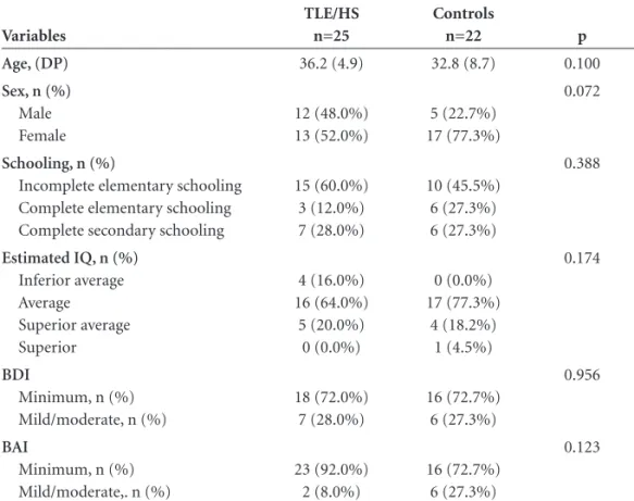
Following selective amygdalo-hippocampectomy, the post-operative performance of patients with TLE/HS in Table 1. Demographic, IQ, and psychiatric fi ndings.
Variables
TLE/HS n=25
Controls
n=22 p
Age, (DP) 36.2 (4.9) 32.8 (8.7) 0.100
Sex, n (%)
Male Female
12 (48.0%) 13 (52.0%)
5 (22.7%) 17 (77.3%)
0.072
Schooling, n (%)
Incomplete elementary schooling Complete elementary schooling Complete secondary schooling
15 (60.0%) 3 (12.0%) 7 (28.0%)
10 (45.5%) 6 (27.3%) 6 (27.3%)
0.388
Estimated IQ, n (%)
Inferior average Average Superior average Superior
4 (16.0%) 16 (64.0%)
5 (20.0%) 0 (0.0%)
0 (0.0%) 17 (77.3%)
4 (18.2%) 1 (4.5%)
0.174
BDI
Minimum, n (%) Mild/moderate, n (%)
18 (72.0%) 7 (28.0%)
16 (72.7%) 6 (27.3%)
0.956
BAI
Minimum, n (%) Mild/moderate,. n (%)
23 (92.0%) 2 (8.0%)
16 (72.7%) 6 (27.3%)
0.123
n, absolute number, BDI, Beck depression inventory; BAI, anxiety inventory; TLE/HS, temporal lobe epilepsy due to hippocampal sclerosis.
Table 2. Performance on the WCST by patients with TLE/HS, and by controls.
WCST parameter
TLE/HS mean (SD)
Controls
mean (SD) p
Number of completed categories 3.1 (2.0) 4.7 (1.6) 0.005
Total number of correct pairings 63.8 (14.9) 74.0 (9.3) 0.100
Total number of errors 56.4 (24.0) 37.8 (18.9) 0.005
Number of perseverative responses 47.4 (34.4) 20.8 (13.7) 0.001
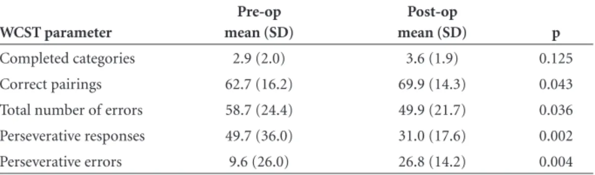
several WCST parameters was significantly better than before operation (Table 3). This included total number of correct pairings (p=0.043), total number of errors (p=0.036), number of perseverative errors (0.004) and also of perseverative responses (p=0.002). Improvement in total number of completed categories had a trend to-ward signifi cance.
Females younger than 35 years at operation, whose epilepsy had started before age 12 and who had recurrent seizures for less than 25 years tended to score higher on the post-operative WCST than males who were older than 35 years at operation and whose seizures had recurred for more than 25 years. Laterality of the epileptogenic zone did not correlate with post-operative scores, although this fi nd-ing should be viewed in the context of the limited patient sample. In contrast, those patients who continued on car-bamazepine or phenytoin monotherapy at the time of re-testing had signifi cantly less post-operative perseverative er-rors in comparison with those who continued on valproate, phenobarbital or clobazan, or on polytherapy regimens.
Discussion
We have shown that patients with unilateral TLE/HS display abnormalities in tests of executive function and that such executive dysfunction can be partially reversed by se-lective resection of the epileptogenic mesial temporal lobe structures. When compared to a control group, TLE/HS patients had a signifi cantly poorer performance in several parameters of the WCST. Other series including patients with TLE of several different etiologies have shown similar results, in that abnormal WCST scores were seen in 42 to 75% of patients.16,17
We believe our fi ndings may prove important for sev-eral reasons. First, we are not aware of similar studies in an etiologically homogeneous population of patients with medically refractory TLE undergoing a selective surgical procedure. Other studies examining executive functions in TLE either included patients with different etiologies or performed more extensive resections of temporal lobe
tissue.18,30 Because distinct etiologies of TLE may interfere with different temporal lobe structures and circuits, the etiological homogeneity of our patients allows more con-sistent considerations regarding the impact of functional and epileptiform abnormalities in the limbic system on the executive functions. Second, our fi ndings may help to explain why patients with TLE/HS often have diffi culties in conducting their lives which appear to extrapolate the negative impact of any recurrent seizures and the memory abnormalities. Admittedly, teasing out the putative disrup-tive effects of the execudisrup-tive dysfunction from those of the epilepsy and of the prevalent mood and memory abnor-malities may be a diffi cult task. However, in view of the pivotal role of the executive functions in problem-solving and decision-making it would hardly be a surprise if execu-tive dysfunction is eventually shown to impact the QoL of patients with TLE/HS. Third, this study adds further neu-ropsychological evidence to a growing body of cognitive, neurophysiological, metabolical, and brain perfusional re-search which consistently points to abnormalities in frontal lobe structures in patients with unilateral TLE/HS, thus suggesting that this disorder may impact the brain in a much more diffuse fashion than previously imagined.19,31-35 Finally, because the post-operative improvement in execu-tive functions reported in this study contrasts with the less favorable results reported in similar patients undergoing non-selective resections, i.e., temporal lobectomy18,30 XX, our findings suggest that sparing temporal neocortical structures may be relevant in this respect.
Traditionally, QoL limitations in patients with TLE/HS have been related to the recurrent seizures and resulting stigmata and deprivations.9 However, the issue of whether the executive dysfunction found in these patients - at least in neuropsychological testing - is also a major contributor to limitations in QoL, warrants further investigation. There is some support for this notion in the recent literature. Getz and colleagues showed a direct association between QoL in patients with TLE/HS and the presence of negative symp-toms, many of which36,37 related to executive dysfunction.
Table 3. Comparison of pre- and post- operative performance of patients with TLE/HS on the WCST.
WCST parameter
Pre-op mean (SD)
Post-op
mean (SD) p
Completed categories 2.9 (2.0) 3.6 (1.9) 0.125
Correct pairings 62.7 (16.2) 69.9 (14.3) 0.043
Total number of errors 58.7 (24.4) 49.9 (21.7) 0.036
Perseverative responses 49.7 (36.0) 31.0 (17.6) 0.002
Clearly, the hypothesis that executive dysfunction contrib-utes to limitations of QoL in patients with TLE/HS needs to be explicitly tested, correlating symptoms with neuro-psychological test scores.
A debated issue is whether the abnormalities in tests of executive function in patients with TLE/HS are due to the fact that the mesial temporal structures anatomically connected to the frontal lobes - and thus part of a fronto-temporal anatomofunctional network - are structurally and functionally abnormal or if such secondary frontal lobe dysfunction derives in a dynamic fashion from the temporo-frontal synaptic transmission of the electrical ab-normalities represented by recurrent seizures and interictal temporal lobe spikes.
It is known that mesial temporal structures contribute heavily toward episodic memory and the emotional rele-vance of incoming stimuli. Coupled with a broader view of working memory as also incorporating previously memo-rized information which is temporarily made conscious to support specifi c decision-making, it can be conceived that hippocampal dysfunction could interfere in the ex-ecutive functions.38,39 Likewise, alterations in the amygdala, often present in patients with TLE/HS, may interfere with emotional tuning thus affecting hierarchization of stimuli and other aspects of decision-making. Thus, theoretically at least, abnormalities in mesial temporal structures related to episodic memory and emotional processing of stimuli may contribute to executive dysfunction, irrespective of the presence or propagation of epileptic discharges into the frontal lobes.
However, irrespective of the debate on the role of the ‘fi xed’ abnormalities of mesial temporal structures in the executive dysfunction of patients with TLE/HS, improve-ment in performance following unilateral resection of these very structures strongly suggests that the dynamic epilepti-form abnormalities in temporo-frontal networks are more relevant to the executive dysfunction than the static histo-logical abnormalities. Following selective resection of me-sial temporal structures, patients committed signifi cantly fewer overall and perseverative errors while augmenting their correct pairings (choices) in comparison to their pre-operative performance. If the ‘fi xed’ amygdalar and hip-pocampal dysfunction were the major determinants of this performance, improvements following resection of these structures would be unlikely. An additional way to prove this point is to correlate the preoperative seizure frequency with performance in the WCST. This may reveal interesting associations and inform which patients with TLE/HS are at a greater risk of developing executive dysfunction with seizure recurrence. These possibilities are to be explored in another study (Tisser et al., in preparation).
Moreover, the results of the present work are in line with those seen in previous neuropsychological PET, SPECT and invasive electrode studies in patients with TLE/ HS, all suggesting a relationship between temporal epilep-tic discharges and seizures and electrical, metabolical, and perfusional disturbances in the frontal lobes.19-31-35 Studying patients with TLE/HS and controls with ictal SPECT, Van Paesschen et al.33 showed signifi cant ictal hypoperfusion in the frontal lobes. The same group furthered these fi ndings by adding interictal FDG-PET analysis of similar patients,34 showing that the most intense interictal hypometabolism was indeed in the frontal lobe ipsilateral to the mesial tem-poral epileptogenic zone, and not, as expected, in the tem-poral lobe itself. Furthermore, during seizures, ictal SPECT showed an extensive ring of hypoperfusion, maximal in the ipsilateral frontal lobe, colocalizing with the frontal areas showing maximal hypometabolism on FDG-PET. These data suggest a dynamic process including signifi cant in-hibition in the frontal lobes in patients with TLE/HS. In another recent study, Takaya et al.35 studied cognitive func-tions and interictal metabolism in 21 patients with mesial TLE. They showed that the 11 with more frequent seizures had more cognitive defi cits than the 10 in whom seizures were infrequent, and that this was associated with prefron-tal hypometabolism.
al.30 did not fi nd signifi cant modifi cations in post- versus pre-operative performance in a group of 174 patients with TLE undergoing (nonselective) anterior temporal lobecto-my. These negative fi ndings were not modifi ed either by the side of resection or the degree of seizure control. In another study on 72 patients with TLE undergoing the nonselec-tive procedure, the same authors18 confi rmed their negative results in terms of changes in post-operative WCST perfor-mance, even when HS was bilateral, and irrespective of the lesion type causing the epilepsies. These diverging results raise the possibility that the additional resection of the lateral temporal neocortex - which was performed in the patients who did not improve18,30 and yet not performed in the series in which many patients improved (41; present study) - somehow prevented a post-operative improvement in the WCST. This is not a universal fi nding19,40 and there-fore this hypothesis needs to be tested prospectively .
A potential limitation of the present study is that we did not re-test the control subjects after a similar 6-month interval. This leaves open the question of whether or not a ‘learning effect’ is operative when re-applying the WCST. However, evidence exists suggesting that the results of re-testing patients with the WCST are not compromised by a learning effect. Data collected for the WCST normative manual26 showed the instrument to produce stable results over time, even when re-testing was performed, in as little as one month following initial exposure, providing, of course, that the rationale of the test was not disclosed to the subjects after the fi rst application. In addition, in one of the few studies directly addressing this issue, Ingram and colleagues provide data supporting the temporal sta-bility of most variables probed with the WCST and suggest that previous contentions suggestive of a learning effect were related to the application of the WCST to non-pa-tient populations.42 Finally, we believe that if a learning curve were operant in our patients, it would be likely that performance during re-testing should have been better than we observed, despite the unequivocal improvements reported here.
As a fi nal point, we found preliminary associations be-tween gender, age at onset, epilepsy duration, and type of antiepileptic drug regimen and post-operative performance in the WCST. The small number of patients included in these analyses precludes bolder statements, but these pre-liminary fi ndings probably warrant further study.
References
1. Engel J Jr. Surgery for seizures. N Engl J Med 1996;334:647-652. 2. Wiebe S, Blume WT, Girvin JP, et al. A randomized controlled
trial of surgery for temporal-lobe epilepsy. N Engl J Med 2001;345:311-318.
3. Semah F, Picot MC, Adam MD et al. Is the underlying cause of epilepsy a major prognostic factor for recurrence ? Neurol-ogy 1998;51:1256-1262.
4. Briellmann RS, Kalnins RM, Berkovic SF, Jackson GD. Hip-pocampal pathology in refractory temporal lobe epilepsy: T2-weighted signal change refl ects dentate gliosis. Neurology 2002;58:265-271.
5. Williamson P, French JA, Thadani VM, et al. Characteris-tics of medial temporal lobe epilepsy: II. Interictal and ictal scalp electroencephalography, neuropsychological testing, neuroimaging, surgical results and pathology. Ann Neurol 1993;34:781-787.
6. Paglioli E, Palmini A, Paglioli E, et al. Survival Analysis of the surgical outcome of temporal lobe epilepsy due to hip-pocampal sclerosis. Epilepsia 2004;45:1383-1391.
7. Paglioli E, Palmini A, Portuguez M, et al. Seizure and memory outcome following temporal lobe surgery: selective compared with nonselective approaches for hippocampal sclerosis. J Neurosurg 2006;104:70-78.
8. McLachlan RS, Rose KJ, Derry PA, et al. Health-related qual-ity of life and seizure control in temporal lobe epilepsy. Ann Neurol 1997;41:482-489.
9. Mikati MA, Comair YG, Rahi A. Normalization of quality of life trhee years afther temporal lobectomy: a controlled study. Epilepsia 2006;47:928-933.
10. Dupont S, Tanguy ML, Clemenceau S, Adam C, Hazemann P, Baulac M. Long-term prognosis and psychosocial outcomes afther surgery for MTLE. Epilepsia 2006;47:2115-2124. 11. Baker GA, Gagnon D, McNulty P. The relationship between
seizure frequency, seizure type, and quality of life: find-ings from three European countries. Epilepsy Res 1998;31: 231-240.
12. Jones-Gotman M. Commentary: Psychological evaluation. Testing hippocampal function. In: Engel J Jr, editor. Surgical Treatment of the Epilepsies. New York: Raven Press; 1987: 203-211.
13. Hermann BP, Seidenberg M, Schoenfeld J, Davis MD. Neu-ropsychological characteristics of the syndrome of mesial temporal lobe epilepsy. Arch Neurol 1997;54:369-376. 14. Helmstaedter C. Neuropsychological aspects of epilepsy
sur-gery. Epilepsy & Behavior 2004;5(suppl 1):S45-S55. 15. Kier EL, Staib LH, Davis LM, Bronen RA. Dissection
tractog-raphy of the uncinate fasciculus, inferior occipital fascicu-lus, and Meyer’s loop of the optic radiation. AJNR 2004;25: 677-691.
16. Giovagnoli AR. Relation of sorting impairment to hippo-campal damage in temporal lobe epilepsy. Neuropsychologia. 2001;39:140-150.
18. Martin RC, Sawrie SM, Gilliam FG, Palmer CA, Faught E. Winsconsin card Sorting performance in patients with tem-poral lobe epilepsy: clinical and neuroanatomical correlates. Epilepsia 2000;41:1626-1632.
19. Hermann B, Seidenberg M. Executive system dysfunction in temporal lobe epilepsy: effects of nociferous cortex versus hippocampal pathology. J Clin Exp Neuropsychol 1995;17: 809-819.
20. Sheehan D, Lecrubeier Y, Sheehan KH, et al. The Mini In-ternational Neuropsychiatric Interview (MINI): The Dvel-opment and Validation of a Structured Diagnostic Psychi-atric Interview for DSM- IV and CID-10. J Clin Psychiatry 1998;59(suppl 20):22-33.
21. Palmini A, Gloor P. The localizing value of auras in partial seizures: a prospective and retrospective study. Neurology 1992;42:801-808.
22. French JA, Williamson P, Thadani VM, et al. Characteristics of medial temporal lobe epilepsy: I. Results of history and physical examination. Ann Neurol 1993;34:774-780. 23. Williamson PD, French JA, Thadani VM, et al.
Characteris-tics of medial temporal lobe epilepsy: II. Interictal and ictal scalp electroencephalography, neuropsychological testing, neuroimaging, surgical results and pathology. Ann Neurol 1993;34:781-787.
24. Wechsler D. Teste de inteligência para adultos WAIS-III. Adap-tação e Padronização Brasileira. São Paulo: Casa do Psicólogo; 2005.
25. Wescheler D. Weschler Memory Scale-revised. San Antonio: The Psychological Corporation; 1987.
26. Cunha JA, Trentini CM, Argimon IL, et al. Wisconsin de Clas-sifi cação de Cartas. Manual revisado e ampliado. Adaptação e Padronização Brasileira. São Paulo: Casa do Psicólogo; 2005. 27. Cunha JA. Manual da versão em português das Escalas Beck.
São Paulo: Casa do Psicólogo; 2001.
28. Chelune GJ, Baer RA. Developmental norms for the Wisconsin Card Sorting test. J Clin Exp Neuropsychol 1986;8:219-228. 29. Niemeyer P. The transventricular amygdalohippocampectomy
in temporal lobe epilepsy. In: Baldwin M, Bailey P, editors. Temporal Lobe Epilepsy. Springfi eld, Il: Charles C. Thomas; 1958:461-482.
30. Martin RC, Sarwrie SM, Edwards R, et al. Investigation of
executive function change following anterior temporal lobec-tomy: selective normalization of verbal fl uency. Neuropsy-chology 2000;14:501-508.
31. Lieb JP, Dasheiff RM, Engel JJr. Role of the frontal lobes in the propagation of mesial temporal lobe seizures. Epilepsia. 1991;32:822-837.
32. Savic I, Altshuler L, Baxter L, Engel JJr. Pattern of interictal hypometabolism in PET scans with fl udeoxyglucose F 18 re-fl ects prior seizure types in patients with mesial temporal lobe seizures. Arch Neurol 1997;54:129-136.
33. Van Paesschen W, Dupont P, Van Driel G, Van Billoen H, Maes A. SPECT perfusion changes during complex partial seizures in patients with hippocampal sclerosis. Brain 2003;126:1103-1111.
34. Nelissen N, Van Paesschen W, Baete K, et al. Correlations of interictal FDG-PET metabolism and ictal SPECT perfusion changes in human temporal lobe epilepsy with hippocampal sclerosis. Neuroimage 2006;32:684-695.
35. Takaya S, Hanakawa T, Hashikawa K, et al. Prefontral hypo-function in patients with intractable mesial temporal lobe epilepsy. Neurology 2006;67:1674-1676.
36. Getz K, Hermann BP, Bell B, et al. Negative symptoms in tem-poral lobe epilepsy. Am J Psychiatry 2002;59:644-651. 37. Getz K, Hermann B, Seidenberg M, et al. Negative symptoms
and psychosocial status in temporal lobe epilepsy. Epilepsy Res 2003;53:240-244.
38. Baddeley A. The episodic buffer: a new component of work-ing memory? Trends Cogn Sci 2000;4:417-423.
39. Palmini A, Haase VG. ‘To do or not to do’? The neurobiology of decision-making in daily life: I: Getting the basics. Dement Neuropsychol 2007;1:10-17.
40. Hermann BP, Wyler AR, Ritchie ET. Wisconsin Card Sort-ing Test performance in patients with complex partial sei-zures of temporal lobe origin. J Clin Exp Neuropsychology 1988;10:467-476.
41. Kim CH, Lee AS, Yoo HJ, Kang JK, Lee JK. Executive perfor-mance on the wisconsin card sorting test in temporal lobe epilepsy. Eur Neurol 2007;57:39-46