Evaluation of oral-motor movements and facial
mimic in patients with head and neck burns by a
public service in Brazil
Dicarla Motta Magnani,IFernanda Chiarion Sassi,ILuiz Philipe Molina Vana,IINivaldo Alonso,II Claudia Regina Furquim de AndradeI,*
IHospital das Clı´nicas da Faculdade de Medicina da Universidade de Sa˜o Paulo (HCFMUSP), Department of Physiotherapy, Speech-language and Hearing
Science and Occupational Therapy, Sa˜o Paulo, SP, Brazil.IIHospital das Clı´nicas da Faculdade de Medicina da Universidade de Sa˜o Paulo (HCFMUSP), Division of Plastic Surgery, Sa˜o Paulo, SP, Brazil.
OBJECTIVES:The purpose of this study was to analyze the characteristics of oral-motor movements and facial mimic in patients with head and neck burns.
METHODS:An observational descriptive cross-sectional study was conducted with patients who suffered burns to the head and neck and who were referred to the Division of Orofacial Myology of a public hospital for assessment and rehabilitation. Only patients presenting deep partial-thickness and full-thickness burns to areas of the face and neck were included in the study. Patients underwent clinical assessment that involved an oral-motor evaluation, mandibular range of movement assessment, and facial mimic assessment. Patients were divided into two groups: G1 – patients with deep partial-thickness burns; G2 – patients with full-thickness burns. RESULTS:Our final study sample comprised 40 patients: G1 with 19 individuals and G2 with 21 individuals. The overall scores obtained in the clinical assessment of oral-motor organs indicated that patients with both second-and third-degree burns presented deficits related to posture, position second-and mobility of the oral-motor organs. Considering facial mimic, groups significantly differed when performing voluntary facial movements. Patients also presented limited maximal incisor opening. Deficits were greater for individuals in G2 in all assessments. CONCLUSION:Patients with head and neck burns present significant deficits related to posture, position and mobility of the oral myofunctional structures, including facial movements.
KEYWORDS: Burn; Head; Face; Post-burn contracture; Speech-language and hearing sciences.
Magnani DM, Sassi FC, Vana LP, Alonso N, Andrade CR. Evaluation of oral-motor movements and facial mimic in patients with head and neck burns by a public service in Brazil. Clinics. 2015;70(5):339-345
Received for publication on December 15, 2014;First review completed on January 16, 2015;Accepted for publication on February 13, 2015
E-mail: [email protected]
*Corresponding author
’ INTRODUCTION
According to the literature, worldwide, burns and fires account for more than 300,000 deaths, and almost 11 million people per year require burn-related medical attention (1). In Brazil, burns remain a significant problem for the public health system (2). Brazil is the fifth largest country in the world, both by geographical area and by population. For this reason, the prevalence rate of burns tends to vary consider-ably across the literature, and reports are often limited to one healthcare setting. A review of the literature published in
2012 indicated that over 4% of the total number of hospitalizations in public hospitals in the country are caused by burns (2). The Brazilian National Health Surveillance Agency (ANVISA) notes that there are approximately 300,000 new cases of burns in children per year in Brazil (3). Studies tend to agree that adult males are often more affected than females (4-6).
The prevalence rates of burns of the head and neck also vary considerably across the literature, with estimates ranging from 6 to 60% of all burns (7). The neck and face regions are exposed to diverse injuries, such as scalds, electrical shocks and splashes. The traction forces caused by contracture may pull and cause insufficient neck extension, incomplete oral occlusion, oromaxillofacial skeletal deformi-ties and tracheal position alterations, resulting in difficult intubation, which can be life-threatening and can result in other severe complications (8). The extrinsic contractile forces from the neck can also cause facial deformities and can adversely affect the maturation of facial scars (9).
DOI:10.6061/clinics/2015(05)06
Copyright& 2015CLINICS –This is an Open Access article distributed under the
terms of the Creative Commons Attribution Non-Commercial License (http:// creativecommons.org/licenses/by-nc/3.0/) which permits unrestricted non-commercial use, distribution, and reproduction in any medium, provided the original work is properly cited.
There have been numerous articles pertaining to burns of the head and neck, mostly devoted to surgical and physiotherapeutic treatments and their results (1,9,10). However, only a very small number of articles describe the disastrous influence of burn sequelae on oral-motor struc-tural morphology, mobility and functions, such as mastica-tion, swallowing and speech. Severe burn injury to the face may result in complications such as facial and labial sensation deficits, poor oral access for oral/dental hygiene, and inadequate oral competence, causing chronic drooling and poor articulation (11). The literature suggests that oral contracture resulting in microstomia may have serious adverse effects on the patient’s ability to perform activities of daily living, including swallowing (12–15). Moreover, facial skeletal deformities are likely to occur at any age if burn contractures are neglected or are not properly and promptly treated (9). There have been reports of the effects of electrical injuries to the lip, cheeks, tongue and hard and soft palates. The sequelae described include severely limited mandibular movement, limited tongue movement due to adhesions to the floor of the mouth, speech problems and difficulty with oral hygiene (16).
Given the complexity of burn care rehabilitation, adequate assessment and monitoring should be undertaken in burn-injured patients. Rehabilitation following severe burn inju-ries requires an individualized approach to achieve the optimum functional outcome possible for every patient (17). Effective rehabilitative technical skills can only be developed if sequelae, i.e., physical and functional, have been well described.
The state of São Paulo, Brazil, has a population of 11.32 million people, comprising approximately 5.7% of the total Brazilian population. There are 13 burn centers registered by the Brazilian Ministry of Health in São Paulo. One of the most important centers is located atHospital das Clı´nicas, the largest public school hospital in all of South America.
Hospital das Clı´nicasadmits approximately 192 patients with acute burns each year. The purpose of this study was to analyze the characteristics of the oral-motor movements and facial mimic of patients who suffered burns to the head and neck and who were seen at the Division of Orofacial Myology of a Brazilian public hospital.
’ MATERIALS AND METHODS
Ethics
The study design was approved by the Ethics Committee for the Analysis of Research Projects (CAPPesq HCFMUSP no. 178.972). Prior to their enrollment, all participants were informed of the purpose and procedures, after which all patients provided written informed consent.
Study design and inclusion criteria
An observational, descriptive, cross-sectional study was conducted with patients who suffered burns to the head and neck and who were referred by the medical team to the Division of Orofacial Myology for assessment and rehabilita-tion. Patients meeting the following criteria were eligible for participation: aged X4 years, referred to the Division of
Orofacial Myology between January 2013 and December 2013, had a mean total burn surface area (TBSA) 44%, presented deep partial-thickness and full-thickness burns to areas of the face and neck, had received or not received skin grafting resulting in potential functional impairment within
a minimum of two months after epithelialization or medical intervention, presented stable medical conditions according to medical records and were fed exclusively by mouth. For characterization purposes, individuals were divided into two groups: G1–patients with deep partial-thickness burns; G2–patients with full-thickness burns (i.e., all participants presented third-degree burns). Importantly, patients who met the inclusion criteria had not yet undergone any form of rehabilitation.
A number of patients were excluded, including those who had cognitive, neurological, hearing and/or communication impairment, a documented diagnosis of facial trauma, previous surgical procedures to the head and neck (i.e., not related to burn wounds), and readmission due to pre-existing burns, as registered in the patient’s medical record.
Oral-motor clinical assessment
Participants underwent clinical oral-motor assessment. Individuals were examined while sitting in a chair in a room with appropriate lighting. The Expanded Protocol of Orofacial Myofunctional Evaluation with Scores (OMES-E) was used for this assessment (18). This protocol was constructed based on previous models of evaluation, with the addition of numerical scales that reflect the physical characteristics and orofacial behaviors of the subjects (individuals can reach a total of 230 points). Although the protocol was initially developed for 6-12-year-olds, its validity has now been reported for young and adult subjects (19).
The clinical protocol used in this study is one of the three validated protocols for orofacial myofunctional evaluation that have been published in the specific literature (19). Because it is based on a scale and requires no special equipment, it can be useful both in clinical practice and in research.
The evaluation was performed according to the OMES-E, and the stomatognathic system components, i.e., lips, tongue, jaw and cheeks, were evaluated in terms of posture and position, mobility and performance during deglutition and mastication functions. The participants were individu-ally evaluated by visual inspection, and the evaluation was later complemented by analyzing images recorded on a digital camera (Sony DSC-W120).
All participants were evaluated by two experienced speech-language pathologists. Inter-rater agreement was verified using Cohen’s Kappa Coefficient. The speech-language pathologists who assigned the scores of the OMES-E presented a high level of agreement (0.87).
Mandibular range of movement
The technique used to measure the mandibular range of movement was based on a methodology already published in the literature (20,21). Using a digital caliper (Digimess Pró-Fono Digital Caliper), the following measurements (in millimeters) were performed:
(1) maximal incisor opening - We measured the distance between the incisive faces of the mandibular and maxillary central incisors.
maxillary central incisors), we used the appropriate adjustment.
(3) mandibular lateralization to the left - The same proce-dure described above was performed to measure mandibular lateralization to the left.
(4) mandibular protrusion - For this measurement, the patient was asked to glide the mandible forward. We then measured the horizontal overlap value between the mandibular central incisors and the maxillary central incisors.
(5) horizontal dental occlusion overlap - We measured the distance between the occlusal face of the maxillary central incisors and the distal face of the mandibular central incisors.
Facial mimic
Facial symmetry and mobility were evaluated using the Clinical Score for Facial Mimic Protocol (22). This protocol was originally developed to perceptually investigate the impact of peripheral facial paralysis on the ability of individuals to produce symmetrical facial movements. To date, there are no specific functional scales available to explore the influence of burn sequelae on facial mimic. This protocol assesses facial functional/cosmetic symmetry.
The muscle groups from each facial side were analyzed under different voluntary facial expressions and scored as follows: zero (0) if there were no movements; one (1) for partial or moderate movement; and two (2) for complete or marked movement. The frontal region was assessed for eyebrow-raising movement, eyelid movement during eye closure, upper lip elevation through the movement of
‘‘frowning the nose’’, oblique traction of the upper lip required for smiling, horizontal traction of the upper lip based on the clinical smile, lip closure by means of lower lip protrusion and depression with the movement for exposing the lower teeth. After this stage, involuntary emotion-related movements were assessed on each side of the face by observing the participants during blinking, talking and spontaneously smiling, using the same scoring criteria mentioned above: zero (0) when absent, one (1) when reduced, and two (2) when normal. Lip and eyelid deformities at rest and the presence of synkinesia or hypertonia were also scored, with negative values: (0) if absent; (-1) if partially or mildly deformed; and (-2) if totally or severely deformed. Finally, the partial sum of the values obtained amounted to the final score, which could range from -6 to 20 points for each evaluated hemiface.
All participants were evaluated by two experienced speech-language pathologists. Inter-rater agreement was verified using Cohen’s Kappa Coefficient. The rates of agreement indicated that the reliability was high: OMES-E 0.87; mandib-ular range of movement 0.85; Facial Mimic Protocol 0.79.
Statistical analysis
Qualitative variables were presented in contingency tables comprising absolute (n) and relative (%) frequencies. All quantitative data were entered into an SPSS 21.0 database. Descriptive analyses of the quantitative data with a normal distribution were performed and presented as the mean values followed by the respective standard deviations (±SD). Data without a normal distribution were presented as the medians and interquartile ranges (IQR25-75%). Normal and homogenous distribution was assessed by the Shapiro-Wilk test.
Student’s t test for independent samples was used to analyze data with a normal distribution, and the non-parametric Mann-Whitney U test was used for data without a normal and homogenous distribution. Categorical data were analyzed using the Chi-squared test and Fisher’s exact test. The adopted significance level was 5% for all analyses.
’ RESULTS
During the study period, 50 of the 52 patients who were eligible for the oral-motor and facial mimic characterization consented to participate in the study. The OMES-E was not completed in 6 patients, and 4 did not complete the mandibular range of motion measurement, leaving 40 patients for the final analysis. Student’s t test did not indicate significant differences between the groups for age (G1 - 28.7±17.6 years-old; G2 - 35.9±19.3 years-old;p=0.23). G1 comprised 19 individuals, including 2 females and 17 males; 8 individuals presented burns restricted to the face, 1 individual presented burns restricted to the neck, and 10 individuals presented burns in both the facial and neck regions. The majority of participants had suffered a thermal burn (n=17), and only 2 patients had burns caused by chemical agents. The mean total body surface area (TBSA) affected was 13.27% (SD=7.06, range=4–28). G2 comprised 21 individuals, including 12 females and 9 males; 8 individuals presented burns restricted to the face, 2 individuals presented burns restricted to the neck, and 11 individuals presented burns in both the facial and neck regions. In this group, 2 individuals had burns caused by chemical agents. All of the other participants had suffered thermal burns. All of the patients included in G2 had suffered surgical procedures due to the presence of sequelae (i.e., skin graft, commissuroplasty, Z-plasty). The mean total body surface area (TBSA) affected was 17.72% (SD=9.18, range=4–35).
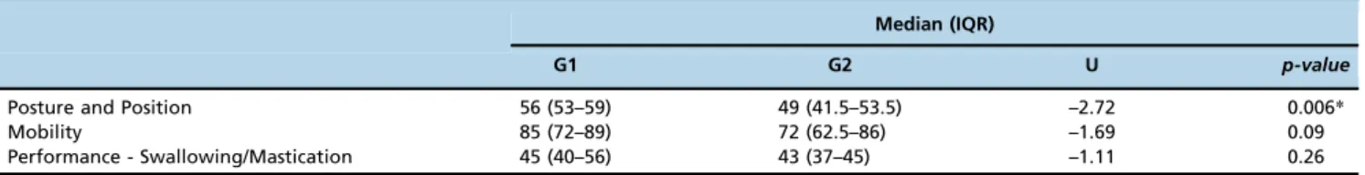
Table 1 presents the results of the Mann-Whitney U test for between-group comparisons, considering the scores obtained on the OMES-E. The results indicate that the groups significantly differed only when considering the static posture and the position of oral-motor organs. Individuals with deep partial-thickness burns presented higher scores in
Table 1 -Evaluation of the Expanded Protocol of Orofacial Myofunctional Evaluation with Scores in deep partial-thickness burns – G1 (n=19) and full-thickness burns – G2 (n=21).
Median (IQR)
G1 G2 U p-value
Posture and Position 56 (53–59) 49 (41.5–53.5) –2.72 0.006*
Mobility 85 (72–89) 72 (62.5–86) –1.69 0.09
Performance - Swallowing/Mastication 45 (40–56) 43 (37–45) –1.11 0.26
Legend: IQR – interquartile range
the clinical protocol compared with individuals with full-thickness burns. The presence of scar contractures and hypertrophic scars were responsible for the lower scores received by patients in G2 (i.e., the presence of facial asymmetry, difficulty maintaining sealed lips, tongue inade-quately positioned inside of the oral cavity). When analyzing the overall OMES-E scores, Student’s t test indicated that the groups also significantly differed (p=0.021); G1 presented higher overall scores (179.68±18.06) compared with G2 (162.1±26.50).
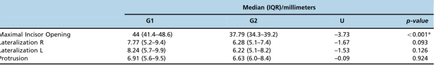
The results for the mandibular range of movement are presented in Table 2. Differences between groups were
significant for the measurement of maximal incisor opening. G1 presented a better range of movement for this parameter. Overall, the results indicate that burns to the head and neck had the same impact on mandibular lateralization and protrusion for both groups of patients.
The analyses of the results obtained for facial mimic are summarized in Tables 3 to 5. Because the data obtained for these analyses are categorical (i.e., a distribution of the indi-viduals among the possible scores), the results were analyzed using the Chi-squared test (number of individuals 45) and Fisher’s exact test (number of individuals o5). According to the protocol for facial mimic assessment, individuals
Table 2 -Evaluation of the mandibular range of movement in deep partial-thickness burns – G1 (n=19) and full-thickness burns – G2 (n=21).
Median (IQR)/millimeters
G1 G2 U p-value
Maximal Incisor Opening 44 (41.4–48.6) 37.79 (34.3–39.2) –3.73 o0.001*
Lateralization R 7.77 (5.2–9.4) 6.28 (5.1–7.4) –1.67 0.093
Lateralization L 8.24 (5.7–9.9) 6.22 (5.1–8.2) –1.53 0.126
Protrusion 6.91 (5.6–9.5) 6.63 (6.0–8.4) –0.09 0.924
Legend: SD – standard deviation R– right; L – left
*– significant results.
Table 3 -Evaluation of voluntary facial movements in deep partial-thickness burns – G1 (n=19) and full-thickness burns – G2 (n=21).
Amplitude of Voluntary Movements
Right Hemiface Left Hemiface
Scores 8 (%) 9 (%) 10 (%) 11 (%) 12 (%) 13 (%) 14 (%) p-value 8 (%) 9 (%) 10 (%) 11 (%) 12 (%) 13 (%) 14 (%) p-value
G1 - 1 (5.3) - 1 (5.3) 3 (15.8) 4 (21.1) 10 (52.5) o0.001* - 1 (5.3) - 2 (10.5) 3 (15.8) 5 (26.3) 8 (42.1) o0.001* G2 - 3 (14.3) 3 (14.3) 6 (28.6) 5 (23.8) 2 (9.5) 2 (9.5) 1 (4.8) 2 (9.5) 3 (14.3) 5 (23.8) 4 (19.0) 3 (14.3) 3 (14.3)
Legend: % - percentage of patients
*- significant results.
Table 4 -Evaluation of involuntary facial movements in deep partial-thickness burns – G1 (n=19) and full-thickness burns – G2 (n=21).
Amplitude of Involuntary Movements
Right Hemiface Left Hemiface
Scores 3 (%) 4 (%) 5 (%) 6 (%) p-value 3 (%) 4 (%) 5 (%) 6 (%) p-value
G1 - 3 (15.8) 1 (5.3) 15 (78.9) 0.415 - 2 (10.5) 3 (15.8) 14 (73.7) 0.696
G2 1 (4.8) 3 (14.3) 4 (19.0) 13 (61.9) 1 (4.8) 2 (9.5) 5 (23.8) 13 (61.9)
Legend: % - percentage of patients
*- significant results.
Table 5 -Evaluation of negative facial findings in deep partial-thickness burns – G1 (n=19) and full-thickness burns – G2 (n=21)
Negative Findings
Right Hemiface Left Hemiface
Scores -4 (%) -3 (%) -2 (%) -1 (%) 0 p-value -4 (%) -3 (%) -2 (%) -1 (%) 0 p-value
G1 - - 1 (5.3) 2 (10.5) 16 (84.2) o0.001* - - 1 (5.3) 3 (15.8) 15 (78.9) 0.002* G2 2 (9.5) 2 (9.5) 5 (23.8) 10 (47.6) 2 (9.5) 2 (9.5) 1 (4.8) 5 (23.8) 10 (47.6) 3 (14.3)
Legend: % - percentage of patients
could score a maximum of 14 points for each hemiface, considering voluntary facial movements (Table 3). The groups significantly differed when producing voluntary facial move-ments. Individuals in G1 presented better scores, with approximately half of the patients obtaining full scores on the analyzed parameters (i.e., symmetrical and preserved move-ments). Individuals in G2, however, due to their more severe burns, presented 11 points or less in this item and less symmetrical movements when comparing the scores obtained for the different hemifaces. The differences were not significant when comparing the groups for involuntary facial movements (Table 4). Individuals could receive a maximum of 6 points for each hemiface when assessed for involuntary facial move-ments. The results indicate that the majority of the participants (i.e., G1 and G2) received a full score for this parameter. Negative findings were indicated by negative scores on the Facial Mimic Protocol (Table 5). Overall, individuals in G1 did not present negative findings (score of 0), whereas individuals in G2 presented moderate or pronounced symptoms.
Given the results obtained for the overall scores on the Facial Mimic Protocol, we decided to assess the scores obtained on the sub-items of the protocol in detail. This
analysis is presented in Table 6 (i.e., Chi-squared test when the number of individuals was 45 and Fisher’s exact test when the number of individuals waso5). Looking closely at the sub-items for voluntary facial movements, we can observe that individuals with more severe burns presented significantly lower scores for movements corresponding to the muscles involved in smiling (i.e., upper lip elevation, upper-lateral traction of the lips and horizontal traction of the lips) and for lip closure. Moreover, individuals in G2 presented poorer scores on the parameter involving negative findings due to the presence of hypertonia.
’ DISCUSSION
To the best of our knowledge, the present study is the first extensive clinical characterization study that has investigated the impact of deep partial-thickness and full-thickness head and neck burns on oral-motor movements and facial mimic. Our results indicated that patients with burns present significant deficits related to oral myofunctional structural posture, position and mobility, including facial movements (i.e., mimic). Moreover, the results indicated that patients Table 6 - Results of the Clinical Score for Facial Mimic Protocol in deep partial-thickness burns – G1 (n=19) and full-thickness burns – G2 (n=21)
Right Hemiface
G1 G2
0(%) 1/-1(%) 2/-2(%) 0(%) 1/-1(%) 2/-2(%) p-value
Amplitude of Voluntary Movements
Eyebrow raise - 4(21.1) 15(78.9) - 2(9.5) 19(90.5) 0.398
Eyelid closure 1(5.3) 1(5.3) 17(89.5) - - 21(100) 0.312
Upper lip elevation - 2(10.5) 17(89.5) - 9(42.9) 12(57.1) 0.022*
Upper-lateral traction of lips - 2(10.5) 17(89.5) - 12(57.1) 9(42.9) 0.003*
Horizontal traction of lips - 3(15.8) 16(84.2) - 15(71.4) 6(28.6) 0.001*
Lip closure - 2(10.5) 17(89.5) - 12(57.1) 9(42.9) 0.003*
Lower lip depression - 2(10.5) 17(89.5) - 7(33.3) 14(66.7) 0.133
Amplitude of Involuntary Movements
Eyelid closure when blinking - 1(5.3) 18(94.7) - 1(4.8) 20(95.2) 0.999
When speaking - 2(10.5) 17(89.5) - 4(19) 17(81) 0.664
When smiling (spontaneous) - 4(21.1) 15(78.9) - 8(38.1) 13(61.9) 0.311
Negative findings
Eyelid deformity at rest 18(94.7) 1(51.3) - 20(95.2) 1(4.8) - 0.999
Lip deformity at rest 18(94.7) - 1(5.3) 15(71.4) 4(19) 2(9.5) 0.104
Synkinesis/hypertonia 18(94.7) 1(5.3) - 3(14.3) 11(52.4) 7(33.3) o0.001*
Left Hemiface
G1 G2
0(%) 1/-1(%) 2/-2(%) 0(%) 1/-1(%) 2/-2(%) p-value
Amplitude of Voluntary Movements
Eyebrow raise - 2(10.5) 17(89.5) - 4(19) 17(81) 0.664
Eyelid closure 1(5.3) 1(5.3) 17(89.5) - - 21(100) 0.312
Upper lip elevation - 3(15.8) 16(84.2) - 5(23.8) 16(76.2) 0.698
Upper-lateral traction of lips - 3(15.8) 16(84.2) - 11(52.4) 10(47.6) 0.022*
Horizontal traction of lips - 5(26.3) 14(73.7) - 14(66.7) 7(33.3) 0.014*
Lip closure - 4(21.1) 15(78.9) - 12(57.1) 9(42.9) 0.027*
Lower lip depression - 2(10.5) 17(89.5) - 8(38.1) 13(61.9) 0.069
Amplitude of Involuntary Movements
Eyelid closure when blinking - 1(5.3) 18(94.7) - 1(4.8) 20(95.2) 0.999
When speaking - 2(10.5) 17(89.5) - 3(14.3) 18(85.7) 0.999
When smiling (spontaneous) - 5(26.3) 14(73.7) - 8(38.1) 13(61.9) 0.511
Negative findings
Eyelid deformity at rest 18(94.7) 1(5.3) - 20(95.2) 1(4.8) - 0.999
Lip deformity at rest 17(89.5) 1(5.3) 1(5.3) 16(76.2) 3(14.3) 2(9.5) 0.531
Synkinesis/hypertonia 18(94.7) 1(5.3) - 4(19) 11(52.4) 6(28.6) o0.001*
with full-thickness burns present poorer performance com-pared with patients with deep partial-thickness burns.
Our results confirm what has already been described in the literature, indicating that contractures and hypertrophic scars have a negative impact on the oral myofunctional system (11-15,16,23). Nevertheless, the overall scores obtained on the OMES-E indicated that patients with deep partial-thickness and full-thickness burns present deficits related to oral-motor organs and movements (i.e., full scores were not observed in either group of patients). According to the literature, differences in wound healing and medical interventions can explain the differences found in our patient groups. Deep partial-thickness burns usually heal with some scarring. After the initial healing with wound closure and complete epithelialization, these patients require careful management and monitoring as they have the potential to develop severe late hypertrophic scars and contractures (8,9). Full-thickness burns are usually excised and skin grafted. Any tension on the neck region may promote early hypertrophic scarring (8,9). In addition to purely physical problems, studies clearly indicate that extensive head and neck scarring from burns may cause permanent alterations to the oral-motor organs (8-11).
Our study also found deficits in the mandibular range of movement in patients with burns. The groups significantly differed only when considering the measurement of the maximal incisor opening, with deep second- and third-degree burns presenting more mandibular movement restriction. This result can be explained by the fact that although some of the participants in G2 had undergone commissuroplasty, the patients still presented restrictions to opening their mouths. According to previous studies (20,21), the expected values for mandibular movements in healthy individuals are as follows, with no distinction between genders and age groups: maximal incisor opening - between 40 mm and 60 mm; mandibular lateralization - between 7 mm and 11 mm (i.e., to each side); and mandibular protrusion - between 7 mm and 11 mm. When more closely analyzing our results, patients with deep partial-thickness and full-thickness burns presented a greater limitation of all mandibular movements compared with the measurements expected for healthy individuals.
Mandibular function requires adaptation to a wide variety of factors related to the stomatognathic system (24). Mandibular movements are responsible for intraoral space modifications. These movements have a strong impact on mastication, swallowing and speech patterns because they are responsible for enabling adequate movements of the tongue and other soft tissues (i.e., amplitude) inside of the oral cavity (25). Maximal incisor opening movement has traditionally been used to evaluate temporomandibular joint (TMJ) functioning (26). Adequate TMJ functioning is therefore reflected by mandibular movements. The literature indicates that functional limitation secondary to burn injury usually results from an anatomical alteration of a major joint (8,9). The degree to which the function of a joint is affected is greatly influenced by the amount of soft tissue loss and the degree of pain associated with the movement (16,27). Full-thickness burns may also result in secondary damage to muscles, bones, tendons and ligaments. Studies have indicated that even when functional and/or structural limitations are present, the orofacial functions are made feasible by means of adaptations that are frequently not perceived by the individual (28). Muscle and structural adaptations arising from numerous conditions with different etiologies may be responsible for restricting muscle function, which can in turn impact mandibular movements (29). This
reduction in muscle activity may cause future structural problems such as atrophy (lack of use), thereby reducing muscle strength, restricting mandibular movements even more and causing structural modifications to the TMJ (30). Likewise, facial muscle pain is a condition that can be associated with physiological alterations such as vascular changes and co-contraction of adjacent muscles if functional muscle imbalances are present (31). The reduction of mandibular movements, either because of muscle atrophy or secondary to pain, may cause changes or compensations in the execution of the stomatognathic functions. Our results strongly suggest that patients with head and neck burns, especially when presenting scar contractures in the perioral region, should be considered at risk for developing future temporomandibular disorders.
As expected, patients also presented deficits in the facial mimic assessment. The effects of severe burns are debilitating and often cause depressive emotional conditions with a variety of possible functional and aesthetic problems (32). Participants with more severe burns (G2) presented greater limitations when performing voluntary facial movements due to hypertrophic scars. Patients, however, did not differ significantly when considering involuntary facial move-ments. We believe that this result can be explained by differences in performing voluntary and involuntary move-ments. Natural facial expressions tend to have a more subtle range of movements. When being assessed for voluntary facial movements, patients were asked to perform the goal movement with the highest range possible. Although the main goal in facial burns is the restoration of normal facial subunits with acceptable or good anatomical balance and symmetry and dynamic facial expressions, the outcomes of treatment are not always successful (12). The management of facial burns remains one of the most argued burn-related topics. The timing, strategy and options for treatment tend to vary considerably across the literature.
Until 2000, the treatment of patients with burns in Brazil was not regulated by the Ministry of Health (33). In 1994, the city of São Paulo was the first to publish a series of technical procedures for the treatment of burns (33). Since 2000, 42 burn centers have been regulated by the Ministry of Health, with new physical and functional structures and specialized multiprofes-sional teams (33). Although rehabilitation is a major emphasis, quality work remains to be performed. It is imperative that burn centers evaluate the functional outcome of thermally injured patients and include the role of all professionals involved the rehabilitation process. This is important not only for assessing disability but also for evaluating multiprofessional teams. Speech-language pathologists, as determined by the Brazilian Ministry of Health, are not part of the multiprofessional team designated to treat patients with burns.
amounts of soft-tissue loss on oral-motor functions. The evaluation of the orofacial myofunctional system is a fundamental step for the diagnosis of oral myofunctional disorders, which are present in several different health problems, including burns (11,16). In our study, we aimed to verify how head and neck burns can affect the oral-motor organs and functions, including facial movements. In this sense, the adopted protocols were demonstrated to be effective instruments to perform this characterization/ diagnosis. Nevertheless, specific protocols to evaluate the impact of head and neck burns on the oral myofunctional system should also be considered in future studies.
Patients with head and neck burns present significant deficits related to oral myofunctional structural posture, position and mobility, including facial movements. The results indicated that patients with full-thickness burns present poorer performance compared with patients with deep partial-thickness burns. Perioral burns that result in microstomia or mouth contracture reduce mandibular movements.
’ AUTHOR CONTRIBUTIONS
All authors have made substantial intellectual contributions to the conception and design of the study and the analysis and interpretation of the data. All authors have been involved in drafting the manuscript and critically revising it for important intellectual content.
’ REFERENCES
1. Peck MD. Epidemiology of burns throughout the world. Part I: Dis-tribution and risk factors. Burns. 2011;37(7):1087-100, http://dx.doi.org/ 10.1016/j.burns.2011.06.005.
2. Cruz BF, Cordovil PBL, Batista KNM. Epidemiological profile of patients who suffered burns in Brazil: literature review. Rev Bras Queimaduras. 2012;11(4):246-50.
3. Brasil. Anvisa–Agência Nacional de Vigilância Sanitária. O álcool na forma de gel é ou não um saneamento? [access: 2014 jan 10]. Available at : http://www.anvisa.gov.br/divulga/noticias/2002/130302.htm. 4. Montes SF, Barbosa MH, Neto ALS. Aspectos clínicos e epidemiológicos
de pacientes queimados internados em um Hospital de Ensino. Rev Esc Enferm USP. 2011;45(2):369-73, http://dx.doi.org/10.1590/S0080-62342011000200010.
5. Coutinho BBA, Balbuena MB, Anbar RA, Almeida KG, Almeida PYNG. Perfil epidemiológico de pacientes internados na enfermaria de queima-dos da Associac¸ão Beneficente de Campo Grande Santa Casa/MS. Rev Bras Queimaduras. 2010;9(2):50-3.
6. Gawryszewski VP, Bernal RTI, Silva NN, Morais Neto OL, Silva MMA, Mascarenhas MDM, et al. Atendimentos decorrentes de queimaduras em servic¸os públicos de emergência no Brasil, 2009. Cad Saúde Pública, Rio de Janeiro. 2012;28(4):629-40.
7. Kara IG, Gok S, Horsanli O, Zencir M. A population-based questionnaire study on the prevalence and epidemiology of burn patients in Denizli, Turkey. J Burn Care Res. 2008;29(3):446-50, http://dx.doi.org/10.1097/ BCR.0b013e3181710807.
8. Makboul M, El-Oteify M. Classification of post-burn contracture neck. Indian Journal of Burns. 2013;21(1):50-4, http://dx.doi.org/10.4103/0971-653X.121883.
9. Güven E, Ugurlu AM, Hocaoglu E, Kuvat SV, Elbey H. Treatment of post-burn upper extremity, neck and facial contractures: report of 77 cases. Ulus Travma Acil Cerrahi Derg. 2010;16(5):401-6.
10. Pallua N, Demir E. Postburn head and neck reconstruction in children with fasciocutaneous supraclavicular artery island flap. Ann Plast Surgery. 2008;60(3):276-82, http://dx.doi.org/10.1097/SAP.0b013e3180db2775.
11. Clayton NA, Ledgard JP, Haertsch PA, Kennedy PJ, Maitz PK. Rehabili-tation of speech and swallowing after burns reconstructive surgery of the lips and nose. J Burn Care Res 2009;30(6):1039-45.
12. Wust KJ. A modified dynamic mouth splint for burns patients. J Burn Care Res. 2006;27(1):86-92, http://dx.doi.org/10.1097/01.bcr.0000192267.55348.dd. 13. Bahnof R. Intra-oral burns: rehabilitation of severe restriction of
mouth opening. Physiotherapy. 2000;86:263-6, http://dx.doi.org/10.1016/ S0031-9406(05)60913-3.
14. Dougherty ME, Warden GD. A 30-year review of oral appliances used to manage microstomia, 1972-2002. J Bur Care Rehabil 2003;24(6):418-31, http://dx.doi.org/10.1097/01.BCR.0000095517.97355.98.
15. Johnson J, Candia J, La Trenta G, Madden MR, Goodwin CW, Finkelstein J. A nasal trumpet orthosis to maintain nares opening and respiratory function for patients with facial burns: a case report. J Burn Care Rehabil 1992;13(6):677-9, http://dx.doi.org/10.1097/00004630-199211000-00012.
16. Hilbert L, Peters WJ, Tepperman PS. Temporomandibular joint destruc-tion after a burn. Burns. 1984;10(3):214-6, http://dx.doi.org/10.1016/ 0305-4179(84)90032-9.
17. Al-Mousawi AM, Mecott-Rivera GA, Jeschke MG, Herdon DN. Burn teams and burn centers: The importance of a comprehensive team approach to burn care. Clin Plast Surg. 2009;36(4)547-54.
18. Felício CM, Folha GA, Ferreira CLP, Medeiros APM. Expanded protocol of orofacial myofunctional evaluation with scores: Validity and reliability. Int J Pediatr Otorhinolaryngol. 2010;74(11):1230-9, http://dx.doi.org/ 10.1016/j.ijporl.2010.07.021.
19. Felício CM, Medeiros APM, Melchior MO. Validity of the‘protocol of oro-facial myofunctional evaluation with scores’for young and adult subjects. J Oral Rehabil. 2012;39(10):744-53, http://dx.doi.org/10.1111/joor.2012.39. issue-10.
20. Celic R, Jerolimov V, Knezovic-Zlataric D, Klaic B. Measurement of mandibular movements in patients with temporomandibular disorders and asymptomatic subjects. Coll Antropol. 2003;27(Suppl 2):43-9. 21. Celic R, Jerolimov V, Knezovic-Zlataric D. Relationship of slightly limited
mandibular movements to temporomandibular disorders. Braz Dent J. 2004;15(2):151-4, http://dx.doi.org/10.1590/S0103-64402004000200012. 22. Salles AG, Toledo PN, Ferreira MC. Botulinum toxin injection in
long-standing facial paralysis patients: improvement of facial symmetry observed up to 6 months. Aesthetic Plast Surg. 2009;33(4):582-90, http:// dx.doi.org/10.1007/s00266-009-9337-9.
23. Rumbach AF, Ward EC, Cornwell PL, Bassett LV, Muller MJ. The chal-lenges of dysphagia management and rehabilitation after extensive ther-mal burn injury: a complex case. J Burn Care Res. 2009;30(5):901-5, http://dx.doi.org/10.1097/BCR.0b013e3181b487e0.
24. Yamada R, Ogawa T, Koyano K. The effect of head posture on direction and stability of mandibular closing movement. J Oral Rehabil. 1999;26(6):511-20, http://dx.doi.org/10.1046/j.1365-2842.1999.00386.x.
25. Bianchini EMG, Paiva G, Andrade CRF. Mandibular movements patterns during speech in subjects with temporomandibular disorders and in asymptomatic individuals. Cranio. 2008;26(1):50-66.
26. Szentpetery A. Clinical utility of mandibular movement ranges. J Orofac Pain. 1993 Spring;7(2):163-8.
27. Fricke N, Omnell M, Dutchere K, Hollender L, Engrav L. Skeletal and dental disturbances in children after facial burns and pressure garment use: a 4 year follow-up. J Burn Care Rehabil. 1999;20(3):239-49, http://dx. doi.org/10.1097/00004630-199905000-00016.
28. Johnson DL, Brand JW, Young SK, Duncanson MG. Adaptation of the temporomandibular joint to altered mandibular function. Int J Prostho-dont. 1995;8(5):446-55.
29. Greene CS. Managing the care of patients with temporomandibular dis-orders: a new guideline for care. J Am Dent Assoc. 2010;141(9):1086-8, http://dx.doi.org/10.14219/jada.archive.2010.0337.
30. Roda RP, Fernandez JMD, Bazan SH, Soriano YJ, Margaix M, Sarrión G. A review of temporomandibular joint disease (TMJD). Part II: Clinical and radiological semiology. Morbidity processes. Med Oral Patol Oral Cir Bucal. 2008;13(2):102-9.
31. Graff-Radford SB, Bassiur JP. Temporomandibular disorders and headaches. Neurol Clin. 2014;32(2):525-37, http://dx.doi.org/10.1016/j.ncl.2013.11.009. 32. Ye E. Psychological morbidity in patients with facial and neck burns. Burns.
1998;24(7):646-8, http://dx.doi.org/10.1016/S0305-4179(98)00081-3. 33. Almeida PCC, Gomez DS. Organizac¸ão de um centro de tratamento de