The relationship between low maternal serum
25-hydroxyvitamin D levels and gestational diabetes
mellitus according to the severity of 25-hydroxyvitamin
D deficiency
Sayid Shafi Zuhur, Rumeysa Selvinaz Erol, Idris Kuzu, Yuksel Altuntas
Endocrinology and Metabolism Department, Sisli Etfal Training and Research Hospital, Istanbul/Turkey.
OBJECTIVE:To assess the relationship between low maternal serum 25-hydroxyvitamin D levels and gestational diabetes mellitus in Turkish pregnant women according to the severity of 25-hydroxyvitamin D deficiency and assess intact parathyroid hormone levels in women with gestational diabetes mellitus and controls with low and sufficient 25-hydroxyvitamin D levels.
METHODS:We analyzed serum 25-hydroxyvitamin D and intact parathyroid hormone levels in 234 women with gestational diabetes mellitus and 168 controls. To define the deficiency status, 25-hydroxyvitamin D levels were further classified into severely deficient, deficient, insufficient and sufficient groups.
RESULTS: Women with gestational diabetes mellitus had significantly lower 25-hydroxyvitamin D levels compared to controls (30.8¡16.3 vs. 36.0¡16.2 nmol/L). However, when subgroups of 25-hydroxyvitamin D were analyzed, gestational diabetes mellitus was significantly more common only in women with severely deficient 25-hydroxyvitamin D levels. After adjusting for covariates, only severely deficient 25-hydroxyvitamin D levels were independently associated with an increased relative risk of gestational diabetes mellitus. The relative risk of gestational diabetes mellitus in women with insufficient and deficient 25-hydroxyvitamin D levels was not statistically significant. Intact parathyroid hormone concentrations were also significantly higher in women with gestational diabetes mellitus compared to the controls (45.3¡26.2vs. 38.7¡27.6 pg/ml). CONCLUSIONS:The results obtained from this study provide novel data indicating that only severely deficient maternal serum 25-hydroxyvitamin D levels are significantly associated with an elevated relative risk of gestational diabetes mellitus, even after adjusting for established risk factors of gestational diabetes mellitus.
KEYWORDS: Gestational Diabetes Mellitus; 25-hydroxyvitamin D; Parathyroid Hormone.
Zuhur SS, Erol RS, Kuzu I, Altuntas Y. The relationship between low maternal serum 25-hydroxyvitamin D levels and gestational diabetes mellitus according to the severity of 25-hydroxyvitamin D deficiency. Clinics. 2013;68(5):658-664.
Received for publication onNovember 20, 2012;First review completed onDecember 17, 2012;Accepted for publication onJanuary 26, 2013 E-mail: [email protected]
Tel.:+90 212 373 50 00
& INTRODUCTION
25-hydroxyvitamin D (25OHD) is well known for its function in maintaining calcium and phosphorus home-ostasis. In addition to its classical actions, a growing body of evidence has linked 25OHD deficiency with an increased risk of cardiovascular disease, some infectious, malignant and autoimmune diseases and diabetes mellitus (DM) (1,2). 25OHD deficiency is common during pregnancy. Bodnar et al. demonstrated that 29.2% of Black and 5% of White
pregnant women residing in a northern United States city had 25OHD deficiency (3). The results of publications investigating the relationship between low maternal serum 25OHD levels and gestational diabetes mellitus (GDM) are controversial (4-7). Some of these publications suggested a relationship between 25OHD deficiency and GDM, while others failed to find such a relationship, despite an association between low 25OHD levels and insulin resis-tance during pregnancy (4-7). Although the results of all of these studies are interesting, none of these studies have addressed the relationship between low maternal serum 25OHD levels and GDM according to the severity of 25OHD deficiency.
Vitamin D and parathyroid hormone (PTH) are both responsible for maintaining extracellular calcium (Ca) homeostasis (8). Vitamin D contributes to intestinal Ca absorption, while low serum Ca levels stimulate PTH secretion from the parathyroid gland to increase the renal Copyrightß2013CLINICS– This is an Open Access article distributed under
the terms of the Creative Commons Attribution Non-Commercial License (http:// creativecommons.org/licenses/by-nc/3.0/) which permits unrestricted non-commercial use, distribution, and reproduction in any medium, provided the original work is properly cited.
No potential conflict of interest was reported.
reabsorption of Ca and the resorption of Ca from bone (8). This condition is known as secondary hyperparathyroidism and has been suggested to increase the risk of DM (9-11).
However, previous studies investigating the relationship between low maternal serum 25OHD levels and GDM were limited by their inability to account for PTH levels. Therefore, this study was designed to assess the relationship between low maternal serum 25OHD levels and GDM according to the severity of 25OHD deficiency and assess PTH levels in women with GDM and controls with low and sufficient 25OHD levels in a large number of Turkish pregnant women with GDM and controls.
& MATERIALS AND METHODS
This cross-sectional study was conducted at the diabetes out-patient clinic of the Sisli Etfal Training and Research Hospital. Four hundred and two consecutive pregnant women referred to our center for an oral glucose tolerance test (OGTT) between January 2010 and April 2011 were included in the study. All of the study participants were residing in Istanbul (41
˚
00’N). According to the month of admission for OGTT, all of the participants were divided into spring-summer (May-October) and autumn-winter (November-April) groups. Pregnant women with pre-gestational diabetes, chronic disease or a history of consumption of drugs that interact with 25OHD metabolism and pregnant women with strict religious clothing were not included in the study. The study was designed in accordance with the Helsinki Declaration of 1975, informed consent was obtained from all participants, and the study was approved by the local ethics committee.A 75 g OGTT was performed in all participants at 24-28 weeks’ gestation. Plasma glucose levels (PGLs) were assessed immediately during OGTT using the glucose oxidase method. GDM was defined if at least one of the following diagnostic criteria was met: fasting, 1-hour or 2-hour PG levels$5.1 mmol/L, 10 mmol/L or 8.5 mmol/L, respectively (12).
Venous blood samples (20 mL) were also collected from each participant into plain (no additive) glass tubes on the day that the OGTT was performed (at 24-28 weeks’ gestation) for the assessment of 25OHD and intact PTH (iPTH). Maternal serum 25OHD levels were measured using an electrochemiluminescence method (Roche Diagnostics GmBH, Mannheim, Germany), with inter-assay and intra-assay coefficients of variation (CVs) of 2.4 and 5.7%, respectively. To define the deficiency status, maternal serum levels of 25OHD were further classified into four groups:
,12.5 nmol/L, severely deficient; 12.5-24.9 nmol/L, defi-cient; 25-49.9 nmol/L, insuffidefi-cient; and$50 nmol/L, suffi-cient. Maternal serum iPTH levels were measured immediately after blood collection using a chemilumines-cence immunoassay method (Roche Cobas, Roche Diagnostics GmBH, Mannheim, Germany), with a mean inter-assay CV of 6%. The experimental design of the study is presented in Figure 1.
Data regarding pre-pregnancy body mass index (BMI), parity, history of type 2 DM in first-degree relatives and the presence of GDM in previous pregnancies were obtained through one-on-one meetings with each participant and from antenatal visit records. BMI was calculated as the weight in kilograms divided by the height in meters squared.
& STATISTICAL ANALYSIS
Data were analyzed using SPSS version 15.0.0 for Windows (SPSS Inc., Chicago, IL). Two-sided tests were used throughout. Categorical data were evaluated using a chi-square analysis or Pearson’s correlation and Fisher’s exact tests as appropriate. Student’s t test and the Mann-Whitney U test were used to compare parametric quantita-tive and non-parametric data, respecquantita-tively. Analysis of variance (ANOVA) was used to compare quantitative variables within groups, followed by post-hoc analyses for multiple comparisons. A logistic regression analysis was used to estimate the odds ratios (ORs) and 95% confidence intervals (95% CIs). GDM was included as a dependent variable in the model. Covariates included in the model as independent variables included maternal serum 25OHD, age, previous history of GDM, history of type 2 DM in first-degree relatives and pre-pregnancy BMI. Scale variables were presented as the mean¡standard deviation (mean¡
SD). A p-value ,0.05 was considered to be statistically significant.
& RESULTS
The participants’ characteristics and baseline biochemical features are summarized in Table 1. Women with GDM had a higher pre-pregnancy BMI, had higher parity, were more likely to have a history of GDM in previous pregnancies and a history of type 2 DM in first-degree relatives, and were older compared with controls. In the current study, 64 (15.9%) pregnant women were classified as severely 25OHD deficient, 79 (19.7%) as deficient, 196 (48.8%) as insufficient and 63 (15.7%) as sufficient. Of the study participants, 230 (57%) were admitted during the autumn-winter months and 172 (43%) during the spring-summer months. The maternal serum 25OHD levels were 28¡16.3 and 39¡14.4 nmol/L during the autumn-winter
and spring-summer months, respectively (p,0.001). As expected, severe 25OHD deficiency and 25OHD deficiency were significantly more common during the autumn-winter months than the spring-summer months (p,0.0001 for both comparisons).
In this study, women with GDM had significantly lower 25OHD levels compared to controls (Table 1). However, when subgroups of 25OHD were analyzed, GDM was significantly more common only in pregnant women with severely deficient 25OHD levels compared to those with deficient, insufficient or sufficient 25OHD levels (p,0.0001 for all comparisons). Nevertheless, the frequency of GDM was not significantly different when pregnant women with deficient 25OHD levels were compared with those with insufficient and sufficient 25OHD levels (p= 0.89 and p= 0.14, respectively) or when pregnant women with
insufficient 25OHD levels were compared with those with sufficient 25OHD levels (p= 0.10). After adjusting for
maternal age, previous history of GDM, history of type 2 DM in first-degree relatives and pre-pregnancy BMI, low maternal serum 25OHD levels were not associated with an increased relative risk of GDM when 25OHD was intro-duced as a single independent variable into the logistic regression analysis (OR = 1.01, 95% CI 1.003–1.03,p= 0.14).
When subgroups of 25OHD were separately introduced into the model, the OR of GDM in women with insufficient and deficient 25OHD levels was also not statistically significant (OR = 1.46, 95% CI 1.27–2.74, p= 0.23, OR = 1.64, 95% CI
1.26–3.43, p= 0.18, respectively). However, after adjusting
for all these factors, pregnant women classified as being severely deficient for 25OHD had a 3.9-fold increased relative risk of GDM compared to those with sufficient, insufficient and deficient 25OHD levels (OR = 3.95, 95% CI 1.68–9.25,p= 0.002).
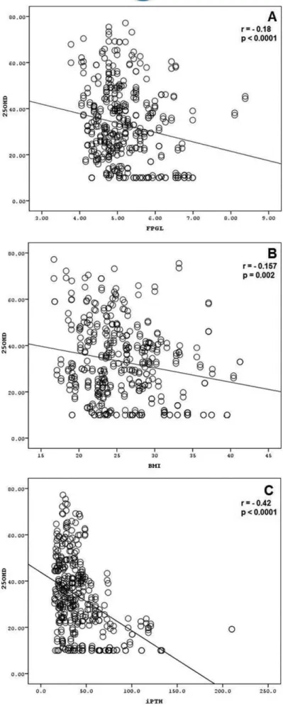
Likewise, as presented in Table 2, the fasting PGL was significantly higher in pregnant women with severely deficient 25OHD levels compared to those with deficient, insufficient and sufficient 25OHD levels (p,0.0001 for all
comparisons). An inverse correlation was also found between the fasting PGL and 25OHD levels (r =20.18,
p,0.0001). In addition, the inverse relationship between 25OHD and fasting PGL became more significant when the 25OHD level of women with severely deficient 25OHD levels was correlated with fasting PGL (r =20.28,p= 0.024).
However, no relationship was found between 2-hour PGL and 25OHD levels (r =20.08,p= 0.10). An inverse
correla-tion was also found between pre-pregnancy BMI and 25OHD levels (r =20.157, p= 0.002). Similar to GDM,
pre-pregnancy BMI was also significantly higher in preg-nant women with severely deficient 25OHD levels com-pared to those with deficient, insufficient and sufficient 25OHD levels (Table 2). The inverse correlation between 25OHD and fasting PGL and BMI in the study participants is presented in Figures 2 A and B.
iPTH levels above the normal laboratory reference ranges (secondary hyperparathyroidism) were present in 26 (40.6%) of 64 pregnant women with severely deficient levels, 26 (32.9%) of 79 pregnant women with deficient levels, 6 (3%) of 196 pregnant women with insufficient levels and none of those with sufficient 25OHD levels. As expected, an inverse association was also found between iPTH and 25OHD levels (r =20.42,p,0.0001). However, no association was found between iPTH concentrations and fasting or 2-hour PGL (r = 0.037,p= 0.45, r =20.01,p= 0.83,
respectively). The inverse correlation between 25OHD and iPTH concentrations is presented in Figure 2 C, and the differences in fasting PGL, 2-hour PGL, pre-pregnancy BMI and iPTH concentrations between pregnant women with severely deficient levels and those with deficient, insuffi-cient and suffiinsuffi-cient 25OHD levels are summarized in Table 2.
& DISCUSSION
As shown in Table 1, only 28 (12%) of 234 women with GDM and 35 (20.8%) of 168 controls had sufficient 25OHD levels, indicating a high prevalence of hypovitaminosis D during pregnancy. Moreover, 51% of women with GDM and 56% of those with normal glucose tolerance were taking a multivitamin supplement that contained 500 IU cholecal-ciferol. Considering the adverse effects of maternal vitamin D deficiency on offspring, such as neonatal hypocalcemia, seizure, impaired development and rickets (13,14), the high prevalence of hypovitaminosis D during pregnancy is a major public health problem, and vitamin D supplementa-tion during pregnancy is of paramount importance.
In the current study, 25OHD levels were significantly lower in pregnant women with GDM compared to controls. However, when subgroups of 25OHD concentrations were analyzed, the difference between 25OHD levels in women with GDM and controls was only significant in pregnant women with severely deficient 25OHD levels, and only severely deficient maternal serum 25OHD levels were associated with an increased risk of GDM after controlling for established risk factors of GDM, including maternal age,
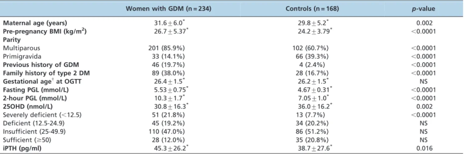
Table 1-Clinical and baseline biochemical characteristics of the study participants.
Women with GDM (n = 234) Controls (n = 168) p-value
Maternal age (years) 31.6¡6.0* 29.8
¡5.2* 0.002
Pre-pregnancy BMI (kg/m2) 26.7¡5.37* 24.2¡3.79*
,0.0001
Parity
Multiparous 201 (85.9%) 102 (60.7%) ,0.0001
Primigravida 33 (14.1%) 66 (39.3%) ,0.0001
Previous history of GDM 46 (19.7%) 4 (2.4%) ,0.0001
Family history of type 2 DM 89 (38.0%) 28 (16.7%) ,0.0001
Gestational age{at OGTT 26.4
¡1.5* 26.2
¡1.5* NS
Fasting PGL (mmol/L) 5.53¡0.75* 4.67¡0.31*
,0.0001
2-hour PGL (mmol/L) 10.3¡1.7* 7.05
¡1.0*
,0.0001
25OHD (nmol/L) 30.8¡16.3* 36.0¡16.2* 0.002
Severely deficient (,12.5) 51 (21.8%) 13 (7.7%) ,0.0001
Deficient (12.5-24.9) 45 (19.2%) 34 (20.2%) NS
Insufficient (25-49.9) 110 (47.0%) 86 (51.2%) NS
Sufficient ($50) 28 (12.0%) 35 (20.8%) NS
iPTH (pg/ml) 45.3¡26.2* 38.7¡27.6* 0.016
25OHD: 25-hydroxyvitamin D,*: mean¡standard deviation.
{: weeks, BMI: body mass index, DM: diabetes mellitus, GDM: gestational diabetes mellitus, NS: non-significant, OGTT: oral glucose tolerance test, PGL: plasma glucose.
Table 2-Differences in fasting and 2-hour PGL levels, pre-pregnancy BMI and iPTH concentrations between pregnant women with severely deficient 25OHD levels and those with deficient, insufficient and sufficient 25OHD levels (post-hoc analysis).
Serum 25OHD*
status
Fasting PGL
(mmol/L) p-value
2-hour PGL
(mmol/L) p-value
Pre-pregnancy BMI
(kg/m2) p-value
iPTH
(pg/ml) p-value
Severely 5.69¡0.76 ,0.0001 9.50¡1.97 NS 27.97¡5.01 0.001 60.21¡29.91 NS Deficient
Deficient 5.04¡0.61 8.73¡2.24 24.88¡4.38 58.27¡40.92
Severely 5.69¡0.76 ,0.0001 9.50¡1.97 NS 27.97¡5.01 0.03 60.21¡29.91 ,0.0001 Deficient
Insufficient 5.14¡0.74 9.04¡2.23 25.91¡4.94 34.68¡13.94
Severely 5.69¡0.76 ,0.0001 9.50¡1.97 NS 27.97¡5.01 0.001 60.21¡29.91 ,0.0001 deficient
Sufficient 4.96¡0.61 8.46¡2.12 23.65¡4.45 29.38¡9.22
*Adjusted for maternal age, previous history of GDM, family history of type 2 DM and pre-pregnancy BMI.
25OHD:25-hydroxyvitamin D,BMI:body mass index,DM:diabetes mellitus,GDM:gestational diabetes mellitus,iPTH:intact parathyroid hormone,
NS:non-significant,PGL:plasma glucose.
a previous history of GDM, a history of type 2 DM in first-degree relatives and pre-pregnancy BMI. In accordance with previous results (5,15), we also found an inverse association between 25OHD and fasting PGL.
Evidence suggests that both type 2 DM and GDM have similar pathophysiologic features characterized by two main metabolic defects: peripheral tissue resistance for insulin and insufficient secretion of insulin by pancreaticb
cells to compensate for this peripheral tissue resistance (16,17). Evidence also suggests a role for vitamin D in maintaining normal glucose homeostasis (2). In both animal and human studies, an association was shown between low vitamin D levels and insulin resistance and impaired insulin secretion (16). Moreover, specific receptors for 1-25 (OH)D3
have been demonstrated in pancreaticbcells, indicating the possible role of vitamin D in the regulation of insulin secretion (19). In a study by Maghbooli et al., maternal serum 25OHD levels were inversely associated with home-ostasis model assessment for insulin resistance (HOMA-IR), demonstrating that 25OHD deficiency may contribute to insulin resistance during pregnancy (4). However, the results of studies evaluating the association between 25OHD and GDM are controversial (4-7). Maghbooli et al. demonstrated that maternal serum levels of 25OHD during 24-28 weeks of pregnancy were significantly lower in women with GDM compared with controls (4). Clifton-Bligh et al. demonstrated an inverse association between maternal serum 25OHD levels and fasting blood glucose, although the association between 25OHD and GDM was not statistically significant (5). In a study performed in Indian pregnant women, no significant association was found between maternal serum levels of 25OHD and GDM risk (6). In a recent study, Makgoba et al. did not find an association between first trimester maternal serum 25OHD levels and subsequent GDM development (7). However, except for the study by Maghbooli et al., data regarding the severity of 25OHD deficiency in subjects included in these studies were insufficient. Therefore, the lack of an associa-tion between low maternal serum 25OHD levels and GDM in these studies may be due to the absence or very few numbers of pregnant women with severely deficient 25OHD levels.
In addition to type 2 DM and insulin resistance, obesity is a well-known risk factor for 25OHD deficiency (20-23). An analysis of the NHANES 2003-2004 data also demonstrated that 25OHD deficiency was highly prevalent in overweight and obese subjects (24). Although a few studies have found a relationship between pre-pregnancy BMI and low serum levels of 25OHD during pregnancy (25), the results from this study also suggest that a pre-pregnancy BMI.25 kg/m2is associated with severe 25OHD deficiency during the late second and third trimesters of pregnancy. In accordance with previous studies (26), this study also suggested that pre-pregnancy BMI was significantly higher in women with GDM compared with controls.
In the current study, iPTH levels were also significantly higher in pregnant women with severely deficient 25OHD levels compared to those with insufficient and sufficient 25OHD levels. Although no association was found between iPTH and fasting or 2-hour PGLs, iPTH concentrations were significantly higher in women with GDM compared to controls. Increased PTH levels, either primary or secondary to other disorders, have been shown to be associated with impaired glucose tolerance (2,27,28), decreased insulin
sensitivity and an increased risk of diabetes in glucose-tolerant subjects (9-11). Studies have suggested a two- to fourfold increased risk of diabetes in subjects with hyper-parathyroidism (9-11). Therefore, in addition to severe 25OHD deficiency, high PTH concentrations may have an additional effect on glucose tolerance during pregnancy. However, further studies are needed to explain the putative role of high PTH concentrations on the pathogenesis of GDM.
As demonstrated in the Hyperglycemia and Adverse Pregnancy Outcome study (HAPO), even a mild increase in fasting and 1-hour PGL corresponded to a significantly higher odds ratio for neonatal birth weight and cord blood c-peptide levels greater than the 90th percentile and caesarean delivery (29). Therefore, if appropriate therapy for severe 25OHD deficiency reduces the frequency or severity of GDM, this could have prominent public health significance.
An important limitation of our study was that maternal serum 25OHD levels were assessed by a single measure-ment during the late stage of pregnancy. Therefore, it may not reflect the maternal 25OHD status during the entire pregnancy. Another limitation was the cross-sectional design of this study; as such, we could not determine the neonatal outcomes of maternal 25OHD deficiency and also could not suggest any causal relationship.
In conclusion, the results obtained from this study provide novel data indicating that only severely deficient maternal serum 25OHD levels during pregnancy are significantly associated with an elevated relative risk of GDM, even after adjusting for established risk factors of GDM. As vitamin D deficiency is a worldwide public health problem and its severe deficiency during pregnancy may contribute to the development of GDM, the investigation of 25OHD deficiency during pregnancy and its appropriate replacement, particularly in patients with severely deficient levels, may contribute to the prevention of GDM. However, although several studies have suggested an association between 25OHD deficiency and DM, including GDM, more evidence is required to determine the effect of vitamin D on pancreaticbcells and peripheral insulin resistance.
& AUTHOR CONTRIBUTIONS
Zuhur SS contributed to the study design, implementation, data analysis and preparation of the manuscript. Erol RS contributed to the study design and recruited and screened the patients. Kuzu I and Altuntas¸ Y contributed to the data analysis and preparation of the manuscript.
& REFERENCES
1. Holick MF. Vitamin D deficiency. N Engl J Med. 2007;357(3):266-81. 2. Danescu LG, Levy S, Levy J. Vitamin D and diabetes mellitus. Endocrine.
2009;35(1):11-7, http://dx.doi.org/10.1007/s12020-008-9115-5. 3. Bodnar LM, Simhan HN, Powers RW, Frank MP, Cooperstein E, Roberts
JM. High prevalence of vitamin D insufficiency in black and white pregnant women residing in the northern United States and their neonates. J Nutr. 2007;137(2):447-52.
4. Maghbooli Z, Hossein-Nezhad A, Karimi F, Shafaei AR, Larijani B. Correlation between vitamin D3 deficiency and insulin resistance in pregnancy. Diabetes Metab Res Rev. 2008;24(1):27-32, http://dx.doi.org/ 10.1002/dmrr.737.
5. Clifton-Bligh RJ, McElduff P, McElduff A. Maternal vitamin d deficiency, ethnicity and gestational diabetes. Diabet Med. 2008;25(6):678-84, http:// dx.doi.org/10.1111/j.1464-5491.2008.02422.x.
6. Farrant HJ, Krishnaveni GV, Hill JC, Boucher BJ, Fisher DJ, Noonan K, et al. Vitamin D insufficiency is common in Indian mothers but is not associated with gestational diabetes or variation in newborn size. Eur J Clin Nutr. 2009;63(5):646-52.
7. Makgoba M, Nelson SM, Savvidou M, Messow CM, Nicolaides K, Sattar N. First-trimester circulating 25-hydroxyvitamin D levels and develop-ment of gestational diabetes mellitus. Diabetes Care. 2011;34(5):1091-3, http://dx.doi.org/10.2337/dc10-2264.
8. Reis JP, von Mu¨hlen D, Kritz-Silverstein D, Wingard DL, Barrett-Connor E. Vitamin D, parathyroid hormone levels, and the prevalence of metabolic syndrome in community-dwelling older adults. Diabetes Care. 2007;30(6):1549-55, http://dx.doi.org/10.2337/dc06-2438.
9. Cheung PS, Thompson NW, Brothers TE, Vinik AI. Effect of hyperpar-athyroidism on the control of diabetes mellitus. Surgery. 1986; 100(6):1039-47.
10. Taylor WH. The prevalence of diabetes mellitus in patients with primary hyperparathyroidism and among their relatives. Diabet Med. 1991;8(7):683-7, http://dx.doi.org/10.1111/j.1464-5491.1991.tb01678.x. 11. Werner S, Hjern B, Sjo¨berg HE. Primary hyperparathyroidism. Analysis
of findings in a series of 129 patients. Acta Chir Scand. 1974;140(8):618-25.
12. Metzger BE, Gabbe SG, Persson B, Buchanan TA, Catalano PA. Damm P. International association of diabetes and pregnancy study groups recommendations on the diagnosis and classification of hyperglycemia in pregnancy. Diabetes Care. 2010;33(3):676-82.
13. Robinson PD, Ho¨gler W, Craig ME, Verge CF, Walker JL, Piper AC, et al. The re-emerging burden of rickets: a decade of experience from Sydney. Arch Dis Child. 2006;91(7):564-8.
14. Munns C, Zacharin MR, Rodda CP, Batch JA, Morley R, Cranswick NE, et al. Prevention and treatment of infant and childhood vitamin D deficiency in Australia and New Zealand: a consensus statement. Med J Aust. 2006;185(5):268-72.
15. Lau SL, Gunton JE, Athayde NP, Byth K, Cheung NW. Serum 25-hydroxyvitamin D and glycated haemoglobin levels in women with gestational diabetes mellitus. Med J Aust. 2011;194(7):334-7.
16. Retnakaran R, Qi Y, Sermer M, Connelly PW, Hanley AJ, Zinman B. Glucose intolerance in pregnancy and future risk of pre-diabetes or diabetes. Diabetes Care. 2008;31(10):2026-31, http://dx.doi.org/10.2337/ dc08-0972.
17. Saisho Y, Miyakoshi K, Tanaka M, Shimada A, Ikenoue S, Kadohira I, et al. Beta cell dysfunction and its clinical significance in gestational diabetes. Endocr J. 2010:57(11):973-80, http://dx.doi.org/10.1507/ endocrj.K10E-231.
18. Cavalier E, Delanaye P, Souberbielle JC, Radermecker RP. Vitamin D and type 2 diabetes mellitus: Where do we stand? Diabetes Metab. 2011;37(4):265-72, http://dx.doi.org/10.1016/j.diabet.2011.01.001.
19. Johnson JA, Grande JP, Roche PC, Kumar R. Immunohistochemical localization of the 1,25(OH)2D3 receptor and calbindin D28k in human and rat pancreas. Am J Physiol. 1994;267(3 Pt 1):E356-60.
20. Ford ES, Ajani UA, McGuire LC, Liu S. Concentrations of serum vitamin D and the metabolic syndrome among U.S. adults. Diabetes Care. 2005;28(5):1228-30, http://dx.doi.org/10.2337/diacare.28.5.1228. 21. Harris MI, Flegal KM, Cowie CC, Eberhardt MS, Goldstein DE, Little RR,
et al. Prevalence of diabetes, impaired fasting glucose, and impaired glucose tolerance in U.S. adults. The Third National Health and Nutrition Examination Survey, 1988-1994. Diabetes Care. 1998; 21(4):518-24, http://dx.doi.org/10.2337/diacare.21.4.518.
22. Wortsman J, Matsuoka LY, Chen TC, Lu Z, Holick MF. Decreased bioavailability of vitamin D in obesity. Am J Clin Nutr. 2000(3);72(3):690-3.
23. Vilarrasa N, Vendrell J, Maravall J, Elio I, Solano E, San Jose´ P, Garcı´a I, Virgili N, Soler J, Go´mez JM. Is plasma 25(OH) D related to adipokines, inflammatory cytokines and insulin resistance in both a healthy and morbidly obese population? Endocrine. 2010;38(2):235-42, http://dx.doi. org/10.1007/s12020-010-9379-4.
24. Gutie´rrez OM, Farwell WR, Kermah D, Taylor EN. Racial differences in the relationship between vitamin D, bone mineral density, and parathyroid hormone in the National Health and Nutrition Examination Survey. Osteoporosis Int. 2011;22(6):1745-53, http://dx. doi.org/10.1007/s00198-010-1383-2.
25. Bodnar LM, Catov JM, Roberts JM, Simhan HN. Pre-pregnancy obesity predicts poor vitamin D status in mothers and their neonates. J Nutr. 2007;137(11):2437-42.
26. Singh J, Huang CC, Driggers RW, Timofeev J, Amini D, Landy HJ, et al. The Impact of Pre-pregnancy body mass Index on the risk of gestational diabetes. J Matern Fetal Neonatal Med. 2012;25(1):5-10, http://dx.doi. org/10.3109/14767058.2012.626920.
27. McCarty MF, Thomas CA. PTH excess may promote weight gain by impeding catecholamine-induced lipolysis-implications for the impact of calcium, vitamin D, and alcohol on body weight. Med Hypotheses. 2003;61(5-6):535-42, http://dx.doi.org/10.1016/S0306-9877(03)00227-5. 28. Wareham NJ, Byrne CD, Carr C, Day NE, Boucher BJ, Hales CN. Glucose
intolerance is associated with altered calcium homeostasis: a possible link between increased serum calcium concentration and cardiovascular disease mortality. Metabolism. 1997;46(10):1171-7, http://dx.doi.org/10. 1016/S0026-0495(97)90212-2.