also discussed.
KEy WoRds: peripheral facial palsy, history.
Paralisia facial periférica nos velhos tempos: as contribuições de avicenna, nicolaus friedreich e charles Bell Resumo – Este estudo apresenta documentos de paralisia facial periférica nas artes plásticas no Egito antigo, Grécia e Roma, Idade Média, Renascimento e também dos últimos 4 séculos. Pensamos que a história da paralisia facial periférica acompanha a história da própria espécie humana. são apresentadas as contribuições de Avicenna e Nicolaus Friedreich, e são mostradas controvérsias sobre a descrição original de Charles Bell. PAlAvRAs-ChAvE: paralisia facial periférica, história.
Botucatu school of Medicine, UNEsP, Botucatu sP, Brazil: 1department of Neurology, Psychology and Psychiatry; 2department of ophthalmology and
otolaryngology,
Received 8 April 2008, received in inal form 9 June 2008. Accepted 1 July 2008.
Dr. Luiz A.L. Resende – Department of Neurology / Psychology and Psychiatry / Botucatu School of Medicine - 18618-970 Botucatu SP - Brasil. E-mail: [email protected]
Charles Bell wrote ...“the human being’s facial expres-sion fascinates me, because it serves the most basic and bestial pleasure and participates in the strongest and most
gentle emotion of spirit”1. With this he deined the
philo-sophical importance of peripheral facial paralysis, which eliminates facial symmetry, one of the attributes of beau-ty, thus creating an antiesthetic effect that minimizes man’s pleasure or increases his suffering. Peripheral facial paralysis has been represented in arts since ancient Egypt. our objective in this work is to present different artis-tic documents of this clinical condition throughout history.
Method
Issues of Index Medicus from 1950 to 2005 were consulted to collect and select scientiic papers on the history of peripheral facial paralysis. The most relevant data mainly from artistic rep-resentations were selected, and presented in chronological order.
results
The results are presented in chronological order as: Ancient times (Fig 1); The Middle Ages (Fig 2);
Pre-Colum-bian America (Fig 3), The Renaissance (Fig 4); Facial paral-ysis in different regions of the world (Fig 5 and 6); The last centuries (Fig 7).
discussion
As be seen, peripheral facial palsy was known to the Egyptians, Greeks, Romans, Incas and other native cultures in Pre-Columbian America. The Egyptian peripheral facial paralysis presented in the Figure 1A is probably one of the irst documents in the history of neurology. Recent works have indicated that the “Mask of Agamemnon”, a gold-face mask from approximately 1550-1500 B.C.10 and
the face on a clay head mask found in smyrne11 probably
show evidence of peripheral facial palsy. Figure 1B and 1C illustrate that peripheral facial paralysis was well docu-mented in the hellenistic period.
Roman doctor Aulus Cornelius Celsius, called “Cicero Medicorum”, gave a summarized description of periph-eral facial paralysis12. Incas in pre-Columbian America have
pe-Fig 1. (A) Clay head from upper Egypt, modeled approximately 4,000 years ago,showing right facial paralysis2. It is one of the oldest documents
in the history of neurology. (B) Chrysaor, the son of Gorgo, with right facial paralysis, Temple of Artemis on the Island of Corfu, from VI to Vth
century B.C. (photography taken by the Doctor of the King Wilhelm II, when traveling to Corfu and visiting the Temple of Artemis)2. Fig A and
B are among the oldest documents of the history of neurology. (C) Marble sculpture found in a tomb from Ancient Greece, probably indicating the disease of the pearson buried there. Left facial paralysis is well represented3. (D) Roman vase found in a tomb from Ancient Greece2.
Fig 2. (A) Frontispiece from the 1593 Roman edition of the Avicenna Canon. Avicenna (Abu-Ali-Al �usayn ibn Abdalla ibn Sina, 979-1037 A.D.)Avicenna (Abu-Ali-Al �usayn ibn Abdalla ibn Sina, 979-1037 A.D.) studied the etiology, treatment and prognosis of peripheral facial paralysis, which he distinguished from central facial paralysis4. (B) Written in
Arabic, differential diagnosis between central and peripheral facial paralysis. (C) Grimaces from Ancient Switzerland with facial paralysis3.
Fig 3. (A) Peruvian jar from the “Chimu” period3. (B) Clay head from the “Mochica” period3. (C and D) Sculptures from Pre-Columbian America
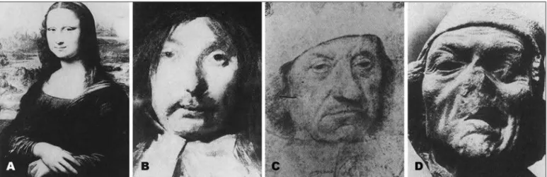
Fig 4. (A) “Mona Lisa” by Leonardo da Vinci. At a meeting of the facial nerve in Zurich, Adour and Jongkees concluded that the “Gioconda” had right peripheral facial paralysis (Adour and Jongkees personal communication, 1987). This is published elsewhere6,7. (B) “The Laughing Knight”,
an anonymous character by Frans �als, with probable left peripheral facial paralysis8. (C) Painting by �ans �olbein the Younger, with left facial
paralysis9. (D) Sculpture by Lucas van Leyden showing probable parotitis and right peripheral facial paralysis3,8.
Fig 5. (A) Eskimo dance mask from Alaska9. (B) Napoleonic style carved bottle stopper from southern France9. (C) Wood mask from Liberia9.
(D) Mask from Ceylon representing the “God of Deafness”, with a snake coming out of the nose3. Peripheral facial paralysis is well
represent-ed in all four images.
Fig 6. (A) The God of Sickness “Naha-Kola-Sanaya” from Ceylon. The “God of Sickness” is accompanied by 18 devils, two of them with facial paralysis (arrows)3. (B) Mask from Java, with left facial paralysis3. (C) Japanese ivory mask from the 17th Century5. (D) Japanese mask carved in
ripheral facial palsy5. Artistic representation of peripheral
facial palsy has become more extensive since the Renais-sance. dutch painters portrayed people with peripheral facial paralysis during and after this period5. old African
masks could have be made for “moral education”, to teach the young not to laugh at human deformity13.
The irst medical study of the disease is attributed to Avicenna (Abu-Ali al husayn ibn Abdalla Ibn sina, 979-1037 A.d.). he was the irst to record differences between central and peripheral facial paralysis: … “If the disease that produces paralysis comes from the middle of the brain, half of the body is paralyzed. If the disease is not in the brain but in the nerve, only that depending on this
nerve is paralyzed”4. Avicenna counted among the causes
of peripheral facial paralysis, compression due to injury, tumor, or nerve sectioning. For treatment, he prescribed medicinal plants for topical application, all of them hav-ing a vasodilator effect. In some cases he recommended cauterization behind the ear in the region of the stylo-mastoid foramen, a procedure that also has a vasodilator effect. he also prescribed face and neck massage. he em-phasized that “If sectioning of the nerve occurs, the only
alternative is stump-to-stump suture”4,8. As to prognosis,
he stated that “no recovery should be expected from any
facial paralysis that lasts more than six months”4. We can
consider that Avicenna had very advanced knowledge on peripheral facial palsy for his time (979–1037 A.d.).
In 1798, Nicolaus A. Friedreich of Würzburg, grandfa-ther of Nicolaus Friedreich of heildelberg who described the ataxia as having been named after him, published a
detailed study on the onset, clinical picture, evolution and treatment of peripheral facial paralysis in three pa-tients14. Exposure to cold drafts had occurred in all three
cases before paralysis onset. Thus Friedreich postulated that paralysis may occur when local causes act on the fa-cial nerve14. he published his study in Germany in 1798 as
…”De paralysis musculorum faciei rheumatica”. An English
review of his paper was published in the journal Annals
of Medicine in 1800 in Edinburgh, where Charles Bell was
a medical student at the time. According to Bird (1979), it is possible that Charles Bell may have read this paper14. his
irst case of peripheral facial palsy was published in 1821, and his most important paper was published in 182814.
however Charles Bell made other contributions to the history of neurology and anatomy. he recognized the dif-ferences between the anterior and posterior division of the spinal nerves; he identiied the thoracic long nerve; singled out the vIIth cranial nerve, separating it from vth and vIIIth, described the Bell sign and was the irst to de-scribed hyperacousis and dysgeusia as symptoms of pe-ripheral facial paralysis after self observation and obser-vations by Professor Roux from Paris8,14. In conclusion, we
think that the pioneers in the medical study of peripheral facial paralysis were: Avicenna, Nicolaus Friedreich and Charles Bell.
Acknowledgements – Figures in this work were kindly provided by Kugler & Ghedini, Kugler publications, P.o. box 20538, 1001 NM Amstelveen, The Netherlands and by George-Thieme verlag, sutt-gart, Germany.
Fig 7. (A) 17th Century: “Jar of Barbed Man”, presented at the baroque art exhibition in Augsburg2. (B) 18th Century: “Facial Gnome” from the “Dwarf
Garden” of the Mirabell Palace, Salzburg (Austria). The sculptor Bernard Mandl by order of Count �arrach9. (C) 19th Century photographs
tak-en by Duchtak-enne. The bald and apprehtak-ensive patitak-ent is suffering8. In A, B and C facial paralysis is presented. (D) Charles Bell, First Professor of