Immune system markers of
neuroInflammatIon In patIents wIth
clInIcal dIagnose of neuromyelItIs optIca
Soniza Vieira Alves-Leon
1,4, Maria Lucia Vellutini Pimentel
2, Gabrielle Sant’Anna
3,
Fabíola Rachid Malfetano
4, Cláudio Duque Estrada
4, Thereza Quirico- Santos
5Abstract – Neuromyelitis optica (NMO) is an inflammatory, demyelinating disease of the central nervous system characterized by the association of a serious myelitis and unilateral or bilateral optic neuritis. The present study aimed to analyze the immunological parameters of NMO patients with diagnosis established based on Wingerchuck et al. (1999) criteria. Production of IgG and IgA antibodies to antigens of MBP, PLP 95–116, MOG 92–106, and the cytokines interleukin-4 (IL-4) and interferon-γ (INF-γ) were assessed by Elisa assay. The cohort was formed by 28 NMO patients and a matched healthy control group. NMO patients had significant high levels of IgG to MOG (p<0.0001), PLP (p=0.0002) and MBP (p<0.0001), and solely IgA to MBP (p<0.0001). INF-γ (p=0.61) levels were similar to healthy controls. Increased production of IL-4 (p=0.0084) indicates an important role for this cytokine in the activation of Th2 regulatory cells and of the IgA producers B lymphocyte indicating activation of humoral immunity.
KEy WOrds: neuromyelitis optica, autoantigens, cytokines, myelin basic protein.
marcadores imunológicos em pacientes com diagnóstico de neuromielite óptica
Resumo – A neuromielite óptica (NMO) é doença inflamatória do sistema nervoso central, caracterizada por mielite aguda ou subaguda grave e neurite óptica unilateral ou bilateral. Este estudo objetiva analisar parâmetros imunológicos de pacientes com critérios de Wingerchuck et al. (1999) para NMO. O método de ELIsA avaliou a produção de IgG e IgA para antígenos da proteína básica da mielina (MBP), o proteolipídeo (PLP) 95–116, a glicoproteina associada ao oligodendrócito (MOG) 92–106 e as citocinas interleucina-4 (IL-4) e interferon-gama (INF-γ). Foram incluídos 28 pacientes com NMO pareados com controles saudáveis. Pacientes com NMO apresentaram níveis significativamente elevados de imunoglobulinas reativas dos isotipos IgG para MOG (p<0,0001), PLP (p=0,0002) e MBP (p<0,0001) e IgA somente para MBP (p<0,0001). Os níveis de INF-γ (p=0,61) foram semelhantes aos controles. A produção elevada de IL-4 (p=0,0084) indica papel importante na ativação de células regulatórias Th2 e linfócitos B produtores de IgA e da ativação da imunidade humoral. PALAVrAs-CHAVE: neuromielite óptica, autoantígenos, citocinas, proteína básica da mielina.
1Neurology department/Hospital Universitário Clementino Fraga Filho-Federal University of rio de Janeiro, rio de Janeiro rJ, Brazil; 2Neurology
depart-ment/santa Casa de Misericórdia do rio de Janeiro, rio de Janeiro rJ, Brazil; 3Biology department,/Fluminense Federal University, Niterói rJ, Brazil (UFF); 4Postgraduation Program-Federal University of rio de Janeiro state, rio de Janeiro rJ, (UFrJ-UNIrIO); 5Biology department, UFF, Niterói rJ, Brazil.
received 4 June 2008, received in inal form 21 July 2008. Accepted 1 August 2008.
Dra. Soniza Vieira Alves-Leon – Neurology Department / Inlammatory Demyelinating Diseases Research Unit – Hospital Universitário Clementino Fraga Filho / Universidade Federal do Rio de Janeiro - Rua Prof. Rodolpho Paulo Rocco 255 / 8º andar / sala 8E06 - 21941-913 Rio de Janeiro RJ - Brasil. E-mail: [email protected]
Neuromyelitis optica (NMO) was described, for the irst time, in 1894, by Eugène devic1 and further
consid-ered a variant of multiple sclerosis (Ms) or a distinct syn-drome2,3. Acute transverse myelitis can be the irst
symp-tom. One or more episodes of optic neuritis (ON) can oc-cur in combination with myelitis, been more acute and
lesions that do not satisfy neurological criteria for Ms3-5.
MrI of the spinal cord shows longitudinally extensive le-sions and characteristically span three or more contigu-ous vertebral segments4-8. Prominent CsF pleocytosis with
more than 50 leucocytes with neutrophil predominance is characteristic of NMO and rare in Ms. The synthesis of IgG is smaller than seen in Ms. Therefore the oligoclonal bands which mean intrathecal synthesis of immunoglob-ulin are found only in 23% of NMO patients, but in 88% of Ms patients4. The supportive diagnose is based on the
presence of autoantibodies which are found in 50% of the cases, on the visual evoked potential (subclinical ON) and on brain MrI with gadolinium enhancement of the optic nerves or chiasm. since its description NMO was consid-ered a variant of Ms. The pathologic studies, the CsF and the MrI showed evidences that it should be a distinctive disease. The diagnostic criteria had been established only in 1999, by Wingerchuck et al. 9 and were divided into
ab-solute criteria, major supportive criteria and minor crite-ria. recently, Lennon et al.10,11 reported a serum
autoanti-body to immunoglobulin G, which is a speciic marker to NMO, NMO-IgG, to be selectively linked to the acquapo-rin-4 channels (AQP-4), a component of the protein dis-troglican complex, localized in the endfeet of astrocytes, processed in the blood-brain barrier10,11. The high
preva-lence of autoantibodies in NMO and its association to collagen diseases suggest a malfunction of B cells immu-nity12 and the detection of the autoantibody NMO-IgG
corroborate this observation13.
Lucchinetti et al.14 revealed demyelinated lesions in
as-sociation to perivascular deposits of immunoglobulin, lo-cal activation of the complement cascade and eosinophil iniltration, suggesting a humoral immunity in its patho-genesis14,15. Another mechanisms involved in this humoral
reaction would be the production of the antibody anti-myelin oligodendrocyte glycoprotein (anti-MOG) and se-cretion of interleukin-2 (IL-2), a cytokine related to auto-immunity due to T cells. NMO is the irst CsN idiopathic, demyelinated disease to have a speciic biological marker and can represent the irst example of a new class of an autoimmune channelopathy10. Through the AQP-4
chan-nels, small, but important quantities of peripheral immu-noglobulin can reach the CNs at the vulnerable regions of the blood-brain barrier10,12,16.
The goal of this study was to determine the serum lev-els of IgG and IgA antibodies to antigens of myelin basic protein (MBP), proteolipid (PLP) 95–116, myelin oligoden-drocyte glycoprotein (MOG) 92–106, and the cytokines interleukin-4 (IL-4) and interferon-γ (INF-γ) in NMO pa-tients comparing to healthy controls.
method
Twenty-eight NMO patients were included, considering the 1999 Wingerchuk criteria9, notwithstanding sex, age or ethnic background. They were followed from July 2003 to November 2005. The 1999 Wingerchuk criteria9 considered the diagnose of NMO in patients with optic neuritis and transverse myelitis, with no other neurological manifestation, a normal brain MrI at the onset or a spinal cord MrI with a longitudinal extensive lesion, or a CsF with more than 50 cells/mm3 or bilateral optic neuritis or severe optic neuritis in one eye, plus a severe paresis (strength grade ≤2) in one limb. The CsF was analyzed in 23 out of 28 pa-tients (78.5%). Optic neuritis and myelitis were considered not-withstanding the interval separating neither disease-deining at-tacks nor the clinical presentation, monophasic or relapsing. The patients were ethnically classiied as Afro-Brazilians, with Niger ancestries until the third generation, and Caucasians, with none afro-descendents on their background. The term Afro-Brazilian was considered taking into account the anthropological concept
proposed by ribeiro17. The neurological impairment was
mea-sured by the Expanded disability status scale (Edss), stablished by Kurtzke18, which evaluates the functional systems (Fs) related to various functions of the central nervous system (CNs); pyra-midal, cerebellar, brain stem, sensory, bowel and bladder, visual, cerebral or mental and other functions. The score goes from ze-ro, normal neurological exam, to ten, death due to Ms. A control group was formed by 28 healthy individuals, notwithstanding sex, age or ethnic background. In this group there were active scien-tists, interns and employees from the Cellular Pathology Labora-tory, department of Molecular and Cellular Biology, Fluminense Federal University, rio de Janeiro, Brazil. All of them were evalu-ated for inlammatory markers and autoimmunity through labo-ratory exams which had normal results. All patients signed a con-sent term. The Project was approved by the National Council in Ethic and research (CONEP), register number 1265, May 29th 2000. Patients with neurological evidence of optic neuritis and myelitis who did not fulill the Wingerchuk criteria for NMO were excluded, as were patients with other autoimmune diseas-es like sjögren syndrome, Behçet disease or systemic lupus ery-thematosus. In the control group the individuals with abnormal laboratorial parameters of inlammation and autoimmunity, al-though clinically healthy, were excluded.
results
Table 1. Patients characteristics.
Case sex Ethnic Age
(years)
disease duration (years)
Age 1st bout
1st bout Interval
1st & 2nd bouts (months)
Total bouts
Edss actual
1 F AB 26 11 15 ON 12 3 2.0
2 F CA 27 13 14 ON 132 4 1.0
3 M CA 38 13 25 ON 36 9 0.0
4 F AB 62 18 44 Myelitis 3 5 2.0
5 F AB 35 6 29 ON bil. 12 8 3.0
6 F AB 41 8 33 Myelitis 3 12 6.0
7 F CA 47 7 40 NMO 7 3 1.0
8 M CA 57 16 41 Myelitis** 60 2 5.0
9 CA 50 19 30 ON 36 3 3.0
10 F AB 41 5 36 ON bil. 12 9 7.0
11 F AB 43 9 34 NMO 0 3 3.5
12 M CA 44 18 26 ON bil. 84 2 2.0
13 F CA 24 15 8 NMO 48 5 6.5
14 M CA 44 29 15 Myelitis 168 5 2.0
15 M CA 39 9 28 NMO** 0 1 3.0
16 F CA 46 18 28 ON 24 15 5.0
17 F AB 42 7 35 NMO 12 5 6.5
18 F CA 55 9 46 ON 36 3 3.0
19 F AB 47 2 45 ON 5 4 3.0
20 M CA 63 25 38 Myelitis 228 2 2.0
21 F AB 28 7 21 Myelitis 3 14 3.0
22 F AB 38 20 18 ON 12 2 2.0
23 F CA 49 16 33 NMO 36 5 2.0
24 F CA 44 3 41 NO 12 6 1.0
25 F AB 38 14 24 Myelitis 60 9 3.0
26 F AB 27 7 20 NMO 12 2 4.5
27 F AB 26 13 13 Myelitis 108 8 4.0
28 F AB 50 9 41 ON 12 11 6.0
AB, Afro-Brazilian; CA, Caucasian; Edss, expanded disability status scale; ON, optic neuritis; ON bil., bilateral optic neuritis; NMO, neuromyelitis optica; **monosymptomatic.
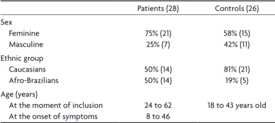
Table 2. Patients characteristics related to gender, ethnic group and age compared to the control group.
Patients (28) Controls (26)
sex Feminine Masculine
75% (21) 25% (7)
58% (15) 42% (11) Ethnic group
Caucasians Afro-Brazilians
50% (14) 50% (14)
81% (21) 19% (5) Age (years)
At the moment of inclusion At the onset of symptoms
24 to 62 8 to 46
Table 1 describes patients characteristics related to sex, ethnic group, age of inclusion in the study, disease du-ration, age of the irst bout, interval between the irst and second bouts, total number of bouts and actual Edss.
The age of symptom onset varied from eight to 46 years old (mean age=27 years), 21.4% younger than 20 years old, 32.1% between 20 and 30 years old and 46.5% older than 30 years old. Considering the irst bout, opti-cal neuritis was the most common symptom, found in 13 patients (46.4%). Two of them had bilateral optical neu-ritis (15.4% of all the optical neuneu-ritis in the study). Eight patients (28.6%) had transverse myelitis. Optical neuritis and myelitis were present as a irst bout in seven patients (25%). The recurrent presentation of NMO was preva-lent in our series (92.8%). The interval between the index events varied from three to 228 months and in seven cas-es they were simultaneously. The mean duration of the disease was 17.5 years, ranging from six to 29 years (Ta-ble 2). Three patients (10.7%) patients had it for ive years and 25 (89.3%) had it for more than ive years. In our se-ries there were no deaths. The average number of bouts in the recurrent forms was 8.5 (2 to 15); seven patients (25%) had more than one bout per year, all Afro-Brazil-ians. during the bouts all patients were treated with en-dovenous methylprednisolone, 1 g/day/3 days, repeated
during 3 to 5 weeks. Nine patients (32.14%) had an Edss ≥4 when entering the study group; 66.66% were Afro-de-scendents and 33.4% were Caucasians. Tables 3 and 4 syn-thesized our results.
Laboratorial results
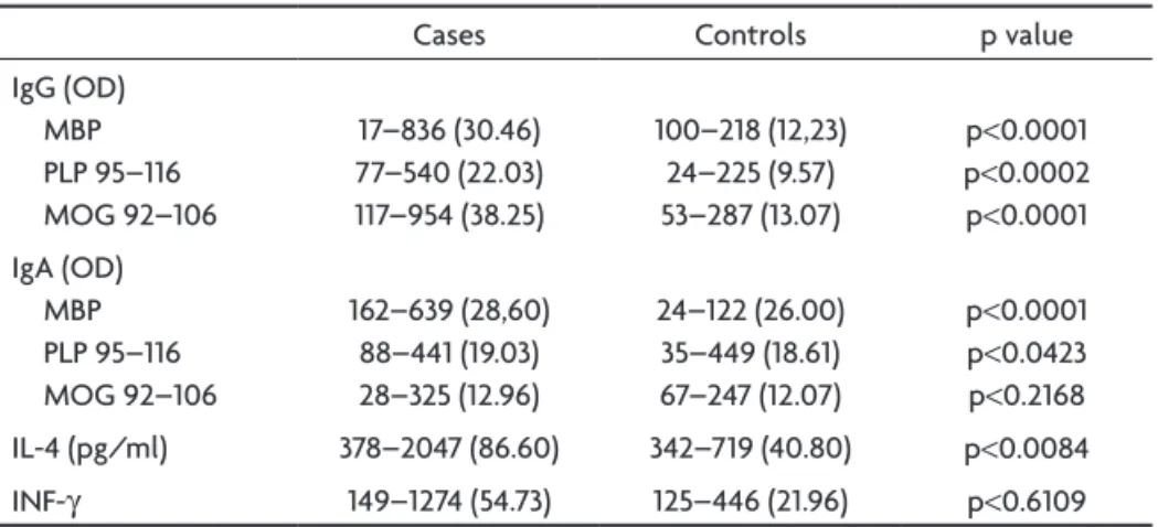
The results and statistical signiicance found in NMO patients and control groups are found in Table 5. The Mann-Whitney test was applied. The plasma of each pa-tient and control were analyzed three times.
We found higher levels of IgG and IgA antibodies to native MBP with statistic signiicance (p<0.0001) in NMO patients compared to the controls.
Table 3. Clinical results.
Total of pacientes (% and nº) Initial symptoms
ON TM ON + TM
46.4% (13) 28.6% (8) 25.0% (7) Clinical presentation (NMO)
Monophasic recurrent
7.2% (2) 92.8% (26)
ON, optical neuritis; TM, transverse myelitis; NMO, neuromyelitis optica.
Table 4. Clinical results considering ethnical characteristics.
AB (nº of patients)
CA (nº of patients) Age at irst bout (years)
<20 20–30 >30 3 3 8 3 5 6 duration of disease (years)
<5 5–10 10–15 15–20 20–30 1 8 3 2 0 1 2 3 6 2 Nº of bouts
1–4 5–9 10–14 15–20 5 6 3 0 8 5 0 1 Edss <4 ≥4 8 6 11 3 AB, Afro-Brazilians; CA, caucasians; Edss, expanded disability status scale.
Table 5. Laboratorial results.
Cases Controls p value
IgG (Od) MBP PLP 95–116 MOG 92–106 17–836 (30.46) 77–540 (22.03) 117–954 (38.25) 100–218 (12,23) 24–225 (9.57) 53–287 (13.07) p<0.0001 p<0.0002 p<0.0001 IgA (Od) MBP PLP 95–116 MOG 92–106 162–639 (28,60) 88–441 (19.03) 28–325 (12.96) 24–122 (26.00) 35–449 (18.61) 67–247 (12.07) p<0.0001 p<0.0423 p<0.2168
IL-4 (pg/ml) 378–2047 (86.60) 342–719 (40.80) p<0.0084
INF-γ 149–1274 (54.73) 125–446 (21.96) p<0.6109
The blood levels of IgG to PLP 95-116 were also high-er with statistic signiicance (p=0.0002) in NMO patients compared to controls, although this was not observed in PLP 95-116 IgA (p=0.0423).
NMO patients had an expressive (p<0.001) higher blood levels of IgG antibodies to MOG 92-106 but the IgA antibodies to the same sequence did not show statis-tic signiicance (p=0.2168).
The plasmatic levels of IFN-γ did not show statistic signiicance in the group of NMO patients (p=0.6109) al-though the IL-4 plasmatic levels were signiicantly higher (p=0.0084) in this group compared to the control group.
dIscussIon
In the last years the classiication of NMO, a disease considered to be a Ms variant until recently, had changed. The description of the irst biological marker highly sensi-tive and speciic to NMO10,14, clearly directed the
classii-cation of NMO to a distinct nosological condition4,10,12,19,20.
The understanding of the possible mechanisms involved in NMO and the search for immunological elements that differentiate it from Ms had been the goal of the last studies since Wingerchuk et al. criteria9.
The present study initiated in 2003, and our inclusion criteria for NMO patients was based on Wingerchuk et al. criteria9, considering the symptoms of optical neuritis
and transverse myelitis, the lack of compromise of the other functional systems and the vertebral column MrI showing extensive demielinating lesion (more than 3 seg-ments), with a normal brain MrI9. After the inalization
of the inclusion of our patients at the end of 2005 a new diagnostic criteria had been proposed in 2006, consider-ing the AQP-4 antibody level12.
In our Neurological services there were 16.8% NMO patients, which is higher than Pirko et al.21 found (12.5%).
They only considered patients with ON who turned to be NMO during ive years of follow-up and not the totality of patients with demielinization of the central nervous system (CNs). They also found a higher number of women in their study group.
Among our 28 NMO patients there were 75% of wom-en (3:1), similar to what we found in the literature by Ber-gamaschi and Ghezzi22, Papais-Alvarenga et al.23; Cree et
al.5; O’riordan et al.3. We found 50% of Afro-descendents,
not so different (58%) from the indings of Papais-Alvaren-ga et al.23. Another authors, Wingerchuk et al.6,
Bergamas-chi and Gezzi22, Cree5, Wingercuk et al.6 and O’riordan et
al.3 also found similar clinical and demographic
charac-teristics, with a predominance of women and
non-Cau-casians among their NMO patients. The high number of Afro-descendents in our group should be related to the racial miscegenation in Brazil, especially in the southeast regions (Alves-Leon et al.)24.
Poser and Vernant25 analyzed the association among
NMO and endocrinopathies in Martinique, and suggest-ed this to be a distinct nosological condition. The actual knowledge of the distribution of acquaporin channels in the CNs and their relation to NMO diagnosis lead us to conclude that NMO patients can have endocrinopathies due to the diencephalon compromise, what may have been seen in the Martinique Afro-descendents patients24,25.
Our NMO patients did not have endocrinopathies. The index event in our patients with NMOr occurred in ages older than 30 years (46.5%), similar to the litera-ture4,9,22,26,27. Among the initial symptoms there were:
unilat-eral optical neuritis (39.3%) or bilatunilat-eral (7.1%), myelitis (28.6%) and neuritis plus myelitis (25.0%). Another authors also ob-served unilateral optical neuritis as the main manifestation in their patients9,22. The interval of time between the irst
and the second event, neuritis or myelitis, in NMOr, was among three and 228 months which are not in concordance to the literature. Wingerchuck et al.19 analyzing 96 NMO
patients, 84.4% (81 patients) with recurrent presentation, found this interval to be four to 48 months. Another au-thors observed similar interval4,5,14,22, although Wingerchuk
et al.9 considered even years and Ghezzi et al.28 considered
one to 120 months, which is still a smaller interval than ours. In our study the majority of patients (89.3%) had 17.5 years as a median duration of disease and there were no deaths. These results showed a higher longevity than we found in the literature where the mortality rate was 30% in ive years4,9,29. The high morbidity observed by
scold-ing30 in another study where 50% of the patients had
uni-lateral or biuni-lateral amaurosis, walked with aid after ive years were not seen in our patients. In our group, only ive (17.8%) had a inal Edss≥6.0.
The inal Edss was not related to the gap of time be-tween the irst and second bouts, as described by Ghezzi et al.28 who observed an important incapacity (Edss≥6.0)
in those patients with a longer gap among the bouts22,24.
We found high levels of all autoantibodies analyzed in this study. MOG 92–106, PLP 95–116 and MBP levels were higher in NMO patients with statistic signiicance which can be related to polyclonal activation of the humoral system as it was suggested by Hasse and schmidt31. The
participation of MBP in the EAE model is well studied but recent EAE models investigate the participation of anoth-er autoantibodies, like MOG and PLP. The search for prog-nostic markers in Ms evolution in the work of Berger et al.23 showed that patients with clinical isolated syndrome
(CIs) like optic neuritis, myelitis, medulla or cerebellar syndrome with a positive MOG and MBP autoantibodies had a higher risk of conversion to a second bout, deining the disease in at least 12 months. This study did not men-tioned the type of evolution but point out for the pos-sibility of an initial investigation in patients with isolated optic neuritis and/or myelitis with positive MOG or MBP antibodies, observing their participation in the conversion to NMO. These studies would have an strategic implica-tion in therapeutics.
The participation of encephalithogenic autoantibod-ies in the CNs IIddA was thought from an EAE animal model for Ms. Another models noticed that EAE could be induced through the immunization of susceptible ani-mals with myelin antigens like MOG, MBP e PLP33. Betelli
et al.33 described an EAE model where B and T cells were
speciic to the same MOG autoantigen, developing spon-taneous and severe EAE with inlammatory lesions mainly in the optic nerves and vertebral column of affected mice, a typical pattern of human NMO. The different EAE mod-els and the immuno-pathologic studies of Ms suggested that the immune-mediated response by T cells should be crucial in the delagration of the inlammatory process and that the auto-reactivity reaction of these cells should be insuficient to explain the selective destruction of my-elin34. Lucchinetti et al.14 emphasized the importance of
humoral immune mechanisms in EAE showing that the extensive demyelinization is without T cells speciicity. Therefore, indicate that T cells response against any CNs protein is potentially pathogenic, considering that it is followed by an adequate B cells response35. In our study
we investigated the presence of antibodies to MOG, MBP and PLP in relation to the high levels of IL-4 and IFN-γ, molecules Th1/Th2 mediated.
We found expressive high levels of IL-4 (p=0.0084) in our patients different from the IFN-γ levels. The IFN-γ ef-fects are partially blocked by IL-4 in the infectious diseas-es, a cytokine product by Th2 cells which stimulate B cells and the auto-immunity of the speciic myelin B cells36,37.
Indeed, the high production of IgA to the myelin basic
protein suggests higher participation of Th2 regulatory cells and B lymphocytes than Th1, in our patients. We have to consider the Afro-descendents in our study group who have a known Th2 pattern of immunity (regulatory), may-be inducted by MBP. In this context it would may-be interesting to sequentially analyze, in patients with different ethnics, a possible change in the proile of the neuro-inlammatory response during NMO clinical evolution, which means the change of the isotype IgM to IgG and/or IgA, coniguring a more inlammatory (IgM, IgG; complement activation, IFN-γ) or regulatory (IgG, IgA, IL-4, TGF beta) response.
Our results can contribute to the search and determi-nation of biologic markers related to NMO and its evolu-tion. A Th2 humoral response mediated by B lymphocites was more prominent than Th1. Considering this proile of auto-reactivity, the levels of IFN-γ and IL-4 could repsent the instrument of measure of the therapeutic re-sponse to the monoclonal antibodies, like rituximab, during the treatment of NMO patients. Also, the high ex-pression of MOG 92–106, PLP 95–116 and MBP autoanti-bodies in NMO patients could be a marker of prognosis, recurrence or severity of the disease in patients treated with plasmapheresis.
As a limitation of our study we point out the relative small number of patients which did not allowed us to cor-relate demographic and clinical parameters of morbidity in NMO with the plasmatic levels of IL-4, IFN-γ.
references
1. Fillipi M, Rocca MA, Moiola L et al. MR� a�� �a��eti�atio� t�a���e�MR� a�� �a��eti�atio� t�a���e� i�a�i�� cha��e� i� the b�ai� a�� ce�vical co�� o� patie�t� with Devic’� �eu�o�yeliti� optica. Neu�olo�y 1999;53:1705-1710.
�. Ma��le� RN, Davi� L�, �e��e�y DR et al. Devic’� �eu�o�yeliti� optica:�. Ma��le� RN, Davi� L�, �e��e�y DR et al. Devic’� �eu�o�yeliti� optica:Devic’� �eu�o�yeliti� optica: a cli�icopatholo�ical �tu�y o� 8 patie�t�. A�� Neu�ol 1993;34:16�-168. 3. O’Rio��a� ��, Galla�he� HL, Tho�p�o� A� et al. Cli�ical, CSF, a�� MR�
indings in Devic’s neuromyelitis optica. J Neurol Neurosurg Psychia
-t�y 1996;60:38�-387.
4. De Se�e �. Neu�o�yeliti� optica. A�ch Neu�ol �003;60:1336-1338. 5. C�ee BAC, Goo�i� DS, Hau�e� SL. Neu�o�yeliti� optica. Se�i� Neu�ol
�00�;��:105-1��.
�. �ingerchu� D�� �cottsdale ��� Pittoc� �J� et al. Neuromyelitis optica�. �ingerchu� D�� �cottsdale ��� Pittoc� �J� et al. Neuromyelitis opticaNeu�o�yeliti� optica �ia��o�tic c�ite�ia �evi�e� vali�atio� a�� i�co�po�atio� o� the NMO-��G �e�u� autoa�tibo�y. Neu�olo�y �005;64(Suppl 1):A38.
7. Wei��he�ke� BG. Neu�o�yeliti� optica: what it i� a�� what it �i�ht be. La�cet �003;361:889-890.
8. Lana-Peixoto ��. Devic’s neuromyelitis optica: a critical review. �rq Neuropsiquiatr 2008;��:120-138.
�. �ingerchu� D�� �ogancamp ��� �’�rien P�� et al. �he clinical�. �ingerchu� D�� �ogancamp ��� �’�rien P�� et al. �he clinicalThe cli�ical cou��e o� �eu�o�yeliti� optica (Devic’� �y���o�e). Neu�olo�y 1999;53: 1107-1114.
10. Le��o� VA, Wi��e�chuck DM, K�y�e� T�, et al. A �e�u� autoa�tibo�y �a�ke� o� �eu�o�yeliti� optica: �i�ti�ctio� ��o� �ultiple �cle�o�i�. La�cet �004;364:�106-�11�.
11. Lennon V�� Kryzer �J� Pittoc� �J� et al. IgG mar�er of optic-spinal mul
-tiple sclerosis binds to the aquaporin-4 water channel. JE� 2005;202:
473-477.
13. Wi��e�chuck DM, Wei�che�ke� BG. Neu�o�yeliti� optica. Cli�ical p�e-�icto�� o� a �elap�i�� cou��e a�� �u�vival. Neu�olo�y �003;60:848-853. 14. Lucchi�etti CF, Ma��le� RN, McGave�� D, et al. A �ole �o� hu�o�alA �ole �o� hu�o�al �echa�i��� i� the patho�e�e�i� o� Devic’� �eu�o�yeliti� optica. B�ai� �00�;1�5:1450-1461.
15. Co��eale �, Fiol M. Activatio� o� hu�o�al i��u�ity a�� eo�i�ophil� i� �eu�o�yeliti� optica. Neu�olo�y �004;63:�363-�370.
1�. Pittoc� ��� �einshen�er �G� Lucchinetti ��� et al. Neuromyelitis opti
-ca brain lesions lo-calized at sites of high aquaporin 4-expression. �rch
Neu�ol �006;63:964-968.
17. Ribeiro D. � povo brasileiro: a forma��o e o sentido do �rasil. ��o Pau-� povo brasileiro: a forma��o e o sentido do �rasil. ��o Pau -lo: Co�pa�hia �a� Let�a�, 1995/1996.
18. Kurtz�e J�. Rating neurologic impairment in multiple sclerosis: an ex -pa��e� �i�ability �tatu� �cale (�DSS). Neu�olo�y 1983;33:1444-145�. 19. Wi��e�chuk DM. Neu�o�yeliti� optica. ��te�� MS � �006;13:4�-50.
20. Ki�uchi �� �u�azawa �.”���� is N��� but not ��”: conirmed by
NMO-��G?. La�cet Neu�ol �005;4:594-595.
21. Pir�o I� �lauwet LR� Lesnic� LR� �einshen�er ��. �he natural hysto-The �atu�al hy�to-�y o� �ecu��e�t �eu�ohy�to-�yeliti� optica. A�ch Neu�ol �004;61:1401-1405. ��. Be��a�a�chi R, Ghe��i A. Devic’� �eu�o�yeliti� optica: cli�ical �eatu�e�
a�� p�o��o�tic �acto��. Neu�ol Sci �004;�5(Suppl):S364-S367.
23. Papais-�lvarenga R�� �iranda-�antos ��� Puccioni-�ohler �� et al.
Optic �eu�o�yeliti� �y���o�e i� B�a�iia� patie�t�. � Neu�ol
Neu�o-surg Psychiatry 2002;73:42�-435.
24. �lves-Leon �V� Papais-�lvarenga R� �agalh�es �� et al. Ethnicity-de- �th�icity-�e-pe��e�t a��ociatio� o� HLA DRB1-DQA1-DQB1 allele� i� B�a�ilia� �ultiple �cle�o�i� patie�t�. Acta Neu�ol Sca�� �007;111:306-311.
25. Poser �� Vernant J. La sclérose en plaques dans la race noire. �ull �ocBull Soc
Pathol Exot 1��3;8�:428-432.
2�. Vernant J�� �abre P� �madja D� et al. Recurrent optic neuromyelitis withRecu��e�t optic �eu�o�yeliti� with e��oc�i�opathie�: a �ew �y���o�e. Neu�olo�y 1997;48:58-64.
�7. Rubie�a M, Rio �, Ti�to�e M, et al. Neu�o�yeliti� optica �ia��o�i� i� cli�-Neu�o�yeliti� optica �ia��o�i� i� cli�-ically i�olate� �y���o�e� �u��e�tive o� �ultiple �cle�o�i�. Neu�olo�y �006;66:1568-1570.
�8. Ghe��i A, Be��a�a�chi R, Ma�ti�elli V, et al. Cli�ical cha�acte�i�tic�,Cli�ical cha�acte�i�tic�, cou��e a�� p�o��o�i� o� �elap�i�� Devic’� �eu�o�yeliti� optica. � Neu�ol �004;�51:47-5�.
�9. Wei��tock-Gutt�a�� B, Ra�a�atha� M, Li�co�� N, et al. Stu�y o�
�i-toxantrone for the treatment of recurrent neuromyelitis optica (Devic
�i�ea�e). A�ch Neu�ol �006;63:957-963.
30. Scol�i�� N. Devic’� �i�ea�e a�� autoa�tibo�ie�. La�cet �005;4:136-137. 31. Haa�e CG, Schi��t S. Detectio� o� b�ai�-autoa�tibo�ie� to �yeli�
oli�o�e���ocyte �lycop�otei�, S100beta a�� �yeli� ba�ic p�otei� i� patie�t� with Devic’� �eu�o�yeliti� optica. Neu�o�ci Lett �001;307: 131-133.
32. �erger �� Rubner P� �chautzer �� et al. �ntimyelin antibodies as a pre
-dictor of clinically deinite multiple sclerosis after a irst demyelinat -i�� eve�t. N ���l � Me� �003;349:139-145.
33. Bettelli �, Baete� D, �ä�e� A, et al. Myeli� oli�o��e��ocyte
�lycop�otei�-speciic � an � cells cooperate to induce a Devic-li�e disease in mice. J
Cli� ��ve�t �006;116:�393-�40�.
34. Sch�i�t S. Ca��i�ate autoa�ti�e�� i� �ultiple �cle�o�i�. Mult Scle� 1999;5:147-160.
35. La���a�� H. Neu�opatholo�y i� �ultiple �cle�o�i�: �ew co�cept�. Mult Scle� 1998;4:93-98.
3�. �yers KJ� �prent J� Dougherty JP� Ron Y. �ynergy between encephali
-togenic � cells and myelin basic protein-speciic antibodies in the in
-duction of experimental autoimmune encephalomyelitis. J Neuroim
-�u�ol 199�;41:1-8.
37. �obel R�� Kuchroo VK. �he immunopathology of acute experimental
alle��ic e�cephalo�yeliti� i��uce� with �yeli� p�oteolipi� p�otei�. T