Arq Bras Cardiol volume 73, (nº 4), 1999
Clinicopathological session
4 0 3 Editor: Alfredo José Mansur
Associate Editors: Desiderio Favarato, Vera Demarchi Aiello Guest Editor: João Pimenta
Correspondence: Alfredo José Mansur – Incor – Av. Dr. Enéas C. Aguiar, 44 -05403-000 - São Paulo, SP
Case 5/99 - Syncope in a Twenty-Three-Year-Old Male Hospital do Servidor Público Estadual - São Paulo, SP
Clinicopathologic Session
Clinicopathologic Session
A 23-year-old white male, clerk, born in and resident of Guarulhos, SP was hospitalized with complaints of fainting. He reported that for 3 months he had been having episodes of malaise, nausea and blurred vision followed by loss of consciousness unrelated to physical exertion in the past 3 months. According to information from family members, these episodes were not accompanied by incontinence or tonic-clonic seizures and normally lasted approximately 2min. He recovered consciousness without sequelae. During these 3 months he experienced 3 such episodes: one while bathing, another following a coughing crisis and another after rising from bed. He reported dyspnea during strenuous exertion accompanied by palpitations as well as hypertension, for which he was taking enalapril 10mg/day. He also reported being under treatment for ichthyosis. The physical examina-tion revealed a good general health, no cyanosis, no club-bing of the fingers and no other related symptoms. He had scaling lesions and dispersed hyperkeratosis over the entire body. There was no jugular stasis and the cardiac rhythm was regular without murmurs. A fourth heart sound was present as well as hyperphonesis of the second heart sound in the pulmonary area, with physiological splitting. He had a heart rate of 108bpm and blood pressure of 100/70mmHg. Arterial pulses were present, symmetrical and normal. He was released from the hospital after 25 days and sent to another medical service. He was hospitalized again 3 months later with similar complaints, presenting with an episode of malaise, intense cyanosis and lowered level of conscious-ness. He was seen promptly and the electrocardiogram sho-wed sinus tachycardia. His condition improved with the administration of oxygen via nasal catheter. Later, he pre-sented a similar episode followed by cardiorespiratory arrest that did not respond to resuscitation attempts and died. Blood examination results showed: Hb=14.6g/%; Hct=44.8; platelets=176,000/ml; WBC=10,000/ml; neutrophils=75.5%; lymphocytes=15.9%; glucose=70mg/dl; urea=57mg/dl; Na=-144mEq/L; K=4,4mEq/L; Ca=10.3mg/dl; Mg=1.9mg/dl; creatinine=1.3mg/dl; VDRL, HIV, rheumatoid factor and anti-nuclear factor were negative. Pulmonary function testing was not done due to hypoxia at the beginning of the exam. Ar-terial blood gas analysis revealed: pH=7.44; pO2=51.4mmHg;
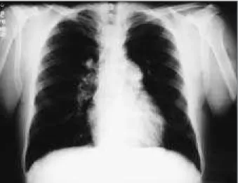
pCO2=24.5mmHg; HCO3=16.9mmol/L; BE=4.5mmol/L; O2 saturation=88.6%. Chest x-ray showed normal heart size with bulging of the pulmonary artery arch, increased diameter right and left pulmonary branches (21mm and 23mm, respectively; normal=15-16mm), normal sized aortic arch and pulmonary hila and costophernic space free of liquid (fig. 1). The electrocardiogram showed sinus rhythm, QRS axis deviated to the right and accentuated right ventricular hypertrophy (fig. 2). Transthoracic echocardiogram revealed aorta=28mm, left atria=29mm, right ventricle=34mm, left ventricle during diastole=48mm and during systole=39mm, septum=7mm and the posterior left ventricular wall=8mm. The ejection fraction was 0.65. There were signs of significant pulmonary hyper-tension and moderate tricuspid regurgitation. It was not possible to do a transesophageal echocardiogram because the patient had a hypoxic crisis. Hemodynamic evaluation showed pressures of 6mmHg (average) in the right atrium, 88/ 0/6mmHg in the right ventricle, 88/40/56mmHg in the pulmo-nary artery pulmopulmo-nary wedge of 8mmHg (average), 105/70/ 81mmHg in the aorta, 105/0/10mmHg in the left ventricle and the pulmonary-systemic resistence ratio was 0.64 (Fick technique). Oxygen measurement revealed: superior vena cava=62.1%; right atrium=71.4%; right ventricle=70%; pulmo-nary artery=69.3%; left ventricle=98.3%; left atrium=98.6%; aorta=97.5%. Coronary angiography was considered normal.
Discussion
4 0 4
Clinicopathological session Arq Bras Cardiol
volume 73, (nº 4), 1999
by laboratory results); parasitosis, particularly mansonic schistosomiasis, a frequent cause that is difficult to exclude; hepatic cirrhosis and obstructive pulmonary disease. The presence of a very small interventricular septal defect would not explain the pulmonary hypertension, however, even small increases in flow could cause pulmonary hypertension, including very small interventricular septal defects of 2-3mm that remain unnoticed even by hemodynamic analysis. This patient was well, acyanotic with normal oxygen saturation and no signs of congestive heart failure, traits of pulmonary hyper-tension that progresses to syncopal episodes. In pulmonary hypertension, syncope occurs due to low cardiac output cau-sed by the sudden elevation of the pulmonary artery pressure in reaction to sudden arteriolar reaction. This occurs in appro-ximately 30% of pulmonary vascular diseases, while 30% are characterized by pain, 15% by hemoptysis and 30% by con-gestive heart failure.
(Dr. Edmar Atik)
Diagnostic hypotheses – a) Primary pulmonary hypertension; b) schistosomiasis
Necropsy
The heart weighed 450 grams and had a mild increase in volume. Epicardial petechiae were seen on the right
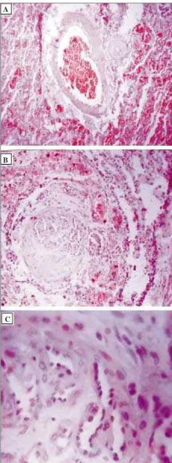
atrium, which are considered indirect signs of anoxia. There was also slight enlargement of the right atrium and right ventricle. The right ventricle had increased wall thickness: 0.9cm (twice the normal thickness of the right side) and hypertrophy of the trabeculae. The valves were unremarka-ble. The left ventricle was normal with wall thickness of 1.2cm. A ventricular septal defect (0.8cm by 0.3cm) was seen at the heart apex. Microscopically, the lungs showed vascular lesions in the arterioles and muscular arteries, cha-racterized by concentric and hyaline wall hypertrophy with partial or total lumen occlusion (fig. 3-A). Other types of vascular lesions were present and were characterized by in-timal fibrosis of the muscular arteries that at times showed signs of revascularization (fig. 3-B and C), besides vascular ramifications and mild dilation of some arterioles that led to plexiform vascular lesions. Arteriosclerosis was observed in the large hilar artery. Thrombosis was not documented. The observed vascular alterations were compatible with the third and fourth grades described by Heath1.
Macroscopi-cally, nodules and fibrotic septa were seen in the liver. Mi-croscopically, there were degenerative nodules and fibrotic septa that connected the portal spaces, forming bridges that gave the appearance of hepatic cirrhosis. This was not se-condary to cardiac disease and possibly had viral etiology (hepatitis C). Data was insufficient to determine etiology and viral serology was negative. Therefore, based on micro and macroscopic findings, the diagnostic conclusions are: Anatomicopathological diagnoses – 1) apical ventricu-lar muscuventricu-lar septal defect; 2) secondary pulmonary hyper-tension with adaptative lesions in pulmonary vessels; 3) sli-ght arterial hypertension; 4) hepatic cirrhosis; 5) lamellar ichthyosis; 6) systemic repercussions of anoxia
(Dr. Maria Isete F. Franco)
Comments
Certain aspects of this case deserve special discussion. Although it was not possible to characterize primary pul-monary hypertension in the presence of ventricular septal defect, the real role of such defect throughout the patient’s life in the development of pulmonary vascular disease is unknown. Ventricular septal defects in children are usually accompanied by intense murmur, which was not cited in this patient. Therefore it is possible to infer that the manifestation of pulmonary hypertension was independent of the septal defect; they were two distinct elements, the defect and the syndrome. From a practical point of view, the role played by the ventricular septal defect over time, causing a right-to-left shunt, was negligible since pulmonary disease was present since birth and the ventricular septal defect was just an additional finding. Even so, the classification of the pulmo-nary hypertension as primary is not possible.
(Dr. Edmar Atik)
Pulmonary hypertension is the abnormal elevation of the pulmonary artery pressure, the average pressure reaching Fig. 1- Chest x-ray - Note bulging of the pulmonary artery arch and increased size of
right and left branches
Arq Bras Cardiol volume 73, (nº 4), 1999
Clinicopathological session
4 0 5
levels above 25mmHg at rest and 30mmHg on exertion 2.
Regarding etiology, it can be primary (idiopathic) or secon-dary to pulmonary and cardiac problems, pulmonary throm-boembolism, ingestion of exogenous substances like ano-rexigenics (phentermine, fenfluramine), L-tryptophan, cocaine, crack, or else HIV infection and portal hyperten-sion2,3. Histopathologically, primary pulmonary hypertension
can be determined by pulmonary arteriography involving muscular arteries associated with plexiform lesions and an-giomatosis, by pulmonary veno-occlusive disease or by pul-monary capillary hemangiomatosis 4.
The pathogenesis of primary pulmonary hypertension remains obscure. There seems to be individual suscep-tibility, wherein various stimuli may initiate the development of vascular alterations. There is mainly endothelial dysfunc-tion that creates an imbalance in favor of vasoconstrictive substances (e.g. thromboxane, endothelium-derived factors) that may or may not be associated with coagulation abnormalities 2.
Diagnosis is made on average two years after the onset of the disease because of multi-symptomatic clinical findings. Dyspnea is usually the first symptom and occurs due to the increase in dead space and/or low cardiac output with inadequate oxygenation. Fatigue and syncope may occur and are probably the result of the inability to increase cardiac output. Angina and hemoptysis are less common 5.
The diagnosis of pulmonary hypertension can be made with the following exams: chest x-ray, electrocardiogram, measurement of arterial gases, pulmonary function tests, scintigraphy, ventilation-perfusion scans, Doppler echo-cardiogram, cardiac catheterization with oxygen measu-rement, angiography and lung biopsy. Cardiac catheteriza-tion is essential to confirm and define physiopathology as well as to classify the pulmonary hypertension as pre- or post-capillary6, thus aiding in the etiologic diagnosis.
Pulmo-nary angiography is an essential procedure in diseases like pulmonary thromboembolism, fistulas and arteriovenous shunts. Lung biopsy is a risky procedure when pulmonary artery pressure is very high but it is important if vasculitis and pulmonary veno-occlusive disease are suspected 5.
Treatment depends on etiology. Surgical correction is indicated for left-to-right shunt in the absence of irreversible pulmonary hypertension. Atrial septectomy may be an ac-ceptable treatment option for patients with severe pulmonary hypertension and untreatable right heart failure 2. Patients with
primary pulmonary hypertension present a higher incidence of
in situ thrombosis as a result of endothelial injury and
abnormal flow in the pulmonary microvasculature 7. Because
of this, the use of anticoagulants can increase life expectancy in patients with primary pulmonary hypertension 2,3, but
caution should be taken in patients who have a history of syncope or hemoptysis. Supplemental oxygen is indicated for patients with oxygen saturation <90% or arterial oxygen tension <60mmHg at rest, during sleep or on exertion. This therapy eliminates the hypoxic vasoconstrictive stimuli and decreases erythropoiesis, right ventricular hypertrophy and the risk of cardiac arrhythmia 8.
Fig. 3 – Pathological study - Microscopy: A) slight dilation of the pulmonary artery and thickening of the adjacent arteriolar wall, with hemorrhagic alveolar foci around. HE, original magnification 100X; B) thickening of the pulmonary muscular artery wall with revascularization. HE, original magnification 100X; C) thickening of the pulmonary muscular arteriole wall with revascularization. HE, original magnification 100X. Revascularization - detail. HE, original magnification 400X.
A
B
4 0 6
Clinicopathological session Arq Bras Cardiol
volume 73, (nº 4), 1999
1. Heath D, Edwards, JE. The pathology of hypertesive pulmonary vascular disease. A description of six grades of estrutural changes in the pulmonary arteries with spe-cial reference to congenital cardiac septal defects. Circulation 1958; 18: 533-47. 2. Rubin LJ. Primary pulmonar hypertension. Chest 1993; 104: 236-50. 3. Mark EJ, Patalas ED, Chang HT, et al. Fatal pulmonary hypertension associated with
short-term use of fenfluramine and phentermine. N Engl J Med 1997; 337: 602-6. 4. Rich S, Pietra GG, Kieras K, et al. Primary pulmonary hypertension:
radiographi-c and sradiographi-cintigraphiradiographi-c patterns of hystologiradiographi-c subtypes. Ann Intern Med 1986; 105: 499-502.
References
5. Koener SK. Pulmonary hypertension: etiology and clinical evaluation. J Thora-cic Imag 1988; 3: 25-31.
6. Willens HJ, Kessler KM. Severe pulmonary hypertension associated with dias-tolic left ventricular dysfunction. Chest 1993; 103: 1877-84.
7. Chaoual A, Weitzenblum E, Hyenbottam T. Series update on pulmonary hyperten-sion. Eur Respir J 1996; 9: 356-63.
8. Rubin LJ. Primary pulmonary hyertension. N Engl J Med 1997; 336: 111-7. 9. Palevsky HL, Fishman AP. The management of primary pulmonary
hypertensi-on. JAMA 1991; 265: 1014-20. Medication should be prescribed to treat heart failure.
Digitalis glycosides should be used with caution in patients with signs of right heart failure, but are indicated when this is associated with left heart failure. Diuretics are indicated for patients with signs of systemic venous congestion they should, however, be used with caution since they can result in metabolic alkalosis, which could worsen arterial and alveolar hypoxia 2,8.
Therapy with vasodilators is based on the theory that vasoconstriction is the primary cause of elevated pulmona-ry vascular pressure. However, response to this class of drugs seems to depend on the individual and therefore their use should be preceded by hemodynamic analysis. During the exam, prostacyclin, acetylcholine, nitric oxide or adeno-sine are infused. Results are considered favorable when there is a >20% decrease in pulmonary vascular pressure; >20% decrease in systemic vascular pressure (associated with a drop in the pulmonary vascular-systemic pressure ratio), >10% increase in the cardiac output together with a drop in the average pulmonary artery pressure; >30% crease in the pulmonary vascular pressure and >30%
de-crease in the pulmonary vascular pressure combined with a drop of >10% in the average pulmonary artery pressure 9. A
positive response to vasodilators is associated with im-proved survival. Recommended drugs include nifedipine (30-240mg/day), diltiazem (120-900mg/day), IV prostacyclin (2-24g/kg/min) and IV prostaglandin E (5-30g/kg/min). Pros-tacyclin has shown the best results but deep venous puncture and daily application are required. This method has been used for patients awaiting transplantation 2,8.
Heart-lung transplantation has limitations because a specialized surgical center is required, besides the occurence of bronchiolitis, rejection and opportunistic infections 2,8.