Universidade de Pernambuco-FESP, Hospital dos Servidores do Estado de Pernambuco, Hospital do Servidor Público Estadual de São Paulo, Universidade Federal de Pernambuco e Centro de Pesquisas Aggeu Magalhães/Fundação Oswaldo Cruz Mailing address: Creso Abreu Falcão, Rua Padre Carapuceiro, 488/603 - 51020-280 Recife, PE - e-mail: [email protected]
Received to publication on 5/22/01 Accepted on 12/11/01
Objective - Lupus anticoagulant and anticardioli-pin antibodies (aCL) have been associated with thrombo-sis, recurrent abortion, and thrombocytopenia in patients with systemic lupus erythematosus (SLE), but their relati-onship with cardiac disease is less clear. The purpose of this study was to evaluate the association between antipho-spholipid antibodies (aPL) and echocardiographic ab-normalities in patients with SLE.
Methods - A total of 70 consecutive patients and 42 control subjects underwent M-mode, 2-dimensional and Doppler echocardiography and tests for lupus anticoa-gulant, aCL IgG, IgM, and IgA. Lupus anticoagulant was assayed with the dilute Russell viper venom time, and aCL IgG, IgM, and IgA were measured by an enzyme-linked immunosorbent assay (ELISA).
Results - Lupus anticoagulant showed a prevalence of 10%. As a whole, aCL had a prevalence of 44.3% and aPL had a prevalence of 50%. Patients with echocardio-graphic abnormalities had a prevalence of 54.3% and showed a trend towards an association with aCL IgG (P=0.06). The presence of pulmonary hypertension (PH) was significantly associated with aCL IgG (p=0.02).
Conclusion - aCL IgG was significantly associated with PH and showed a strong trend towards an association with echocardiographic abnormalities taken together. These findings suggest a role for aCL IgG in the develop-ment of lupus cardiovascular disease.
Key words: lupus erythematosus, antiphospholipid
syndrome, lupus coagulation inhibitor
Arq Bras Cardiol, volume 79 (nº 3), 285-91, 2002
Creso Abreu Falcão, Izabel Cristina Alves, Wiliam Habib Chahade, Ângela Luzia Branco Pinto Duarte, Norma Lucena-Silva
Recife, PE - São Paulo, SP - Brazil
Echocardiographic Abnormalities and Antiphospholipid
Antibodies in Patients with Systemic Lupus Erythematosus
A number of relevant studies in patients with systemic lupus erythematosus (SLE) have evidenced an association of antiphospholipid antibodies (aPL) with cardiopathy, par-ticularly in the case of Libman-Sacks endocarditis, valvar dysfunction, and valvar thickening 1-5.Specific associa-tions of aPL with pulmonary hypertension (PH) 6, myocar-dial dysfunction 3 , and ischemic cardiopathy 7,8 in lupus patients have also been reported. These findings suggest that aPLs are responsible for basic immunological events related to the development of cardiopathy in SLE, and that some cardiac abnormalities may be considered as real com-ponents of primary or secondary aPL syndrome (APS) 9. However, other authors were not able to demonstrate an as-sociation between cardiac disease in SLE and aPL 10-12.
The similarity between valvar lesions detected in pa-tients with primary aPL syndrome and valvar lesions of lu-pus patients has reinforced the statistical associations ob-served 9. Also, the detection of selective deposition of im-munoglobulins and complement at valve vegetations of Libman-Sacks endocarditis 9, as well as the more recent fin-dings concerning the deposition of IgG isotype ACL in the subendothelial layer of heart valves in patients with APS 13, have consistently suggested a role for immunological dis-turbances in the development of valvar lesions in SLE.
In the present study, we evaluated the prevalence of 2-dimensional and Doppler echocardiographic abnormalities and aPL [lupus anticoagulant (LA) and anticardiolipin anti-bodies (aCL)] in patients with SLE in Recife, Brazil, and ana-lyzed the association between the presence of antibodies and cardiac lesions detected.
Methods
re-vised criteria of the American College of Rheumatology 14. Patients with a history of cancer, drug abuse, previous rheu-matic fever, coagulation disturbances, or that were currently receiving anticoagulant drugs were excluded.
In addition, 42 female volunteers being treated as out-patients in the Gynecological Cancer Prevention Center of the Hospital das Clínicas-UFPE served as controls. All were premenopausal women with no clinical evidence of cardiac or systemic complaints, and no history of cancer, drug abu-se, SLE, APL syndrome, or any immunological distur-bances. A document of consent was signed by all patients and controls.
The tests for the lupus anticoagulant were carried out with the dilute Russell viper venom time (dRVVT) 15. Blood was collected in one-tenth volumes of 3.8% trisodium ci-trate and was centrifuged at 1500 g for 30 minutes at room temperature to obtain platelet-poor plasma. All the samples were frozen in small aliquots at -70° C before testing. The values considered normal for the test varied between 26.6 and 28.8 seconds and were obtained from a pool of plasma samples from normal patients, industrially fabricated (IL Test TM Plasma Control Normal, Instrumentation Laborato-ry Company, Lexington, MA/USA). Two reagents were uti-lized for gauging the dRVVT and to confirm the existence of phospholipid inhibitors, respectively: a reagent mixture containing Russell viper venom (< 8mg/kg), antiheparin agent (polybrene) and calcium (IL Test TM lupus anticoa-gulant Screen, Instrumentation Laboratory Company, Lexington, MA/USA), and another reagent mixture of identical content, except for an additional quantity of phos-pholipids (IL Test TM LA Confirm, Instrumentation Labora-tory Company, Lexington, MA/USA).
The test was considered to indicate an alteration when the time for coagulation of the patient’s plasma sam-ple was at least 20% greater than the time obtained for the coagulation of the pool of plasma from normal patients (dR VVT/ dRVVTn ratio > 1.2). In the tests considered to show alteration, the presence of the lupus anticoagulant as a coa-gulation inhibitor and a cause of the increase in coacoa-gulation time was confirmed through the reagent with a higher concentration of phospholipids 16-18. A ratio between the time using the low-phospholipid reagent and that using the high-phospholipid reagent equal to or greater than 1.2 was considered positive.
The aCL test was carried out on patients and controls as described previously 19. Briefly, increasing dilutions of serum samples in a 10% solution of FCS/PBS were applied in duplicate to ELISA plates that had been previously sensi-tized with 25mg/mL of cardiolipin in ethanol, treated with a 10% solution of skimmed milk, then incubated at 4oC. After washing the plates with a 0.05% Tween 20/PBS solution, the anti-IgG, anti-IgM, or anti-IgA antibody, together with pero-xidase or phosphatase alkaline diluted 1:5000, was added separately for 1 hour at ambient temperature. The reaction was revealed by the addition of ABTS substrate for peroxi-dase or pNNN for phosphatase alkaline as recommended by the manufacturer. The absorption reading was at 405 nm.
The analysis of the mean optical density of each dilution of patient and control serum enabled the identification of the best dilution for detection of each immunoglobulin isotope, which was 1:128 for IgA and 1:256 for IgG and IgM. For each class of antibodies, samples with optical density values hi-gher than the mean (by 5 standard deviations of 6 controls considered negative) were considered positive for the presence of the isotopes IgG, IgM, or IgA 20.
Transthoracic Doppler echocardiographic examina-tions were carried out by a single examiner, usually within a period of up to 7 days following the interview and blood collection for antiphospholipid antibody tests. The equip-ment used for all Doppler echocardiographic examinations was an Interspect Apogee model with a 2.75 MHz transdu-cer, capable of performing unidimensional and bidimensio-nal studies using continuous and pulsed Doppler. The echocardiographic measurements were made in accordance with the norms suggested by the American Echocardiogra-phic Society 21. The images for bidimensional studies were also obtained according to the usual standardization 22. Doppler flow studies and bidimensional imaging were car-ried out simultaneously. The presence of pericardial effu-sion, global or segmental contractile deficit of the left ventri-cle (LV), diastolic dysfunction of the LV, left ventricular hy-pertrophy (LVH), pulmonary arterial hypertension, valvar thickening, and valvar regurgitation were registered. Mitral valve prolapse was not considered an alteration pertaining to the pathological spectrum in this study.
In bidimensional images, mild pericardial effusion was defined as the presence of isolated posterior effusion, mo-derate pericardial effusion as the presence of anterior and posterior effusion, and effusion of great magnitude as the presence of anterior and posterior effusion and abnormal excessive cardiac movements (“swimming heart”).
The ejection fraction (EF) of the left ventricle (LV) was used as an indicator of the magnitude of the systolic dys-function of the LV. Myocardial damage was defined as the presence of global contractile deficit of the LV (EF < 0.60), in the absence of clinical and/or electrocardiographic data of myocardial ischemic disease, or both, with mild systolic dysfunction reported when EF was 0.50 – 0.60, moderate systolic dysfunction reported when EF was 0.40 – 0.49, and systolic dysfunction of a great magnitude reported when EF < 0.40.
Left ventricular hypertrophy was defined as a left ven-tricular wall thickness of more than 12 mm in diastole. Valvar thickness was measured from the parasternal long axis view and valvar thickening was reported if thickness was 3 mm or more.
31-50 mmHg, the moderate form as the presence of PSAP 51-70 mmHg, and the severe form as the presence of PSAP > 51-70 mmHg.
The results were analyzed with Epi Info software, ver-sion 6. The data obtained were expressed as mean + stan-dard deviation. The Dopplerechocardiographic morpholo-gical findings were related to the presence or absence of antiphospholipid antibodies with the chi-square test. Variance analysis was used to estimate the difference between means. A value of P lower than 0.05 was considered significant.
Results
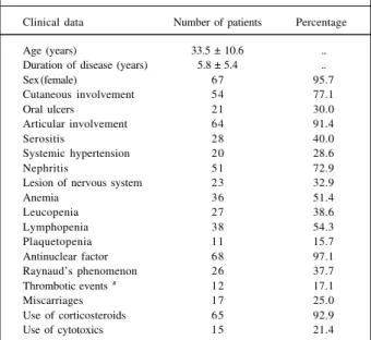
In the group of lupus patients, the mean age was 33.5±10.6 years (range of 13-65 years), 3 patients being male and 67 female. The mean disease duration time was 5.8±5.4 years (range of 1-32). Sixty-five patients (92.9%) were using corticosteroids. The mean duration of corticosteroid use was 3.4±4.2 years (range of 0.08 to 18 years). Fifteen patients (21.4%) used cytotoxins in the 30-day period prior to inter-view. The control group, composed of 42 patients, had a mean age of 32.8±6.5 years (range of 19-44 years), all being female. The clinical characteristics of patients with SLE are summarized in Table I.
Doppler echocardiographic abnormalities considered compatible with cardiopathy were registered in 38 of the 70 patients with SLE (54.3%).
Pericardial effusion was observed in 8 of 70 (11.4%) patients with SLE and in none of the controls (P=0.02). Mild effusions were found in 5 of 8 (62.5%) patients, 2 of 8 (25%) having effusion considered moderate and 1 of 8 (12.5%) ef-fusion of great magnitude. No echocardiographic evidence
of tamponade or pericardial constriction was found in the patients studied.
Myocardial damage (EF<0.6) was present in 14 of 70 (20%) patients with SLE and in none of the controls (P=0.005). In the lupus patients with myocardial damage, anemia (serohemoglobin < 10 mg/100 mL) in 78.5% of cases (P=0.04) and systemic arterial hypertension (blood pressure > 140 x 90 mmHg) in 35.7% of patients (P=0.51) were detec-ted. Findings suggesting heart failure was detected in 2.86% of patients with SLE. Eight of the 70 lupus patients (11.4%) were considered to have mild systolic dysfunction (EF 0.50-0.60), whilst 5 of 70 (7.1%) displayed moderate systolic dysfunction (EF= 0.40 – 0.49) and 2 of 70 (2.9%) systolic dysfunction of a great magnitude (EF < 0.40). The mean measurement of EF of LV of lupus patients was 0.66 + 0.09, whilst the mean for control group patients was 0.69±0.03 (P=0.04). Segmental contractile deficit of LV was found in the echocardiogram to be present in 4 of 70 (5.7%) patients with SLE and in none of the controls (P=0.44).
LVH was present in 21 of 70 (30%) patients and in none of the controls (P<0.001). Twelve of the 21 lupic patients with LVH (57.1%) displayed systemic hypertension (PR= 3.33; CI 95% = 1.67 to 6.66; P=0.001).
PH was detected in the Doppler echocardiogram of 15 of 70 (21.4%) patients with SLE and in 3 of 42 (7.1%) control group individuals (P=0.03). PSAP was used as an indicator of the magnitude of PH. Twelve of the 15 patients with SLE and PH (80%) displayed the mild form of PH (PSAP 31-50 m mHg), 2 of 15 (13.3%) the moderate form (PSAP 51-70 m m Hg), and 1 of 15 (6.6%) the severe form (PSAP > 70 mmHg). The 3 individuals of the control group with raised PSAP displayed forms of PH considered mild, with figures of 31, 34, and 37 mmHg. The mean measurements of PSAP in the group of patients with SLE was 40.5±14.1 mmHg, detecting a value of 30.8 + 5.1 mmHg in the individuals of the control group (P<0.001).
Valvar alterations not related to mitral valve prolapse were found in 5 of 70 patients (7.1%) with SLE and in none of the controls (P=0.07). Minimal aortic insufficiency was observed in 1 of 5 patients (20%), in whom hypertension and LVH were detected. Mitral insufficiency (MI) was found in 4 of 5 (80%) lupus patients, in all cases of a small degree. One of these cases of MI was associated with calci-fication of the mitral ring, 1 was associated with thickening of the mitral valve, and in 2 other cases the regurgitation was considered functional, associated with dilation of the left ventricle. MI of minimal degree was detected in 2 lupus patients with normal leaflets, and among the control group patients, 4 cases of mild MI in patients with normal mitral leaflets were also registered. These cases of minimal MI wi-th normal mitral leaflets were not considered pawi-thological 4 and were not included in statistical analysis. None of the patients or controls displayed any clinical data that might indicate hemodynamic repercussion of valvar alterations detected on the Doppler echocardiogram.
Lupus anticoagulant was present in 7 of 70 (10%) patients with SLE and in none of the controls (P=0.03). The Table I - Clinical characteristics of 70 patients with SLE *
Clinical data Number of patients Percentage
Age (years) 33.5 ± 10.6 ..
Duration of disease (years) 5.8 ± 5.4 ..
Sex (female) 67 95.7
Cutaneous involvement 54 77.1
Oral ulcers 21 30.0
Articular involvement 64 91.4
Serositis 28 40.0
Systemic hypertension 20 28.6
Nephritis 51 72.9
Lesion of nervous system 23 32.9
Anemia 36 51.4
Leucopenia 27 38.6
Lymphopenia 38 54.3
Plaquetopenia 11 15.7
Antinuclear factor 68 97.1
Raynaud’s phenomenon 26 37.7
Thrombotic events # 12 17.1
Miscarriages 17 25.0
Use of corticosteroids 65 92.9
Use of cytotoxics 15 21.4
*Present anytime during evolution of SLE; # Myocardial infarction,
mean Russell time obtained on sampling for the lupus anti-coagulant test in patients with SLE was 32.2±12 seconds and in the controls was 25.6±2.1 seconds (P<0.0 01). The results of the sampling and confirmatory tests of lupus anticoagulant-positive patients with SLE are shown in Table II.
Among the lupus patients with cardiopathy, 4 of 38 (10.5%) displayed a positive test for lupus anticoagulant (P=0.81). In the 3 lupus anticoagulant-positive lupus pa-tients without cardiopathy, aPL syndrome findings were present, that is plaquetopenia, episodes of miscarriage, and evident livedo reticularis of the hands, respectively.
The lupus anticoagulant did not show itself to be signi-ficantly associated with the presence of pericardial effusion, myocardial dysfunction, myocardial ischemic disease, LVH, PH, or valvar disease. Similarly, no statistically significant as-sociation of the presence the lupus anticoagulant with pla-quetopenia, history of miscarriage, or thrombotic events (ce-rebrovascular accident, deep venous thrombosis, peripheral arterial occlusion, or myocardial infarction at some time in clinical evolution of patients) was found.
The aCL IgG were present in 13 of (18.6%) patients with SLE and in 4 of 42 (9.5%) control group individuals (P=0.30). In the group of lupus patients, aCL IgG were de-tected in 10 of 38 (26.3%) patients with Doppler echocardio-graphic alterations, a trend towards statistical significance being observed (PR=1.57; IC 95% = 1.05 to 2.33; P=0.06). Meanwhile, the aCL IgG were detected in 6 of 15 (40%) PH cases, with statistical significance (PR=2.92; CI 95% = 1.26 to 6.77; P=0.02). Positive results for aCL IgG, IgM, and IgA are shown in Table III.
Only 1 patient (1.4%) positive for aCL IgA was obser-ved among the lupus patients and 1 of 42 (2.3%) among the controls (P= 0.71).
When aCL IgG, IgM, and IgA were evaluated as a whole, it was observed that aCL were detected in 31 of 70 (44.3%) patients with SLE and in 14 of 42 (33.3%) control group individuals (P=0.34). The aCL were present in 17 of 38 (44.7%) lupus patients with Doppler echocardiographic al-terations and in 14 of 32 (43.7%) of lupus patients without these alterations (P=0.87).
Patients with lupus anticoagulant and/or positive for aCL were considered positive for the presence of aPL. The frequency of aPL in patients with SLE was 35 of 70 (50%) and 14 of 42 (33.3%) in the controls (P=0.12).The aPL were present in 19 of 38 (50%) lupus patients with Doppler echo-cardiographic alterations and in 16 of 32 (50%) lupus pati-ents without these alterations. No statistically significant association was found between aPL evaluated as a whole and the Doppler echocardiographic alterations, whether evaluated together (P=0.81) or separately.
With respect to thrombotic events, it was verified that aCL were present in 9 of 12 (75%) lupus patients with cur-rent or previous thrombotic events, and in 22 of 58 (37.9%) lupus patients without these alterations, with statistical sig-nificance (PR=3.77; CI 95% =1.12 to 12.77; P=0.04). In turn, aPL were present in 10 of 12 (83.3%) lupus patients with thrombotic events and in 25 of 58 (43.1%) lupus patients wi-thout these alterations, also with statistical significance (PR=5; CI 95% = 1.18 to 21.19; P=0.02). No significant asso-ciation was found between aCL or aPL and plaquetopenia or a history of miscarriage.
Discussion
Lupus anticoagulant was detected in 7 of 70 (10%) pa-tients, showing a frequency comparable to that of other reports in which similar methods were used 27,28. In the pre-sent study, the presence of lupus anticoagulant did not show itself to be significantly associated with Doppler echo-cardiographic findings, whether evaluated together or sepa-rately. Among 7 lupus anticoagulant-positive lupus patients (10%), 4 (57%) demonstrated Doppler echocardiographic alterations, 2 of them (28.5%) showing PH and another 2 (28.5%) having myocardial damage.
The literature includes reports of occasional observati-ons of lupus anticoagulant-positive lupus patients displa-ying mitral valvopathy and thrombosis in heart chambers 29,30. An association between cardiac damage in SLE specifically Table II - Values of dRVVT for seven 7 lupus anticoagulant-positive
lupus patients and their relationship both with dRVVT of the normal pool and with the time found using the high-phospholipid reagent
dRVVT DRVVT/ DRVVT/ Presence Cardiopathy (seconds) dRVVTn dRVVTc of LA
85.9 2.98 2.27 High Mild PH
50.2 1.74 1.52 Moderate LV systolic dysfunction 45.7 1.59 1.54 Moderate LV dilation and LVH 71.1 2.47 1.97 Moderate Absent 68,9 2.51 1.65 Moderate Absent 63.4 2.31 1.84 Moderate Moderate PH
42.1 1.50 1.30 Low Absent
DRVVT- dilute Russell viper venom time; dRVVTn- dRVVT of the normal pool; dRVVTc=dRVVT with the use of high-phospholipid reagent (confirmatory); PH- pulmonary arterial hypertension; LV- left ventricle; LVH-left ventricular hypertrophy.
Table III - Doppler echocardiographic and aPL syndrome findings in 70 patients with SLE. Comparison between positive results for aPL
and aCL IgG and IgM
Findings No patients aPL(+)# aCL(+) ACL IgG (+) aCL IgM(+)
Pericardiopathy 8 3 3 0 3 LVH 21 8 7 5 4 LV dysfunction
Global 14 4 2 1 2
Segmental 4 3 3 2 2
Valvopathy
MI 4 2 2 2 1
AI 1 1 1 1 0
P H 15 10 9 6* 6
Thrombotic events 12 10* 9* 4 4
Miscarriages 17 8 8 5 5
Plaquetopenia 11 6 5 1 4
with lupus anticoagulant has been researched by Jouhikai-nem et al 31 who studied data on 74 patients with SLE follo-wing an evolution period averaging 22 years. The authors observed 5 cases (7%) of valvar disease in the group of 37 lupus anticoagulant-positive patients and no cases in the same-sized group of lupus anticoagulant-negative lupus patients (P=0.05). No indication of any statistically signifi-cant difference existed in relation to the number of cases of pericarditis, myocardial infarction, angina and heart failure, considering a tendency for lupus anticoagulant to be asso-ciated with cor pulmonale, a condition observed in 2 lupus anticoagulant-positive patients (3%). Although the lupus patients in the present study had not been selected on the basis of being lupus anticoagulant-positive or not, 2 lupus anticoagulant-positive cases (285%) with concurrent PH were also observed.
Among the patients demonstrating lupus anticoagu-lant and Doppler echocardiographic abnormalities, 2 also showed alterations potentially associated with aPL syndro-me. One of them was suffering from acute arterial occlusion of the right upper member, and another related an isolated episode of recurrent miscarriage. Furthermore, livedo reti-cularis, plaquetopenia, and another report of isolated mis-carriage were registered in the patients with lupus anticoa-gulant but without Doppler echocardiographic abnormali-ties. Although no statistically significant association for these findings was found, it should be considered that the relatively low prevalence of lupus anticoagulant among lu-pus patients results in only relatively large groups of lulu-pus anticoagulant patients being able to consistently demons-trate significant associations of the latter with detection of aPL syndrome 28. Nevertheless, thrombotic events signifi-cantly associated with lupus anticoagulant were observed in the groups of Petri et al 27, in which lupus anticoagulant was detected in 4 of 60 (6.7%) patients with SLE.
The association of aPL with cardiopathy in SLE has been most commonly reported in studies that include tests for lupus anticoagulant carried out in conjunction with re-search into aCL, or only the latter. The majority of signifi-cant associations found have been related to valvar dys-functions 2,3,5. In the present study, an association of aPL with valvar dysfunction was not found. Although the low frequency of valvar dysfunctions found here (7.14%) sho-uld be considered, other authors did not detect an associa-tion between these findings and aPL 10-12,32.
Li et al 32 evaluated 50 Chinese patients with SLE and the association of their Doppler echocardiographic alterati-ons with aPL, which were measured with ELISA for aCL IgG. The authors did not find a statistically significant associati-on between the presence and gravity of valvar lesiassociati-ons and aCL, and considered that racial differences might be implica-ted in the disparity between their results and those of other studies in which patients of Caucasian origin were involved. Although it is not possible to establish a perspective of ra-cial homogeneity for the patients in the present study, who are from a country where racial mixing is a social reality, it is pertinent to consider that racial factors may have an
influen-ce on the association of these valvopathies with aPL, as well as on the prevalence of valvar damage itself, in SLE 33.
The frequency of cardiopathy in lupus patients in the present study (54.3%) was similar to that found in other re-search 5,11,34-36, with the exception of valvar dysfunctions. We consider it possible that the low frequency (7.1%) and the reduced magnitude of valvar alterations found in the present study are a consequence of an underestimation of the true frequency and magnitude of valvopathy in patients with SLE in the population evaluated, which presents a high prevalence of rheumatic cardiopathy. The possibility of lu-pus patients affected by Libman-Sacks endocarditis being confused with those with rheumatic disease or subacute bacterial endocarditis has already been pointed out by Gross 37. In this way, patients previously diagnosed as suf-fering from chronic rheumatic valvar disease, and being in fact lupus patients with valvar disease, could have inadver-tently been excluded from the evaluation. In another study carried out in Brazil, Macêdo 38 observed a higher frequen-cy of valvopathy in patients with SLE, but was unable to find a statistically significant difference between the fre-quencies of valvopathy in patients with SLE (28.3%) and those of the control group (26%), this being attributed to a possibly high prevalence of subclinical rheumatic cardio-pathy in the study’s target population.
It may also be supposed that the prolonged use of corticosteroids in 65 (92.9%) of the lupus patients, 51 (72.8%) of them having been using corticosteroids for 12 consecutive months or more, may have contributed to the reduced frequency of echocardiographic detection of Lib-man-Sacks endocarditis in this study. Through necropsy, Bulkley and Roberts 39 observed a frequency of 18% of Libman-Sacks endocarditis in 36 patients with SLE using corticosteroids, verifying that endocardial lesions were considerably smaller in number and size than those found in necropsy of patients with SLE carried out before the advent of corticosteroid use.
In the present study, a tendency for aCL IgG to be as-sociated with all Doppler echocardiographic alterations taken together (P=0.06) was demonstrated. Nihoyannopou-los et al 4 found an association between aPL and valvar dys-functions in 93 patients with SLE, and also observed an as-sociation of aCL IgG and IgM with all the Doppler echocar-diographic alterations taken together. The authors comment on the possibility of cardiac involvement in SLE being mea-sured by aPL, possibly owing to the primary stimulation of the vascular endothelium. A direct lesion of the endothe-lium produced by aPL, leading to the activation of coagula-tion factors, consumpcoagula-tion of platelets and the formacoagula-tion of thrombi, in conjunction with alterations in the interaction between platelets, circulating proteins, and endothelium in the cascade of coagulation, have been suggested as possi-ble mechanisms for explaining Libman-Sacks endocarditis and other cardiac lesions in SLE 40.
References
other basic immunological disturbances. Furthermore, aPL could also be activated secondarily by exposed antigens in valve tissue previously damaged by other autoantibodies 41. In the present study, a significant association betwe-en PH and aCL IgG was also found. PH has bebetwe-en observed in Doppler echocardiograms and related to aPL in various studies 6,25,42. The relation between aPL and PH does not ap-pear to be absolute, and its pathogenic mechanisms are un-clear 9. The pathogenesis of PH in SLE has been related to the lesion of vascular endothelium by aPL 43, and also to vasospasm and Raynaud’s phenomenon rather than to aPL 24. Nevertheless, even though the thromboembolic form of PH may be considered less frequent in SLE than the vasospastic form, in the latter aPL would also be concur-rently observed 43.
IgG class antibodies have been more frequently asso-ciated with thrombotic events than those of IgG and IgA classes 44, it being possible that aCL IgM and IgA in the ab-sence of IgG and/or lupus anticoagulant do not represent a risk of thrombosis 42. Even though it should be considered that PH in SLE might not necessarily be of thromboembolic origin, the detection in this study of an association of PH with aCL IgG, but not with aCL IgM or IgA, is in agreement with the above-mentioned observations.
A limitation of this study could be related to the nonu-tilization of lupus activity indices for the evaluation of the patients, given that lupus activity has been related to higher frequencies of cardiopathic findings in some studies 10,45,46. However, no association was found between the activity of the disease and a higher percentage of Doppler echocardio-graphic alterations in other groups of patients with SLE 11,35. Another limitation could be linked to the fact that certain marginal forms of PH may be present without backflow through the tricuspid valve, preventing the evaluation of PASP by Doppler. Nevertheless, Murata et al 23 believe that the absence of tricuspid regurgitation on Doppler implies normal pulmonary pressures.
At the moment, no clear explanation exists for the high percentage of aCL-positive control group patients (33.3%) detected in this study. aPL would have a frequency of 2 to 10% of the general population 47 and can be present in various other conditions, such as cancer, drug abuse, acquired immunodeficiency syndrome, and syphilis 48-50. In the present study, cancer or drug abuse were not present in the population studied, but syphilis and AIDS, whose identification usually requires serological tests, were not tested for. Future research, in which samples are obtained from a population evaluated for aPL, could also include these tests.
Harris 19 and Harris et al 51 observed that patients with syphilis may be positive on solid-phase immunological tests for the study of aCL, even though the antibodies of syphilitic patients may show a greater affinity for the anti-genic complex (cardiolipin, cholesterol and lecithin) utilized in VDRL than for isolated cardiolipin used in solid-phase im-munological tests.
In summing up, although no statistically significant association between aCL or aPL evaluated in conjunction with Doppler echocardiographic alterations taken together or separately was observed in the present study, aCL IgG showed a strong trend towards association with the Dop-pler echocardiographic alterations taken together (PR=1.57; CI 95% =1.05 to 2.33; P=0.06), and were significantly asso-ciated with PH (PR=2.92; CI 95% =1.26 to 6.77; P=0.02). The findings support the hypothesis of a primary pathogenic role of aCL IgG in relation to cardiac alterations in general and PH in particular, in SLE and in aPL syndrome.
Acknowledgments
We wish to acknowledge the help of Dr. Paula Loureiro, Dr. Paulo Miranda, Dr. Moacir Novaes, Dr. Emanuelli Tenó-rio, Umbelina Raposo, Wayner de Souza, and Wlademir Gomes de Melo.
1. Chartash EK, Lans DM, Paget AS, et al. Aortic insufficiency and mitral regurgi-tation in patients with systemic lupus erythematosus and the antiphospholipid syndrome. Am J Med 1989; 86: 407-12.
2. Khamashta MA, Cervera R, Asherson RA, et al. Association of antibodies against phospholipids with heart valve disease in systemic lupus erythemato-sus. Lancet 1990; 335: 1541-4.
3. Leung W-H, Wong K-L, Lau C-P, et al. Association between antiphospholipid antibodies and cardiac abnormalities in patients with systemic lupus erythema-tosus. Am J Med 1990; 89: 411-9.
4. Nihoyannopoulos P, Gomez PM, Joshi J, et al. Cardiac abnormalities in systemic lupus erythematosus: association with raised anticardiolipin antibodies. Circulation 1990; 89: 369-75.
5. Cervera R, Font J, Paré C, et al. Cardiac disease in systemic lupus erythematosus: prospective study of 70 patients. Ann Rheum Dis 1992; 51: 156-9. 6. Asherson RA, Higenbottam TW, Dihn Xuan AT, et al. Pulmonary hypertension in a
lupus clinic: experience with twenty-four patients. J Rheumatol 1990; 17: 1291-8. 7. Morton KE, Gavaghan TP, Krilis SA, et al. Coronary artery bypass graft failure
-an autoimune phenomenon? L-ancet 1986; 1: 1353-7.
8. Hamstem A, Bkörkholm M, Norberg R, et al. Antibodies to cardiolipin in young
survivors of myocardial infarction: an association with recurrent cardiovascular events. Lancet 1986, 1: 113-6.
9. Asherson RA, Cervera R. Antiphospholipids and the heart - lessons and pitfalls for the cardiologist. Circulation 1991; 84: 920-3.
10. Alves JA, Hydalgo L, Rolim LF, et al. Avaliação clínica e laboratorial da cardio-patia no lúpus eritematoso sistêmico. Arq Bras Cardiol 1997; 68: 79-83. 11. Ong ML, Veerapen K, Chambers JB, et al. Cardiac abnormalities in systemic lupus
erythematosus: prevalence and relationship with disease activity. Int J Cardiol 1992; 34: 69-74.
12. Roldan CA, Shively BK, Lau CC, et al. Systemic lupus erythematosus valve di-sease by transesophageal echocardiography and the role of antiphospholipid antibodies. J Am Coll Cardiol 1992; 20: 1127-34.
13. Cervera R, Font J, Ingelmo M. Cardiac manifestations in the antiphospholipid syndrome. In: Asherson R, Cervera R, Piette J-C et al. (Eds). The Antiphospholi-pid Syndrome. Boca Raton: CRC Press, 1996: 151-60.
14. Tan EM, Cohen AS, Fries J, et al. Criteria for the diagnosis of systemic lupus ery-thematosus. Arthritis Rheum 1982; 25: 1271-7.
16. Tripplet DA, Brandt J. Laboratory identification of the lupus anticoagulant. Br J Haematol 1989; 73: 139-42.
17. Rauch J, Tannembaum M, Janoff A. Distinguishing plasma lupus anticoagulants from anti-factor antibodies using hexagonal (II) phase phospholipids. Thromb Haemostas 1989; 62: 892-6.
18. Exner T, Tripplet DA, Taberner D, et al. Guidelines for testing and revised criteria for lupus anticoagulants. Thromb Haemostas 1991; 65: 320-2.
19. Harris EN. Antiphospholipid antibodies. Br J Haematol 1990; 74: 1-9. 20. Loizou JDS, McCrea AC, Rudge R, et al. Measurement of anti-cardiolipin
antibo-dies by an enzyme-linked immunosorbent assay (ELISA): standardization and quantitation of results. Clin Exp Immunol 1985; 62: 738-45.
21. Sahn DJ, DeMaria A, Kisslo J, et al. Recommendations regarding quantitation in M-Mode echocardiography: results of a survey of echocardiographic measure-ments. Circulation 1978; 58: 1072-83.
22. Tajik A J, Seward JB, Hagler DJ, et al. Two-dimensional real-time ultrasonic ima-ging of the heart and great vessels - technique, image orientation, structure iden-tification, and validation. Mayo Clin Proc 1978; 53: 271-303.
23. Murata I, Takenaka K, Toshinoya S, et al. Clinical evaluation of pulmonary hy-pertension in systemic sclerosis and related disorders: a Doppler echocardio-graphic study of 135 Japanese patients. Chest 1997; 111: 36-43.
24. Simonson JS, Schiller NB, Petri M, et al. Pulmonary hypertension in systemic lupus erythematosus. J Rheumatol 1989; 16: 918-25.
25. Sturfelt G, Eskilsson J, Nived O, et al. Cardiovascular disease in systemic lupus erythematosus - a study of 75 patients from a defined population. Medicine 1992; 71: 216-23.
26. Winslon TM, Ossipov MA, Fazio GP, et al. Five-year follow-up study of the pre-valence and progression of pulmonary hypertension in systemic lupus erythe-matosus. Am Heart J 1995; 129: 510-5.
27. Petri M, Rheinschmidt BAM, Whiting-O’Keefe Q, et al. The frequency of lupus an-ticoagulant in systemic lupus erythematosus. Ann Int Med 1987; 106: 524-31. 28. Feng C-S, Tsang SSF, Li EK. Evaluation of laboratory tests for lupus
anticoagu-lant in a group of Chinese lupus patients. Pathology 1994; 26: 40-2. 29. Lubbe WL, Asherson RA. Intracardiac thrombus in systemic lupus
erythemato-sus associated with lupus anticoagulant. Arthritis Rheum 1988; 31: 1453-4. 30. Ford PM, Ford SE, Lillicrap DP. Association of lupus anticoagulant with severe
valvular heart disease in systemic lupus erythematosus. J Rheumatol 1988; 15: 597-600.
31. Jouhikainem T, Pohjola-Sintonen S, Stephansson E. Lupus anticoagulant and cardiac manifestations in systemic lupus erythematosus. Lupus 1994; 3: 167-72. 32. Li E, Crozier IG, Milne MJ, et al. Lack of correlation between anticardiolipin an-tibodies and heart valve disease in Chinese patients with systemic lupus erythe-matosus. Lancet 1990; 336: 504-5.
33. Crozier I, Li E, Milne M, et al. Valvular disease in systemic lupus erythematosus. N Engl J Med 1989; 320: 739-40.
34. Leung W-H, Wong K-L, Lau C-P, et al. Cardiac abnormalities in systemic lupus
erythematosus: a prospective M-Mode, cross-sectional and Doppler echocar-diographic study. Int J Cardiol 1990; 27: 367-75.
35. Roldan CA, Shively BK, Crawford MH. An echocardiographic study of valvular heart disease associated with systemic lupus erythematosus. N Engl J Med 1996; 335: 1424-30.
36. Galve E, Candell-Riera J, Pigrau C, et al. Prevalence, morphologic types, and evo-lution of cardiac valvular disease in systemic lupus erythematosus. N Engl J Med 1988; 319: 817-23.
37. Gross L. The cardiac lesions in Libman-Sacks disease - with a consideration of its relationship to acute diffuse lupus erythematosus. Am J Pathol 1940; 16: 375-407.
38. Macêdo TB. Avaliação ecocardiográfica no lúpus eritematoso sistêmico: um es-tudo transversal de 53 pacientes (dissertation). Recife: Universidade Federal de Pernambuco, 1998: 100 p.
39. Bulkley HB, Roberts WC. The heart in systemic lupus erythematosus and the changes induced in it by corticosteroid therapy - a study of 36 necropsy patients. Am J Med 1975: 58: 243-64.
40. O’Rourke RA. Antiphospholipid antibodies: a marker of lupus carditis? Circu-lation 1990; 82: 636-8.
41. Hojnik M, George J, Ziporen L, et al. Heart valve involvement (Libman-Sacks en-docarditis) in the antiphospholipid syndrome. Circulation 1996; 93: 1579-87. 42. Miyata M, Suzuki K, Sakuma F, et al. Anticardiolipin antibodies are associated with pulmonary hypertension in patients with mixed conective tissue disease or systemic lupus erythematosus. Int Arch Allergy Immunol 1993; 100: 351-4. 43. Asherson RA, Cervera R. Antiphospholipid antibodies and the lung. J
Rheu-matol 1995; 22: 62-6.
44. Petri M. Pathogenesis and treatment of the antiphospholipid antibody syndrome. Med Clin North Am 1997; 81: 151-77.
45. Leung W-H, Wong K-L, Lau C-P, et al. Doppler echocardiographic evaluation of left ventricular diastolic function in patients with systemic lupus erythemato-sus. Am Heart J 1990; 120: 82-7.
46. Castier MB, Meneses ME, Albuquerque EM, et al. O envolvimento cardíaco no lúpus eritematoso sistêmico: uma avaliação ecocardiográfica. Arq Bras Card 1994; 62: 407-12.
47. Lockshin MD. Which patients with antiphospholipid antibody should be treated and how? Rheum Dis Clin North Am 1993; 19: 235-47.
48. Bick RL. The antiphospholipid-thrombosis syndromes: fact, fiction, confusion and controversy. Am J Clin Pathol 1993; 100: 477-80.
49. Bick RL, Kaplan H. Syndromes of thrombosis and hypercoagulability - congeni-tal and acquired causes of thrombosis. Med Clin North Am 1998; 82: 409-58. 50. Harris EN. Antiphospholipid syndrome. In: Klippel JH, Dieppe PA. (Eds).
Rheumatology (2nd ed.). London: Mosby, 1998; sec.7: 35.1-35.7.