AR
TIGO ORIGINAL / ORIGINAL AR
TICLE
EFFECT OF AGE ON PROXIMAL
ESOPHAGEAL RESPONSE TO SWALLOWING
Roberto Oliveira
DANTAS
, Leda Maria Tavares
ALVES
,
Juciléia
DALMAZO
, Carla Manfredi dos
SANTOS
,
Rachel de Aguiar
CASSIANI
and Weslania Viviane do
NASCIMENTO
ABSTRACT – Context – It has been demonstrated that the ageing process affects esophageal motility. Objectives - To evaluate the effect of the age on the proximal esophageal response to wet swallows. Method - We measured the proximal esophageal response to swallows of a 5 mL bolus of water in 69 healthy volunteers, 20 of them aged 18-30 years (group I), 27 aged 31-50 years (group II), and 22 aged 51-74 years (group III). We used the manometric method with continuous perfusion. The proximal esophageal contractions were recorded 5 cm from a pharyngeal recording site located 1 cm above the upper esophageal sphincter. The time between the onset of the pharyngeal and of the proximal esophageal recording (pharyngeal-esophageal time) and the amplitude, duration and area under the curve of the proximal esophageal contraction were measured. Results - The pharyngeal-esophageal time was shorter in group I subjects than in group II and III subjects (P<0.05). The duration of proximal esophageal contractions was longer in group I than in groups II and III (P<0.001). There was no differences between groups in the amplitude or area under the curve of contractions. There were no differences between groups II and III for any of the measurements. Conclusion - We conclude that the age may affects the response of the proximal esophagus to wet swallows.
HEADINGS – Aging. Esophagus, physiology. Deglutition, physiology. Muscle contraction, physiology.
INTRODUCTION
There are demonstrations that the ageing process
cause loss of esophageal myenteric plexus neurons(8, 15,
17, 25), with consequent possible changes in esophageal
motility(1, 2, 9, 18, 21). Most of the time the ageing process
does not cause symptoms but may be associated with
dysphagia(2) or gastroesophageal relux disease(2, 10).
Changes in esophageal physiology with age were
described many years ago(24), when the concept of
presbyesophagus was irst reported. Although the concept of presbyesophagus was later ruled out by many, changes in the esophagus deinitely occur with
ageing(2).
The alterations caused by age are more frequently seen in the distal esophageal body. The proximal and distal parts of the esophageal body have different muscles
and neural controls(14), but they have contractions with
similar manometric features(19).
Our aim in this investigation was to evaluate the inluence of the age on the proximal esophageal response to swallows in healthy subjects. Our hypothesis was that, although without symptomatic impairment of swallowing, some changes may occur in the proximal esophageal response to swallowing with the age.
METHODS
We studied 69 healthy volunteers who did not have dysphagia, gastrointestinal disease or previous surgery on the upper gastrointestinal tract, and with normal lower esophageal sphincter relaxation and pressure and peristaltic contractions in the esophageal body, seen in the esophageal manometric examination. To evaluate the inluence of the age on proximal esophageal contractions we divided the subjects into three groups: group I (20 subjects, 8 men, aged 18-30 years; mean:
24.9 ± 3.6 years), group II (27 subjects, 7 men, aged
31-50 years; mean: 40.6 ± 6.0 years), and group III (22
subjects, 10 men, aged 51-74 years; mean: 58.1 ± 5.5
years). We also divided group II into A (31-39 years, n = 12) and B (40-50 years, n = 15), and group III into C (51-59 years, n = 14) and D (60-74 years, n = 8).
The manometric examination was performed with a round eight-lumen silicone catheter with and outer diameter of 4.5 mm and an inner diameter of 0.8 mm for each lumen. The four proximal lateral openings of the catheter were spaced 5 cm apart at 90º angles. They were connected to external pressure transducers (pvb Medizintechnik G mb H, Kirchseeon, Germany), which in turn were connected to a PC Polygraph HR (Synectics Medical, Stockholm,
Department of Medicine, Medical School, Ribeirão Preto University of São Paulo, Ribeirão Preto, SP, Brazil.
FIGURE 1. Pharyngeal-esophageal time (PET) (A) and duration of proximal esophageal contractions (B) of subjects of group I (18-30 years, n = 20), group II (A: 31-39 years, n = 12; B: 40-50 years, n = 15) and group III (C: 51-59 years, n = 14; D: 60-74 years, n = 8) after wet swallows. The results
are means and SEM. * P<0.05. Group I vs groups II and III
0 0,2 0,4 0,6 0,8 1
*
GROUP I
A
PET
(s)
GROUP III GROUP II
B C D 0
0,5 1 1,5 2 2,5 3
*
DUR
A
T
ION
(s)
GROUP I
A
GROUP III GROUP II
B C D
A
B
Sweden). The manometric signals were stored in a computer. During the manometric recording, a minimally compliant pneumohydraulic pump (JS Biomedicals, Ventura, CA, USA) perfused distilled water at 0.5 mL/min through each lumen.
The individuals were studied in the supine position after 12 hours of fasting. The catheter was introduced through the nose. The response of the proximal esophagus to a 5 mL bolus of water at room temperature was analyzed with the proximal opening of the catheter located 1 cm above the upper esophageal sphincter (UES) and the other opening located at 5 cm from the proximal opening. Two swallows were performed with an interval of at least 30 seconds between them. The results for each subject were the mean of the two measurements.
Using the Polygram Upper GI software version 6.4 (Gastrosoft, Stockolm, Sweden) we measured the time between the onset of the pharyngeal contraction 1 cm above the UES and the onset of the esophageal contraction 5 cm from the pharyngeal contraction, and the amplitude, duration and area under the curve (AUC) of the esophageal contraction,
as previously demonstrated(5, 6, 20).
The study was approved by the Human Research Committee of the University Hospital of Ribeirão Preto. Written informed consent was given by all volunteers. Data were analyzed statistically by analysis of variance (ANOVA) and the Tukey multiple comparison test. The results are reported as mean and standard deviation unless stated otherwise.
RESULTS
The pharyngeal-esophageal time (PET) was shorter in
group I subjects than in groups II and III (P<0.05, Table 1).
The duration of proximal esophageal contraction was longer in younger subjects (group I) compared with groups II and
III (P<0.001, Table 1).
There was no difference between groups in the amplitude
and AUC of contractions (P>0.05, Table 1). Table 2 shows
the results of the statistical analysis of all measurements. The results of groups I, IIA, IIB, IIIC and IIID are shown in Figures 1 and 2. Although it is possible to see the differences in PET and duration between group I and group II, and III subjects, there was no signiicant differences between groups IIA, IIB, IIIC and IIID (Figure 1). However, the results
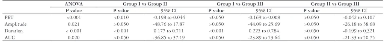
TABLE 2. Results of the statistical analysis (ANOVA and Tukey’s multiple comparison test) of the pharyngeal-esophageal time (PET) and of amplitude, duration and area under the curve (AUC) of the proximal esophageal contractions of normal volunteers aged 18-30 years (Group I, n = 20), 32-50 years (Group II, n = 27) and 51-74 years (Group III, n = 22)
ANOVA Group I vs Group II Group I vs Group III Group II vs Group III P value P value 95% CI P value 95% CI P value 95% CI
PET <0.001 <0.010 -0.198 to-0.044 <0.050 -0.169 to-0.008 >0.050 -0.042 to 0.107 Amplitude 0.021 >0.050 -48.76 to 17.87 >0.050 -44.09 to 25.69 >0.050 -26.18 to 38.68 Duration < 0.001 <0.001 0.177 to 0.711 <0.001 0.225 to 0.784 >0.050 -0.199 to 0.321 AUC 0.020 >0.050 -36.85 to 37.19 >0.050 -23.89 to 53.64 >0.050 -21.33 to 50.75
CI – conidence interval of differences
TABLE 1. Results of evaluation of the pharyngeal-esophageal time (PET) and of the amplitude, duration and area under the curve (AUC) of the proximal esophageal contractions of normal volunteers aged 18-30 years (Group I, n =20), 31-50 years (Group II, n = 27) and 51-74 years (Group III, n = 22). The results are shown as mean (SD)
Group I Group II Group III
PET (seconds) 0.74 (0.13)* 0.86 (0.17) 0.83 (0.18) Amplitude (mm Hg) 94.6 (31.0) 111.0 (41.9) 103.7 (52.3) Duration (seconds) 2.61 (0.69)* 2.16 (0.44) 2.10 (0.59) AUC (mm Hg x s) 125.4 (57.8) 125.2 (43.7) 102.6 (60.8)
suggested the possibility that subjects of group IIID (older
subjects) had a lower amplitude (P = 0.08) and AUC of
contractions (P = 0.10) than the younger subjects (Figure 2).
the ageing process are: reduced resting UES pressure, reduced lower esophageal sphincter (LES) relaxation, upward LES displacement into the chest, delayed esophageal emptying with tertiary contractions, decrease contraction velocity and duration, reduced myenteric ganglion cells, increased amplitude of distal contractions, and thickening of the smooth muscle
layer(11). Further changes of esophageal motility in elderly
subjects are: frequent nonpropulsive contractions, reduction in the amplitude of contractions in subjects older than 80
years, and week esophageal body(2). However, the esophageal
transit seems to be normal and these esophageal motility changes have not been shown to correlate with esophageal
symptoms(2).
The inluence of age is also seen on the pharyngeal phase of swallowing. Ageing is associated with a signiicant decrease in the level of negative pressure resulting from the opening of
the UES, and with incomplete relaxation of this sphincter(7).
Hypopharyngeal amplitude and duration of contractions are
increased in the elderly(23). Ageing prolongs the pharyngeal
transit time and the pharyngeal clearance time and causes
an increase in the amount of pharyngeal residues(4). Healthy
old persons have low resting UES pressure and delayed UES
relaxation after swallowing than young subjects(12). The
duration of oropharyngeal transit is longer in older than in younger subjects, possibly as a consequence of a delay in the
initiation of hypolaryngeal excursion(22).
In the esophageal body, edrophonium chloride does not increase pressures readily in older compared to younger
subjects(13), suggesting a weakening of the muscle itself
in addition to the possibility of neurological dysfunction. A decrease in the number and density of striated muscles has been described in the proximal esophagus of elderly
subjects(16).
In the proximal esophageal body, striated muscle ibres predominate cranially and are gradually replaced by smooth
muscle cells caudally(14). The control of contraction in the
proximal esophagus is done by the enteric co-innervation, which exerts an inhibitory modulation of esophageal motility
at the motor endplate level(14). The manometric behavior of
the striated proximal esophageal muscle is more similar to that of the distal esophageal smooth muscle and not similar
to that of the striated pharyngeal muscle(19).
The explanation for the alteration in esophageal response to swallows may be the impairment of the sensitivity of the
proximal esophagus(25) which may cause the delay in the
esophageal response to swallowing, or may cause alteration
of striated muscle contraction in the proximal esophagus(16).
Sensory deicits in the pharyngeal region may provide clues that a particular loss of intrinsic sensory neurons could
be a factor in the dysfunctions of the presbyesophagus(25).
Enteric sensory neurons of the submucosal plexus are more
susceptible to neurodegeneration with age(25).
The results suggested that the group of subjects over the age of 60 years may have a more important alteration of proximal esophageal contraction, but the number of individuals of this age was not suficient to reach a clear conclusion. However, it is reasonable to think that older
FIGURE 2. Amplitude (A) and area under the curve (AUC) (B) of proximal esophageal contractions of subjects of group I (18-30 years, n = 20), group II (A: 32-39 years, n = 12; B: 40-50 years, n = 15) and group III (C: 51-59 years, n = 14; D: 60-74 years, n = 8) after wet swallows. The results are means and SEM
0 20 40 60 80 100 120 140 160 0 20 40 60 80 100 120 140 160 A U C ( m m H g ) A M PLITUDE 9 mmHg) GROUP I A GROUP III GROUP II
B C D
GROUP I
A
GROUP III GROUP II
B C D
A
B
DISCUSSION
We found that the age may be associated with a delay in the esophageal response to wet swallows and that this response has a shorter duration. We also suggest that older subjects had a decrease in amplitude of contraction, although this result was inconclusive due to the small number of subjects included in group IIID.
Dantas RO, Alves LMT, Dalmazo J, Santos CM, Cassiani RA, Nascimento WV. Inluência da idade na resposta do esôfago proximal à deglutição. Arq Gastroenterol. 2010;47(4):339-43
RESUMO – Contexto - O processo natural de envelhecimento altera a motilidade do esôfago. Objetivo - Estudar o efeito da idade na resposta da parte proximal do esôfago à deglutição de água. Método - Mediu-se a resposta do esôfago proximal à deglutição de 5 mL de água em 69 voluntários saudáveis, 20 com idades de 18 a 30 anos (grupo I), 27 com idades de 31 a 50 anos (grupo II) e 22 com idades de 51 a 74 anos (grupo III). Utilizamos o método manométrico com perfusão contínua. As contrações do esôfago proximal foram medidas 5 cm distal a um registro das contrações em faringe, localizado 1 cm acima do esfíncter superior do esôfago. Foram medidos o tempo entre o início da contração em faringe e o início da contração em esôfago proximal (tempo faringoesofágico), e a amplitude, duração e área sob a curva da contração proximal. Resultados - O tempo faringoesofágico teve menor duração nos sujeitos do grupo I do que naqueles dos grupos II e III (P<0,05). A duração da contração em esôfago proximal foi maior nos sujeitos do grupo I do que naqueles dos grupos II e III (P<0,001). Não houve diferenças entre os grupos na amplitude e na área sob a curva das contrações, e não houve diferenças entre os grupos II e III em todas as medidas. Conclusão – Observou-se que a idade pode alterar a resposta do esôfago proximal à deglutição de água.
DESCRITORES - Envelhecimento. Esôfago, isiologia. Deglutição, isiologia. Contração muscular, isiologia.
subjects may have alterations of amplitude and duration of proximal esophageal contractions.
In diseases that cause loss of the myenteric plexus neurons such as Chagas’ disease and idiopathic achalasia, the results of the evaluation of proximal esophageal contractions are
similar(6, 20), suggesting that the results seen may be consequent
to the impairment of the myenteric plexus.
The observed alterations of esophageal physiology are not clinically relevant. None of the subjects had problems with swallowing and all were able to eat all kinds of food. It is possible that with ageing the subject makes adaptations in swallowing, or changes the ingested food in order to perform a safe swallow. The described alterations may be important when a disease that impairs the swallowing process affects an older individual. Since they already have an alteration
of swallowing, a further alteration caused by a disease has a greater chance to cause dysphagia. We should take into consideration that the swallowing of an older subject, even a healthy one, does not show the same behavior as in
younger subjects(2). We do not know what age is the time to
see modiications in swallowing, but certainly it is not the same for all individuals.
Recent publications have focused on the proximal
esophagus(3, 14). During a gastroesophageal relux episode the
proximal extent of relux along the esophagus appears to be
one of the main determinants of symptoms perception(3).
Esophageal sensitivity is decreased in older subjects(2), a situation
that impairs their perception of gastroesophageal relux(10).
In conclusion, the age may affects the response of the proximal esophagus to wet swallows.
REFERENCES
1. Achem AC, Achem SR, Stark ME, DeVault KR. Failure of esophageal peristalsis in older patients: association with esophageal acid exposure. Am J Gastroenterol. 2003;98:35-9.
2. Achem SA, DeVault KR. Dysphagia in aging. J Clin Gastroenterol. 2005;39:357-71.
3. Cicala M, Habib FI, Emerenziani S. Proximal esophagus: the added value in understanding GORD symptoms. Neurogastroenterol Motil. 2009;21:790-5. 4. Cook IJ, Weltman MD, Wallace K, Shaw DW, McKay E, Smart RC, Butler
SP. Inluence of aging on oral-pharyngeal bolus transit and clearance during swallowing: scintigraphic study. Am J Physiol. 1994;266:G972-7.
5. Dantas RO, Alves LM, Cassiani, RA. Gender differences in proximal esophageal contractions. Arq Gastroenterol. 2009;46:284-7.
6. Dantas RO, Alves LMT, Cassiani RA, Santos CM. Clinical measurement of swallowing and proximal esophageal contractions in Chagas’ disease. Esophagus. 2009;6:231-6.
7. Dejaeger E, Pelemans W, Bibau G, Ponette E. Manuluorographic analysis of swallowing in the elderly. Dysphagia. 1994;9:156-61.
8. Eckardt VE, LeCompte PM. Esophageal ganglia and smooth muscle in the elderly. Am J Dig Dis. 1978;23:443-8.
9. Ferriolli E, Dantas RO, Oliveira RB, Braga FJ. The inluence of ageing on oesophageal motility after ingestion of liquids with different viscosities. Eur J Gastroenterol Hepatol. 1996;8:793-8.
10. Ferriolli E, Oliveira RB, Matsuda NM, Braga FJHN, Dantas RO. Aging, esophageal motility and gastroesophageal relux. J Am Geriatr Soc. 1998;46:1534-7.
11. Firth M, Prather CM. Gastrointestinal motility problems in the elderly patients. Gastroenterology. 2002;122:1688-700.
12. Fulp SR, Dalton CB, Castell JA, Castell DO. Aging-related alterations in human upper esophageal sphincter function. Am J Gastroenterol. 1990;85:1569-72. 13. Hollis JB, Castell DO. Esophageal function in elderly men: a new look at
presbyesophagus. Ann Intern Med. 1974;80:371-4.
14. Kallmünzer B, Sörensen B, Neuhuber WL, Wörl J. Enteric co-innervation of striated muscle ibres in human oesophagus. Neurogastroenterol Motil. 2008:20:597-610.
15. Köberle F. Chagas’ disease and Chagas’ syndrome: the pathology of American trypanosomiasis. Adv Parasitol. 1968;6 63-116.
16. Leese G, Hopwood D. Muscle ibre typing in the human pharyngeal constrictors and oesophagus: the effect of ageing. Acta Anat. 1986;127:77-80.
17. Meciano Filho J, Carvalho VC, Souza RR. Nerve cell loss in the myenteric plexus of the human esophagus in relation to age: a preliminary investigation. Gerontology. 1995;41:18-21.
18. Meshkinpour H, Haghighat P, Dutton C. Clinical spectrum of esophageal aperistalsis in the elderly. Am J Gastroenterol. 1994;89:1480-3.
19. Peghini PL, Pursnani KG, Gideon MR, Castell JA, Nierman J, Castell DO. Proximal and distal esophageal contractions have similar manometric features. Am J Physiol. 1998;274:G325-30.
20. Ramos RI, Varrica LMM, Dantas RO. Differences in response of the proximal esophagus to wet swallows in patients of Chagas’ disease and idiopathic achalasia. Dis Esophagus. 2006;19:401-5.
22. Robbins J, Hamilton JW, Lof GL, Kempster GB. Oropharyngeal swallowing in normal adults of different ages. Gastroenterology. 1992;103:823-9.
23. Shaker R, Ren J, Podvrsan B, Dodds WJ, Hogan WJ, Kern M, Hoffmann R, Hintz J. Effect of aging and bolus variables on pharyngeal and upper esophageal sphincter motor function. Am J Physiol. 1993;264:G427-32.
24. Soergel KH, Zboralske FF, Amberg JR. Presbyesophagus: esophageal motility
in nonagenarians. J Clin Invest. 1964;43:1472-9.
25. Wade PR, Cowen T. Neurodegeneration: a key factor in the ageing gut. Neurogastroenterol Motil. 2004;16:19-23.