Prone position in patients with acute respiratory
distress syndrome
INTRODUCTION
Acute respiratory distress syndrome (ARDS) occupies a great deal of attention in intensive care units (ICU), not only because of its mortality rates but also due to the high resource consumption and the long-term functional and neuro-psychological consequences. In the ICU, the focus largely consists of life-supporting treatments and avoiding the side-efects of invasive treatments such as mechanical ventilation (MV), sedation, neuromuscular blocks, and the administration of high oxygen concentrations.(1) Although great advances
in MV with a signiicant impact on mortality have occurred in the past 20 years,(2,3) the incidence continues to be high.(3-8)
Mariano Setten1,2, Gustavo Adrián Plotnikow1,3, Matías Accoce1,4,5
1. Respiratory Therapists Committee, Sociedad Argentina de Terapia Intensiva - Ciudad Autónoma de Buenos Aires, Argentina. 2. Centro de Educación Médica e Investigaciones Clínicas - CEMIC - Ciudad Autónoma de Buenos Aires, Argentina.
3. Sanatorio Anchorena - Ciudad Autónoma de Buenos Aires, Argentina.
4. Hospital de Quemados - Ciudad Autónoma de Buenos Aires, Argentina.
5. Sanatorio Mater Dei - Ciudad Autónoma de Buenos Aires, Argentina.
Acute respiratory distress syndrome occupies a great deal of attention in intensive care units. Despite ample knowledge of the physiopathology of this syndrome, the focus in intensive care units consists mostly of life-supporting treatment and avoidance of the side efects of invasive treatments. Although great advances in mechanical ventilation have occurred in the past 20 years, with a signiicant impact on mortality, the incidence continues to be high. Patients with acute respiratory distress syndrome, especially the most severe cases, often present with refractory hypoxemia due to shunt, which can require additional treatments beyond mechanical ventilation, among which is mechanical ventilation in the prone position. his method, irst recommended to improve oxygenation in 1974, can be easily implemented in any intensive care unit with trained personnel.
Conflicts of interest: None.
Submitted on July 11, 2016 Accepted on July 18, 2016
Corresponding author: Mariano Setten
Coronel Díaz 2423, Ciudad Autónoma de Buenos Aires (C1425DQW), Argentina
E-mail: [email protected]
Responsible editor: Gilberto Friedman
Decúbito prono en pacientes con síndrome de distrés respiratorio
agudo
ABSTRACT
Keywords: Prone position; Respiratory distress syndrome, acute/ complications; Refractory hypoxemia/ etiology; Mechanical ventilation
Prone position has extremely robust bibliographic support. Various randomized clinical studies have demonstrated the efect of prone decubitus on the oxygenation of patients with acute respiratory distress syndrome measured in terms of the PaO2/FiO2 ratio, including its efects on increasing patient survival.
he members of the Respiratory herapists Committee of the Sociedad Argentina de Terapia Intensiva performed a narrative review with the objective of discovering the available evidence related to the implementation of prone position, changes produced in the respiratory system due to the application of this maneuver, and its impact on mortality. Finally, guidelines are suggested for decision-making.
Patients with ARDS, especially the most severely afected, often present with refractory hypoxemia due to shunt, which can require additional treatments beyond MV, including MV in the prone position (PP). his method, irst recommended to improve oxygenation in 1974,(9) is easily implemented in any ICU(10) and
has extremely robust bibliographic support. Various randomized clinical trials (RCTs) have demonstrated the beneicial efect of PP on the oxygenation of patients with ARDS,(11,12) including its efects on increasing
patient survival.(11-14)
METHODS
A bibliographic search was performed in the PubMed, SciELO, Cochrane, and Lilacs databases using the following MeSH term and keyword combinations: “randomized controlled trial” OR “controlled clinical trial” OR “random” OR “trial” OR “groups” AND “prone position” (MeSH) OR “supine position” (MeSH) OR “patient positioning” (MeSH) OR “prone” OR “proning” OR “prone position” OR “supine” AND “respiratory distress syndrome, adult” (MeSH) OR “acute lung injury” OR “ARDS” OR “respiratory distress syndrome” OR “respiratory failure”. Also included was an unpublished abstract (reference 63) due to its inclusion in one of the meta-analyses.
his narrative review attempts to summarize the physiological modiications associated with PP and to review the clinical trials, meta-analyses, and most relevant systematic reviews from recent years, with special emphasis on the impact on mortality. Finally, this review will establish suggested guidelines and working protocols for decision-making and implementation of MV in PP.
Physiological modifications associated with prone position
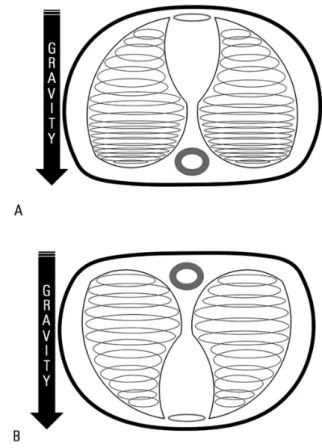
In the lungs of patients with ARDS, alveoli in relatively normal condition coexist with others that are collapsed but recruitable, together with other non-recruitable alveolar sectors. his situation produces an increase in lung weight due to edema, generating over-pressure four to ive times greater than normal, which precipitates collapse of the most dependent lung regions (compression atelectasis) and increased distension of non-dependent regions due to traction(8,15,16) (Figure 1A).
he displacement of gases into and out of the lungs is determined by a pressure gradient. Elastance of the respiratory system (ERS = ECW + EL) is a function of the elastance values of the chest wall (ECW[ET]) and the lungs (EL[EP]). We can deine EL as the diference in transpulmonary pressure over tidal volume (Vt):
- [PAo - esophageal pressure at the end of inspiration] - [PAo - esophageal pressure at the end of expiration]/Vt
(PAo = open airway pressure)
ET can be deined as the diference in esophageal pressure over Vt:
that is, for any given Vt, the plateau pressure can increase, decrease, or remain constant due to the interaction between the chest wall and the lungs.(17,18)
Lung elastance behavior
In a patient on MV and without diaphragmatic activity, during inspiration, air is directed to non-dependent regions due to collapse of the dependent regions. In the prone position, the availability of the pulmonary parenchyma increases. Collapsed alveoli, potentially recruitable, are reopened, and the inferior lobes (which have a higher quantity of alveoli than the superior lobes) ofer higher surface area for difusion, at once improving ventilatory pressures and decreasing the deformation of ibers (strain) and tension (stress) (Figure 1A and 1B). Prone position varies the pressure gradient distribution in relation to the redistribution of the iniltrated areas, the weight of the cardiac mass (the supine position compresses the left lower lung lobe), variations in EL, and cephalic displacement of the abdomen, which results in more homogenous alveolar ventilation.(8,12,16,19-26)
When there is a net provoked alveolar recruitment, EL decreases proportional to the degree of recruitment. If the decrease in EL is similar to the increase in ECW, respiratory system elastance is maintained with no changes. In contrast, if the decrease in EL associated with recruitment is greater than the increase in ECW, the inal result will be a decrease in respiratory system elastance.
Increases in stress and strain produce structural changes in the alveoli, including cellular damage, surfactant dysfunction, edema and increased capillary permeability, and biological alterations, such as increased proinlammatory mediators.(22) Decreases in stress and
strain produced by PP can have some inluence on these
mechanisms and decrease the risk of ventilator-induced damage.(27)
Mentzelopoulos et al. have demonstrated that, in patients with severe ARDS, implementation of PP combined with optimization of the positive-end expiratory pressure (PEEP) level post-procedure improves lung volume at the end of expiration, increasing it by approximately 30%, with reductions in elastance and pulmonary resistance. At the same time, PP reduces pulmonary stress (relected by reduced transpulmonary pressure) and strain (relected by the relationship between
Vt/lung volume at the end of expiration, decreasing from 27% to 33%) compared to Fowler’s position.(28)
Cornejo et al. evaluated the responses to PP combined with high levels of PEEP (15cmH2O) in 24 patients with ARDS. hey found that using this strategy improved pulmonary recruitment, as evidenced by decreases in unventilated lung tissue from 501 to 322 grams (p < 0.001) when using 15cmH2O for PEEP and from 322 to 290 grams (p = 0.028) when combined with PP. Likewise, this strategy (PP + PEEP 15cmH2O), in patients with high recruitment potential, decreased alveolar instability from 4.1 ± 1.9% to 2.9 ± 0.9% (p = 0.003).(25)
Chest wall elastance behavior
he dorsal region of the chest wall is more rigid than the ventral region due to the presence of the spinal column and para-vertebral muscle masses. When a patient is placed in the PP, thoracic expansion is produced mainly in the direction of the abdominal and dorsal regions. In addition to these changes, we may assume that the ventral wall becomes more rigid as a result of the position per se, with a resulting increase in ECW. Recalling the foregoing explanation, if EL does not change, the result is an increase in respiratory system elastance secondary to increased ECW.(29)
Prone position and intra-abdominal pressure
Although their behaviors may be unique, we can describe the thoracic and abdominal cavities as two compartments
of diferent volume.(29) he two compartments are
occupied by organs of diferent densities and are separated by the diaphragm. With respect to the diference in chest wall rigidity (the dorsal wall is more rigid than the ventral), both pleural and intra-abdominal pressures would be modiied by a change in body position, inluenced by the increase in abdominal wall rigidity. Increases in intra-abdominal pressure inluence the curvature and position of the diaphragm.(30)
In the supine position, the abdominal cavity hydrostatic pressure can be as much as ive times higher than that in the thoracic cavity,(31) a diference that increases
signiicantly in obese patients.(32) he causes of ARDS
intra-abdominal pressures in supine decubitus correspond to the dorsal regions, where pressure is inexorably transmitted to the pleural space, generating extrinsic compression to the postero-basal pulmonary region. Prone position modiies this situation, and some authors have reported decreased intra-abdominal pressure;(34) in the end, the abdominal
wall becomes more rigid, with a resulting increase in intra-abdominal pressure.(35-37)
Changes in the ventilation/perfusion ratio
Describing a lung model in vertical position suggests a ventilation/perfusion ratio (V/Q) based on a “gravitational” hypothesis, which may explain why perfusion is greater in the more dependent regions of the lungs. Studies of PP, both human and experimental, conirm the hypothesis in which the distribution of perfusion shows a non-gravitational gradient. Upon making the non-dependent zones the more perfused and increasing the ventilated lung volume in PP, a notable improvement in the V/Q ratio is produced.(38-40) Other factors inluencing this type
of perfusion distribution are the fractal architecture of the vessels, greater production of nitric oxide in dorsal zones compared to the ventral, and lower vascular resistance in dorsal zones.(41-45)
Effects of the prone position on hemodynamics
We could suppose that the mere fact of changing the mediastinum’s position in the thoracic cavity by placing patients in PP has some hemodynamic efect. In a study of patients without ARDS, the elimination of the weight of the heart from ventral lung zones showed a freeing of a small portion of the lung parenchyma.(46) However, this efect
is diferent in patients with cardiomegaly and congestive heart failure, situations often associated with ARDS, and the improvement in oxygenation upon adopting the PP position is immediate,(47) possibly explained by a greater
portion of lung parenchyma freed by this maneuver.(48)
However, the speciic efects on hemodynamic changes have also been studied via impacts on the right-ventricular ejection fraction,(49) favored due to a decrease in the load
and explained by PP. Another study(50) demonstrated an
increase in preload and a decrease in postload of the right ventricle and an increase in preload of the left ventricle.
During PP, the pulmonary artery occlusion pressure was also increased, with a decrease in the transpulmonary pressure gradient (diference between the average pulmonary artery pressure and its occlusion pressure),
which was associated with “pulmonary vascular dysfunction” and may be associated with an increase in mortality in patients with ARDS.(51,52)
Prone position also has an impact on the extravascular lung water index, although its clinical relevance has not been observed.(53,54) While large studies on PP in patients
with ARDS have excluded those with hemodynamic instability, patients with myocardial ischemia can be more susceptible to cardiac dysfunction during PP.(55,56)
Studies included for analysis
For the review, the ive RCTs considered most relevant were selected (Table 1), in which the attempt was made to show that ventilation in PP in patients with hypoxemia decreases mortality, in addition to six reviews and meta-analyses. Below, we will analyze the results.
Results of clinical trials
he irst RCT, published in 2001 by the Prone-Supine Study Group,(11) randomized 304 patients with a wide
range of severity of acute lung injury. he patients were kept prone for an average of seven hours/day for a maximum of 10 days, but there was no efect on survival. hree years later, Guerin et al.(12) executed a similar multicentric study:
patients were kept in PP for approximately eight hours/day until compliance with clinical improvement criteria was met. his study also showed no reduction in mortality.
Two subsequent multicentric RCT attempted to correct some of the deiciencies of the prior studies: they only included patients with ARDS and kept them prone for approximately 20 hours/day. he study performed by Mancebo et al.(13) was ended prematurely, after including
only 142 patients, due to recruitment diiculties. he most recent RCT by Taccone et al.(57) (Prone-Supine II
Study) included 342 patients and showed a signiicantly higher frequency of adverse events in patients receiving PP. Neither of the two studies mentioned showed improvements in survival, not even in patients with severe ARDS (Figure 2).
In 2013, the French multicentric RCT PROSEVA
Study Group(14) showed a marked improvement in
mortality at day 28 (Figure 2): 16% in the prone group (38/237 patients) versus 32.8% (75/229 patients) in the supine group (p < 0.001). he study’s design had these novel characteristics:
Table 1 - Comparative description of the five most relevant randomized clinical studies selected for review
Gatinnoni et al., 2001(11) Guerin et al., 2004(12) Mancebo et al., 2006(13) Taccone et al., 2009(57) Guerin et al., 2013(14)
Number of patients 304 791 136 342 466
Prone/Supine 152/152 413/378 76/60 168/174 237/229
ALI/ARDS** 6/94 21/31/others ARDS ARDS ARDS
PaO2/FiO2 127 153 145 113 > 150
Prone duration (hours/day) 7 ± 1.8* 8# (IR 7.7; 9.8) 17# 18 ± 4* 17 ± 3*
Days of pronation 4.7* 4# (IR 2 - 6) 10.1# (IR 0 - 54) 8.4 ± 6* 4 ± 4*
Protective ventilation No No Yes Yes Yes
Weaning protocol No Yes Yes --- Yes
Primary result** Mortality 10 days Mortality 28 days Mortality ICU Mortality 28 days Mortality 28 days
21.1/25 32.4/31.5 43/58 31/32.8 16/32.8
Mortality ICU** 50.7/48 --- --- 38.1/42
---Mortality day 90** --- 43.3/42.2 --- --- 23.6/41
Mortality in hospital** --- --- 50/62 ---
---Mortality at six months** 62.5/58.6 --- --- 47/52.3
---ALI - acute lung injury; ARDS - acute respiratory distress syndrome; IR - interquartile range; ICU - intensive care unit. * Average ± standard deviation; # Median and interquartile ranges (IR); ** %.
Figure 2 - Comparison of the results from various randomized clinical studies related to mortality at day 28 with respect to the use of prone position.
NS - not significant.
< 30cmH2O).
- Inclusion with patients with severe ARDS, deined as PaO2/FiO2 < 150mmHg with PEEP ≥ 5cmH2O and FiO2 ≥ 60%.
- Stabilization period of 12 - 24 hours prior to randomization, which allowed the selection of patients with ARDS who did not improve solely
with recruitment, discarding those with atelectasis or hydrostatic pulmonary edema as signiicant contributors to the acute hypoxemia.
- Use of neuromuscular blocks on continuous infusion during the initial 48 hours.
Results of reviews and meta-analyses
As previously mentioned, some reviews and meta-analyses have been published with the objective of analyzing the data from RCTs published on the topic with more representative samples, efecting stratiications according to PaO2/FiO2 and number of hours/day in the prone position, to elucidate whether subgroups with diferences in mortality exist.
Abroug et al.(58) published a meta-analysis that included
six RCTs with data from 1372 patients to analyze mortality in the ICU at 28 days as the primary variable. A total of 713 patients were ventilated in PP, with 659 ventilated in the supine position. he secondary variables included changes in PaO2/FiO2 and the incidence rates of ventilator-associated pneumonia (VAP) and adverse efects of PP. Also analyzed was the duration of stay in the ICU. Ventilation in PP was not associated with an improvement in survival, with a change in mortality of 3% (odds ratio [OR] 0.97, 95% conidence interval [CI] 0.77 - 1.22). Regarding oxygenation, ventilation in PP in this meta-analysis showed a signiicant improvement in PaO2/FiO2 (95%CI: 15 - 35, p < 0.00001, I2 = 56%). he results of this meta-analysis
do not justify the routine use of PP during MV in patients with acute hypoxemic respiratory insuiciency, including acute lung injury and ARDS.
In the same year, another meta-analysis was published by Sud et al.(10) that included more studies with a small
number of patients. heir objectives were to evaluate mortality, oxygenation, VAP, duration of MV, and adverse efects. In the primary analysis (10 clinical studies,(11-13,59-65)
n = 1486), ventilation in PP did not decrease mortality (relative risk [RR] 0.96, 95%CI: 0.84 - 1.09; p = 0.52). he duration of the prone position was up to 24 hours over one to two days in the short-term studies(63-65) and
up to 24 hours a day over more than two days in longer-term studies.(11-13,59-62) In subgroup analysis (short- and
long-duration PP), signiicant mortality diferences were likewise not found (RR 0.77, 95%CI: 0.46 - 1.28 and OR 0.97, 95%CI: 0.85 - 1.11, respectively; p = 0.39 for comparison of the two ORs). Ventilation in PP increased the PaO2/FiO2 by 23 - 34% in the irst three days after randomization, measured at the end of the prone period. Post-hoc analysis revealed that the major part of this improvement was produced during the irst hour of the prone position. In six studies(12,13,59-61,66) (n = 1026),
ventilation in PP reduced the risk of VAP (RR 0.81, 95%CI: 0.66 - 0.99, p = 0.04), without heterogeneity
(I2 = 0%). In six studies(11,59-63) (n = 504), ventilation in the
prone position increased the risk of pressure ulcers (RR 1.36, 95%CI: 1.07 - 1.71; p = 0.01; I2 = 0%).
In 2010, Sud et al.(67) published a systematic review
and meta-analysis focused on the impact on mortality, hypothesizing that ventilation in PP might reduce mortality in severely hypoxemic patients (PaO2/FiO2 < 100mmHg), but not in patients with moderate hypoxemia (100mmHg
≤ PaO2/FiO2 ≤ 300mmHg). he primary variable was
mortality in the patient subgroup with PaO2/FiO2 < 100 mmHg versus patients with PaO2/FiO2 ≥ 100 and ≤ 300mmHg. For each study, mortality was determined on discharge from the hospital or on later follow-up. Secondary results included mortality stratiied according to PaO2/ FiO2 but were limited to patients with acute lung injury/ ARDS; in all patients, the results also included duration of MV, days of of MV up to day 28, and adverse events. he review included 10 studies(1,11-13,57,59-62,66) (n = 1867;
one study(62) included 102 children). Seven(1,11-13,57,61,62) of
the ten studies reported mortality stratiied by PaO2/FiO2 and were included for the primary variable. Ventilation in PP signiicantly reduced mortality in patients with PaO2/FiO2 < 100mmHg (RR 0.84, 95%CI: 0.74 - 0.96, p = 0.01, n = 555), but not in patients with PaO2/FiO2 ≥ 100mmHg (RR 1.07, 95%CI: 0.93 - 1.22, p = 0.36, n = 1169). In the severely hypoxemic subgroup, the number of patients necessary to pronate to avoid one death was 11 (95%CI 6 - 50). Post-hoc analyses with variation of cut-ofs for PaO2/FiO2 suggested a decrease in mortality in the most severe subgroup, using a PaO2/FiO2 cutof limit of up to approximately 140mmHg. In the irst three days after randomization, prone ventilation improved PaO2/ FiO2(1,11-13,57,61,62,66) between 27 and 39% in seven studies.
In spite of these improvements, there were no efects on the duration of MV (average diference -0.70 days, 95%CI -2.01 to 0.62 days, p = 0.3; eight studies,(1,11,12,57,59,60,62,66)
n = 1588) or in days of of MV up to day 28 (average diference -0.88 days, 95%CI: -2.14 to 0.37 days, p = 0.17; 5 studies(1,11,57,60,62) n = 771). According to this
meta-analysis, PP increases the risk of pressure ulcers (RR 1.29, 95%CI: 1.16 - 1.44, p < 0.00001; seven studies,(11,13,59-62)
n = 1279), endotracheal tube obstruction (RR 1.58, 95%CI 1.24 - 2.1, p = 0.0002; seven studies(1,12,57,59,60,62,64)
n = 1351), and accidental chest tube removal (RR 3.14, 95%CI 1.02 - 9.69, p = 0.05; eight studies,(1,11,57,59-62,64)
n = 886, of which only two studies(11,57) reported events).
In the same year, a meta-analysis by Gattinoni et al.(68)
variable and found, as in the meta-analysis by Sud et al.,(67)
diferences in favor of the prone patient group with severe hypoxemia (PaO2/FiO2 < 100mmHg).
In 2011, Abroug et al.(69) published a new meta-analysis
that focused on a subanalysis of studies prior to 2005 and included seven works(1,11-13,57,59,61) (n = 1675; 862 ventilated
in prone from seven to 24 hours/day). Studies published prior to 2006(11,12,59) included 1135 patients with acute
lung injury/ARDS, with a short prone duration (less than 17 hours/day), and without protective ventilation. he four most recent studies(1,13,57,61) included only patients
with ARDS (n = 540) and applied the prone position for longer times (17 - 24 hours/day), using protective ventilation. In only the most recent four studies(1,13,57,61)
(n = 540) that included only patients with ARDS, PP signiicantly reduced mortality in the ICU (OR 0.71, 95%CI 0.5 - 0.99, p = 0.048; number required to treat = 11; I2 = 0%).
In 2014, Beitler et al.(70) published a meta-analysis
whose primary variable was mortality at 60 days. his study included seven RCTs(1,11-13,57,59) (n = 2119); 1088
patients were ventilated in the prone position and 1031 in the supine position. To test the a priori hypothesis that PP reduces mortality only when high and harmful tidal volumes are avoided, the analysis was stratiied according to high Vt (more than 8mL/kg predicted body weight)
versus low Vt (less than or equal to 8mL/kg). After the
stratiication, PP was associated with a signiicant decrease in mortality in the studies that used low tidal volumes (RR = 0.66, 95%CI 0.50 - 0.86, p = 0.002), but not for those using high tidal volumes (RR = 1.00; 95%CI 0.88 - 1.13, p = 0.949). Stratiication by Vt substantially reduced heterogeneity (I2: from 64% to 11% and 25%
in high- and low-Vt models, respectively). he meta-regression demonstrated a dose-response relationship between average basal Vt (mL/kg predicted body weight) and the relation of mortality risk at 60 days in PP. A decrease in the average basal Vt of 1mL/kg was associated with a mortality risk decrease of 16.7% (95%CI 6.1 - 28.3, p = 0.001). Analysis stratiied by long- or short-duration prone positioning showed a signiicant reduction in mortality with long duration (RR = 0.71, IC95% 0.56 - 0.90, p = 0.004). his meta-analysis demonstrates that PP signiicantly reduces mortality in patients with ARDS when it is used with low Vt.
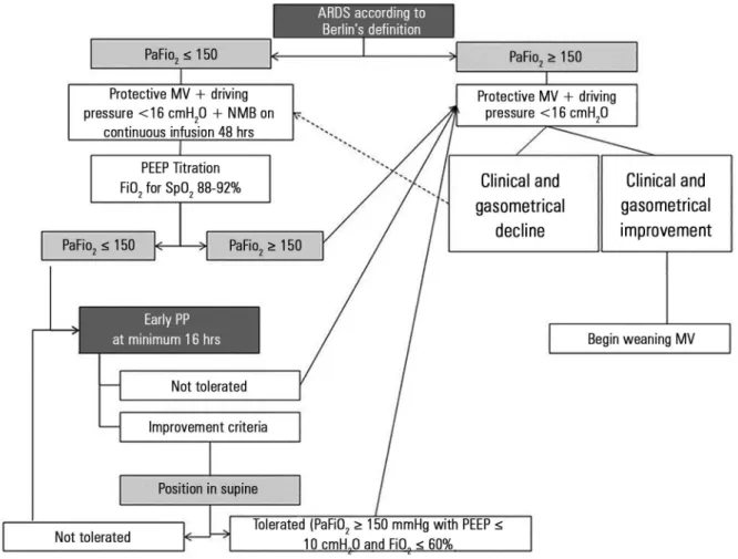
Recommendations (Figure 3)
- Deine ARDS according to Berlin’s deinition. - In ARDS, early intervention with PP is efective
(irst 24/36 hours from initiation of MV).
- Prior to PP, deine the severity of ARDS with a sedated patient, adapted to the MV (RASS - 4/-5), with muscle relaxers (if necessary) on continuous infusion, ventilated using a protective strategy of 6 - 8mL/kg predicted weight of Vt, PEEP ≥ 5cmH2O, plateau pressure < 30cmH2O, working pressure < 16cmH2O and FiO2 with a saturation target of 88 - 92%.
- PP ofers advantages in terms of survival of patients with relatively severe ARDS (PaO2/FiO2 ≤ 150mmHg).
- In the majority of cases, a minimum of four people is required to implement PP.
- Protect the areas most subject to decubitus lesions: hips, knees, shoulders, and face.
- Once the maneuver is performed, re-evaluate the PEEP level required.
- PP sessions should ideally be maintained from 16 to 20 hours. During this time, the patient should alternate positions (swimmer’s pose).
- PP may be suspended due to positive or negative efects. Positive: PaO2/FiO2 > 150mmHg for at least four hours in the supine position following
the last PP period (with PEEP ≤ 10 cmH2O
and FiO2 ≤ 60%). Negative: deterioration of oxygenation (decrease in PaO2/FiO2 > 20%) in PP
versus supine decubitus.
- Consider some of the unanticipated events that may occur during the maneuver and require it to be halted:
- Accidental extubation.
- Sustained desaturation (< 85%) or PaO2 < 55mmHg with FiO2 100% sustained over ive minutes.
- Cardiac arrest or sustained bradycardia (≤ 30 beats per minute for one minute).
- Hypotension (< 60mmHg) sustained for ive minutes.
Figure 3 - Decision-making algorithm for implementing prone position in patients with acute respiratory distress syndrome. ARDS - acute respiratory distress syndrome; MV - mechanical ventilation; NMB - neuromuscular blockade; PEEP - positive-end expiratory pressure; PP - prone position.
Maneuver for placing the patient in prone position
At least four operators will be required. One is in charge of the airway, two are in charge of rotating the patient, and one more gives direction and checks catheters, tubes, lines, and probes. he maneuver begins by placing the patient in the lateral decubitus position. Once it is decided which position will be used, the length of any guides, probes, catheters, and tubes the patient has in place should be checked. Shut of nutrition and re-evaluate the hemodynamic situation. he ability to apply protective patches to areas prone to decubitus lesions is also necessary (knees, shoulders, face).
First, move the patient toward the edge of the bed opposite the side onto which they will be turned. he hand on the rotational side should be placed in contact with the ipsilateral buttock (palm-buttock).
For the second step, place the patient in the lateral decubitus position. Check catheters, probes, and tubes, and control hemodynamics.
In the third step, place the patient in the prone position and recheck everything mentioned in the previous step. It is recommended that leg and arm positions be alternated (swimmer’s position) to avoid decubitus lesions. his precaution also applies to the face.
CONCLUSIONS
El síndrome de distrés respiratorio agudo ocupa gran aten-ción en la unidad de cuidados intensivos. A pesar del amplio conocimiento alcanzado sobre la isiopatología de éste síndro-me, el enfoque en la unidad de cuidados intensivos consiste, en gran parte, en un tratamiento de soporte vital y en evitar los efectos secundarios de las terapéuticas invasivas. Si bien, durante los últimos 20 años, se generaron grandes avances en ventilación mecánica con un impacto importante sobre la mortalidad, ésta continúa siendo elevada. Una característica de los pacientes con síndrome de distrés respiratorio agudo, sobre todo los más seve-ros, es la presencia de hipoxemia refractaria debido a la existencia de shunt, pudiendo requerir tratamientos adicionales a la venti-lación mecánica, entre ellos la ventiventi-lación mecánica en decúbito prono. Este método, recomendado para mejorar la oxigenación por primera vez en 1974, puede ser implementado fácilmente en cualquier unidad de cuidados intensivos con personal entrenado.
El decúbito prono tiene un sustento bibliográico sumamen-te robusto. Varios ensayos clínicos randomizados han demostra-do el efecto del decúbito prono sobre la oxigenación en pacien-tes con síndrome de distrés respiratorio agudo medida a través de la relación PaO2/FiO2 e incluso su impacto en el aumento de la sobrevida de estos pacientes.
Los integrantes del Comité de Kinesiología Intensivista de la Sociedad Argentina de Terapia Intensiva realizaron una revisión narrativa con el objetivo de exponer la evidencia disponible en relación a la implementación del decúbito prono, los cambios producidos en el sistema respiratorio por la aplicación de la ma-niobra y su impacto sobre la mortalidad. Por último, se sugeri-rán lineamientos para la toma de decisiones.
RESUMEN
Descriptores: Posición prona; Síndrome de distrés respiratorio agudo/complicaciones; Hipoxemia refractaria/ etiología; Ventilación mecánica
REFERENCES
1. Fernandez R, Trenchs X, Klamburg J, Castedo J, Serrano JM, Besso G, et al. Prone positioning in acute respiratory distress syndrome: a multicenter randomized clinical trial. Intensive Care Med. 2008;34(8):1487-91. 2. Ventilation with lower tidal volumes as compared with traditional tidal
volumes for acute lung injury and the acute respiratory distress syndrome. The Acute Respiratory Distress Syndrome Network. N Engl J Med. 2000;342(18):1301-8.
3. Chang SY, Dabbagh O, Gajic O, Patrawalla A, Elie MC, Talmor DS, Malhotra A, Adesanya A, Anderson HL 3rd, Blum JM, Park PK, Gong MN; United States Critical Illness and Injury Trials Group: Lung Injury Prevention Study Investigators (USCIITG-LIPS). Contemporary ventilator management in patients with and at risk of ALI/ARDS. Respir Care. 2013;58(4):578-88. 4. ARDS Definition Task Force, Ranieri VM, Rubenfeld GD, Thompson BT,
Ferguson ND, Caldwell E, Fan E, et al. Acute respiratory distress syndrome: the Berlin Definition. JAMA. 2012;307(23):2526-33.
5. Amato MB, Meade MO, Slutsky AS, Brochard L, Costa EL, Schoenfeld DA, et al. Driving pressure and survival in the acute respiratory distress syndrome. N Engl J Med. 2015;372(8):747-55.
6. Papazian L, Forel JM, Gacouin A, Penot-Ragon C, Perrin G, Loundou A, Jaber S, Arnal JM, Perez D, Seghboyan JM, Constantin JM, Courant P, Lefrant JY, Guérin C, Prat G, Morange S, Roch A; ACURASYS Study Investigators. Neuromuscular blockers in early acute respiratory distress syndrome. N Engl J Med. 2010;363(12):1107-16.
7. Villar J, Sulemanji D, Kacmarek RM. The acute respiratory distress syndrome: incidence and mortality, has it changed? Curr Opin Crit Care. 2014;20(1):3-9.
8. Kacmarek RM, Villar J. Management of refractory hypoxemia in ARDS. Minerva Anestesiol. 2013;79(10):1173-9.
9. Bryan AC. Conference on the scientific basis of respiratory therapy. Pulmonary physiotherapy in the pediatric age group. Comments of a devil’s advocate. Am Rev Respir Dis. 1974;110(6 Pt 2):143-4.
10. Sud S, Sud M, Friedrich JO, Adhikari NK. Effect of mechanical ventilation in the prone position on clinical outcomes in patients with acute hypoxemic respiratory failure: a systematic review and meta-analysis. CMAJ. 2008;178(9):1153-61.
11. Gattinoni L, Tognoni G, Pesenti A, Taccone P, Mascheroni D, Labarta V, Malacrida R, Di Giulio P, Fumagalli R, Pelosi P, Brazzi L, Latini R; Prone-Supine Study Group. Effect of prone positioning on the survival of patients with acute respiratory failure. N Engl J Med. 2001;345(8):568-73. 12. Guerin C, Gaillard S, Lemasson S, Ayzac L, Girard R, Beuret P, et al. Effects
of systematic prone positioning in hypoxemic acute respiratory failure: a randomized controlled trial. JAMA. 2004;292(19):2379-87.
13. Mancebo J, Fernández R, Blanch L, Rialp G, Gordo F, Ferrer M, et al. A multicenter trial of prolonged prone ventilation in severe acute respiratory distress syndrome. Am J Respir Crit Care Med. 2006;173(11):1233-9. 14. Guérin C, Reignier J, Richard JC, Beuret P, Gacouin A, Boulain T, et al
Mercier E, Badet M, Mercat A, Baudin O, Clavel M, Chatellier D, Jaber S, Rosselli S, Mancebo J, Sirodot M, Hilbert G, Bengler C, Richecoeur J, Gainnier M, Bayle F, Bourdin G, Leray V, Girard R, Baboi L, Ayzac L; PROSEVA Study Group. Prone positioning in severe acute respiratory distress syndrome. N Engl J Med. 2013;368(23):2159-68.
15. Pelosi P, D’Andrea L, Vitale G, Pesenti A, Gattinoni L. Vertical gradient of regional lung inflation in adult respiratory distress syndrome. Am J Respir Crit Care Med. 1994;149(1):8-13.
16. Gattinoni L, Marini JJ, Pesenti A, Quintel M, Mancebo J, Brochard L. The ‘‘baby lung’’ became an adult. Intensive Care Med. 2016;42(5):663-73. 17. Benditt JO. Esophageal and gastric pressure measurements. Respir Care.
2005;50(1):68-75; discussion 75-77. Review.
18. Zin WA. Elastic and resistive properties of the respiratory system. In: Lucangelo U, Pelosi P, Zin WA, Aliverti A, editors. Respiratory system and artificial ventilation. Milan: Springer; 2008. p. 15-26.
19. Broccard A, Shapiro RS, Schmitz LL, Adams AB, Nahum A, Marini JJ. Prone positioning attenuates and redistributes ventilator-induced lung injury in dogs. Crit Care Med. 2000;28(2):295-303.
20. Martínez O, Nin N, A Esteban. Evidencias de la posición en decúbito prono para el tratamiento del síndrome de distrés respiratorio agudo: una puesta al día. Arch Bronconeumol. 2009;45(6):291-6.
21. Albaiceta GM, Blanch L. Beyond volutrauma in ARDS: the critical role of lung tissue deformation. Crit Care. 2011;15(2):304.
22. Slutsky AS, Ranieri VM. Ventilator-induced lung injury. N Engl J Med. 2013;369(22):2126-36. Erratum in N Engl J Med. 2014;370(17):1668-9. 23. Pelosi P, Brazzi L, Gattinoni L. Prone position in acute respiratory distress
24. Terragni P, Ranieri VM, Brazzi L. Novel approaches to minimize ventilator-induced lung injury. Curr Opin Crit Care. 2015;21(1):20-5.
25. Cornejo RA, Díaz JC, Tobar EA, Bruhn AR, Ramos CA, González RA, et al. Effects of prone positioning on lung protection in patients with acute respiratory distress syndrome. Am J Respir Crit Care Med. 2013;188(4):440-8.
26. Santini A, Protti A, Langer T, Comini B, Monti M, Sparacino CC, et al. Prone position ameliorates lung elastance and increases functional residual capacity independently from lung recruitment. Intensive Care Med Exp. 2015;3(1):55.
27. Gattinoni L, Pesenti A, Carlesso E. Body position changes redistribute lung computed-tomographic density in patients with acute respiratory failure: impact and clinical fallout through the following 20 years. Intensive Care Med. 2013;39(11):1909-15.
28. Mentzelopoulos SD, Roussos C, Zakynthinos SG. Prone position reduces lung stress and strain in severe acute respiratory distress syndrome. Eur Respir J. 2005;25(3):534-44.
29. Pelosi P, Tubiolo D, Mascheroni D, Vicardi P, Crotti S, Valenza F, et al. Effects of the prone position on respiratory mechanics and gas exchange during acute lung injury. Am J Respir Crit Care Med. 1998;157(2):387-93. 30. Hedenstierna G, Strandberg A, Brismar B, Lundquist H, Svensson L,
Tokics L. Functional residual capacity, thoracoabdominal dimensions, and central blood volume during general anesthesia with muscle paralysis and mechanical ventilation. Anesthesiology. 1985;62(3):247-54.
31. Froese AB. Gravity, the belly, and the diaphragm: you can’t ignore physics. Anesthesiology. 2006;104(1):193-6.
32. Froese AB, Bryan AC. Effects of anesthesia and paralysis on diaphragmatic mechanics in man. Anesthesiology. 1974;41(3):242-55.
33. De Keulenaer BL, De Waele JJ, Powell B, Malbrain ML. What is normal intra-abdominal pressure and how is it affected by positioning, body mass and positive end-expiratory pressure? Intensive Care Med. 2009;35(6):969-76.
34. Malbrain ML. Abdominal pressure in the critically ill: measurement and clinical relevance. Intensive Care Med. 1999;25(12):1453-8. Review. 35. Fletcher SJ. The effect of prone ventilation on intra-abdominal pressure.
Clin Intensive Care. 2006;17(3-4):109-12.
36. Hering R, Wrigge H, Vorwerk R, Brensing KA, Schröder S, Zinserling J, et al. The effects of prone positioning on intraabdominal pressure and cardiovascular and renal function in patients with acute lung injury. Anesth Analg. 2001;92(5):1226-31.
37. Hering R, Vorwerk R, Wrigge H, Zinserling J, Schröder S, von Spiegel T, et al. Prone positioning, systemic hemodynamics, hepatic indocyanine green kinetics, and gastric intramucosal energy balance in patients with acute lung injury. Intensive Care Med. 2002;28(1):53-8.
38. Michelet P, Roch A, Gainnier M, Sainty JM, Auffray JP, Papazian L. Influence of support on intra-abdominal pressure, hepatic kinetics of indocyanine green and extravascular lung water during prone positioning in patients with ARDS: a randomized crossover study. Crit Care. 2005;9(3):R251-7. Erratum in Crit Care. 2005;9(4):308.
39. Glenny RW, Lamm WJ, Albert RK, Robertson HT. Gravity is a minor determinant of pulmonary blood flow distribution. J Appl Physiol (1985). 1991;71(2):620-9.
40. Lamm WJ, Albert RK. Effect of zonal conditions and posture on pulmonary blood flow distribution to subpleural and interior lung. J Appl Physiol (1985). 2000;88(1):120-5.
41. Richter T, Bellani G, Scott Harris R, Vidal Melo MF, Winkler T, Venegas JG, et al. Effect of prone position on regional shunt, aeration, and perfusion in experimental acute lung injury. Am J Respir Crit Care Med. 2005;172(4):480-7.
42. Glenny RW, Robertson HT. Fractal properties of pulmonary blood flow: characterization of spatial heterogeneity. J Appl Physiol (1985). 1990;69(2):532-45.
43. Rimeika D, Nyrén S, Wiklund NP, Koskela LR, Tørring A, Gustafsson LE, et al. Regulation of regional lung perfusion by nitric oxide. Am J Respir Crit Care Med. 2004;170(4):450-5.
44. Wenz M, Hoffmann B, Bohlender J, Kaczmarczyk G. Angiotensin II formation and endothelin clearance in ARDS patients in supine and prone positions. Intensive Care Med. 2000;26(3):292-8.
45. Hlastala MP, Glenny RW. Vascular structure determines pulmonary blood flow distribution. News Physiol Sci. 1999;14:182-6.
46. Glenny RW, Bernard S, Robertson HT, Hlastala MP. Gravity is an important but secondary determinant of regional pulmonary blood flow in upright primates. J Appl Physiol (1985). 1999;86(2):623-32.
47. Albert RK, Hubmayr RD. The prone position eliminates compression of the lungs by the heart. Am J Respir Crit Care Med. 2000;161(5):1660-5. 48. Nakos G, Tsangaris I, Kostanti E, Nathanail C, Lachana A, Koulouras V, et al.
Effect of the prone position on patients with hydrostatic pulmonary edema compared with patients with acute respiratory distress syndrome and pulmonary fibrosis. Am J Respir Crit Care Med. 2000;161(2 Pt 1):360-8. 49. Wiener CM, McKenna WJ, Myers MJ, Lavender JP, Hughes JM. Left lower
lobe ventilation is reduced in patients with cardiomegaly in the supine but not the prone position. Am Rev Respir Dis. 1990;141(1):150-5.
50. Vieillard-Baron A, Charron C, Caille V, Belliard G, Page B, Jardin F. Prone positioning unloads the right ventricle in severe ARDS. Chest. 2007;132(5):1440-6.
51. Jozwiak M, Teboul JL, Anguel N, Persichini R, Silva S, Chemla D, et al. Beneficial hemodynamic effects of prone positioning in patients with acute respiratory distress syndrome. Am J Respir Crit Care Med. 2013;188(12):1428-33.
52. Repessé X, Charron C, Vieillard-Baron A. Acute cor pulmonale in ARDS: rationale for protecting the right ventricle. Chest. 2015;147(1):259-65. 53. Bull TM, Clark B, McFann K, Moss M; National Institutes of Health/
National Heart, Lung, and Blood Institute ARDS Network. Pulmonary vascular dysfunction is associated with poor outcomes in patients with acute lung injury. Am J Respir Crit Care Med. 2010;182(9):1123-8. 54. Langer M, Mascheroni D, Marcolin R, Gattinoni L. The prone position in
ARDS patients. A clinical study. Chest. 1988;94(1):103-7.
55. Chatte G, Sab JM, Dubois JM, Sirodot M, Gaussorgues P, Robert D. Prone position in mechanically ventilated patients with severe acute respiratory failure. Am J Respir Crit Care Med. 1997;155(2):473-8.
56. Jolliet P, Bulpa P, Chevrolet JC. Effects of the prone position on gas exchange and hemodynamics in severe acute respiratory distress syndrome. Crit Care Med. 1998;26(12):1977-85.
57. Taccone P, Pesenti A, Latini R, Polli F, Vagginelli F, Mietto C, Caspani L, Raimondi F, Bordone G, Iapichino G, Mancebo J, Guérin C, Ayzac L, Blanch L, Fumagalli R, Tognoni G, Gattinoni L; Prone-Supine II Study Group. Prone positioning in patients with moderate and severe acute respiratory distress syndrome: a randomized controlled trial. JAMA. 2009;302(18):1977-84. 58. Abroug F, Ouanes-Besbes L, Elatrous S, Brochard L. The effect of prone
positioning in acute respiratory distress syndrome or acute lung injury: a meta-analysis. Areas of uncertainty and recommendations for research. Intensive Care Med. 2008;34(6):1002-11.
59. Voggenreiter G, Aufmkolk M, Stiletto RJ, Baacke MG, Waydhas C, Ose C, et al. Prone positioning improves oxygenation in post-traumatic lung injury--a prospective randomized trial. J Trauma. 2005;59(2):333-41, discussion 341-3. 60. Beuret P, Carton MJ, Nourdine K, Kaaki M, Tramoni G, Ducreux JC. Prone
position as prevention of lung injury in comatose patients: a prospective, randomized, controlled study. Intensive Care Med. 2002;28(5):564-9. 61. Chan MC, Hsu JY, Liu HH, Lee YL, Pong SC, Chang LY, et al. Effects of prone
position on inflammatory markers in patients with ARDS due to community-acquired pneumonia. J Formos Med Assoc. 2007;106(9):708-16. 62. Curley MA, Hibberd PL, Fineman LD, Wypij D, Shih MC, Thompson JE, et
63. Leal RP, Gonzalez R, Gaona C, Garcia G, Maldanado A, Dominguez-Cherit G. Randomized trial compare prone vs supine position in patients with ARDS [abstract]. Am J Respir Crit Care Med. 1997;155:A745.
64. Papazian L, Gainnier M, Marin V, Donati S, Arnal JM, Demory D, et al. Comparison of prone positioning and high-frequency oscillatory ventilation in patients with acute respiratory distress syndrome. Crit Care Med. 2005;33(10):2162-71.
65. Demory D, Michelet P, Arnal JM, Donati S, Forel JM, Gainnier M, et al. High-frequency oscillatory ventilation following prone positioning prevents a further impairment in oxygenation. Crit Care Med. 2007;35(1):106-11. 66. Watanabe I, Fujihara H, Sato K, Honda T, Ohashi S, Endoh H, et al. Beneficial
effect of a prone position for patients with hypoxemia after transthoracic esophagectomy. Crit Care Med. 2002;30(8):1799-802.
67. Sud S, Friedrich JO, Taccone P, Polli F, Adhikari NK, Latini R, et al. Prone ventilation reduces mortality in patients with acute respiratory failure and severe hypoxemia: systematic review and meta-analysis. Intensive Care Med. 2010;36(4):585-99.
68. Gattinoni L, Carlesso E, Taccone P, Polli F, Guérin C, Mancebo J. Prone positioning improves survival in severe ARDS: a pathophysiologic review and individual patient meta-analysis. Minerva Anestesiol. 2010;76(6):448-54. 69. Abroug F, Ouanes-Besbes L, Dachraoui F, Ouanes I, Brochard L. An updated
study-level meta-analysis of randomised controlled trials on proning in ARDS and acute lung injury. Crit Care. 2011;15(1):R6.