UNIVERSIDADE FEDERAL DE MINAS GERAIS
FACULDADE DE MEDICINA
M
OLECULARM
EDICINEP
OSTGRADUATE PROGRAMMariana Costa Rossette
THE ANTICANCER PROPERTIES OF FLAVOKAWAIN B, A KAVA-KAVA
COMPOUND, TOWARDS OVARIAN CANCER CELLS AND ITS
ANTIANGIOGENIC ACTION
IN VIVO
AND
IN VITRO
Belo Horizonte
Mariana Costa Rossette
THE ANTICANCER PROPERTIES OF FLAVOKAWAIN B, A KAVA-KAVA
COMPOUND, TOWARDS OVARIAN CANCER CELLS AND ITS
ANTIANGIOGENIC ACTION
IN VIVO
AND
IN VITRO
Doctoral thesis submitted to the Postgraduate program of Molecular Medicine at Universidade Federal de Minas Gerais, as a partial requirement for obtaining the title of PhD in Molecular Medicine.
Concentration area: Molecular Medicine
Supervisor: Prof. Luiz Armando De Marco
To my parents, sister, fiancé and dearest friends,
ACKNOWLEDGEMENTS
I wish to express my sincere gratitude to my advisor Professor Luiz Armando De Marco for
the great opportunity to learn about science and to go beyond the books and guidelines
seeking for new knowledge about this challenging disease, called cancer. Thank you for
being an example of researcher, professor and human being.
I am very grateful to Dr. Hanna Caldas for giving me the opportunity to work and learn in her
laboratory in 2009, where I started to study about Flavokawain B. Thank you for being so
kind, teaching me valuable skills and encouraging me to continue this research.
I also sincerely thank Dr. Maria José Campagnole for teaching me about research for the first
time, inspiring me to love it and for the great opportunity to go to Wake Forest University as
an exchange student.
To Dr. Eitan Friedman and Dr. Luciana Bastos Rodrigues, I express my appreciation for being
a constant source of motivation, helping me with new ideas and precious advice for this
work.
I am extremely thankful and hugely indebted to Débora Chaves Moraes, my collaborator and
close friend. I couldn’t have done this project without you. Thank you for being present,
incredibly hard-working and believing in this research as I do.
I would also like to thank Erika Kelmer, my collaborator, who has brilliantly made the
I also acknowledge Dr. Luiz Alexandre Magno, Dr. Daniela Valadão, Dr. Alexandre Barros, Dr.
Luiza Martins, Dr. Karen Torres, Dr. Carolina Aguiar, Dr. Vitor Rezende, Dr. Juliana Carvalho
and Dr. Dawidson Gomes for the support and advice during the experiments.
To all our current and past laboratory staff, I extend my deepest gratitude for the research
suggestions, words of motivation and friendship, especially Flávia Melo, Helena Sarubi,
Patrícia Couto, Cristina Sábato and Nayra Soares.
I want to express my gratitude to Dr. José Renan and all his laboratory staff for being so kind,
receptive and allowing me to use their cell culture room, so I could finish my in vitro studies. To the Coordenação de Aperfeiçoamento de Pessoal de Nível Superior (CAPES) for the
scholarship in the CAPES-FIPSE and PhD programs.
To my parents, Elizabete and Altair, I’m eternally grateful for giving me the opportunity to
follow my dreams and making them a reality.
I thank my sister, Raquel and nephew, Arthur for being a constant source of happiness.
Hálisson Flamini, my fiancé, your understanding, love and words of encouragement have
made this possible.
“Learn from yesterday, live for today, hope for tomorrow. The important
thing is not to stop questioning.”
ABSTRACT
Natural products have been utilized for centuries as novel compounds for the
treatment and prevention of many diseases. The anticancer potential of chalcones in the
Kava-kava extract, a plant grown in the Pacific Islands, have been inferred from the
correlation with kava consumption and lower incidence of cancer in these populations.
Flavokawain B, a naturally occurring chalcone of kava-kava, has shown impressive
anti-cancer effects in several studies, besides other significant anti-inflammatory and
antinociceptive activities. Epithelial ovarian cancer (EOC) is the most lethal gynecologic
malignancy and the fifth most common cause of cancer-related deaths in woman. Current
treatment with surgery and chemotherapy has improved survival; however recurrence and
disease progression occurs in approximately 80% of patients in advanced stage. The
development of more effective treatment strategies is urgent and many studies are being
done in order to develop target chemotherapeutics against ovarian cancer molecular
pathogenesis pathways, such as antiangiogenic drugs, PARP and PI3K inhibitors. In the
present work we identified, for the first time, flavokawain B as causing strong
antiproliferative and apoptotic effects against ovarian cancer cells with less cytocidal action
against normal cells. Flavokawain B results in downregulation of Bcl-2 anti-apoptotic protein
expression, increased Bax:Bcl-2 ratio and a significant dose-dependent inhibition of Akt
activation on OVCAR-3 cells. These are important findings, since Bcl-2 overexpression and
Akt overactivation are frequently associated with poor prognosis and resistance to
chemotherapy in ovarian cancer. In addition, this work has demonstrated the strong
believe that FKB might be a promising weapon in the arsenal of treatment options against
RESUMO
Produtos naturais têm sido utilizados por séculos no tratamento e prevenção de
muitas doenças. O potencial anti-carcinogênico das chalconas obtidas do extrato da planta
Kava-kava, comumente cultivada nas Ilhas do Pacífico, tem sido inferido a partir da
correlação do consumo de kava e a baixa incidência de câncer nestas regiões. A
Flavokawaína B (FKB), uma chalcona natural presente no extrato de kava, demonstrou
efeitos anti-carcinogênicos, anti-inflamatórios e anti-nociceptivos significativos em diversos
estudos. O câncer de ovário epitelial é a neoplasia ginecológica mais letal e quinta causa de
morte relacionada ao câncer entre mulheres. O tratamento atual que consiste em cirurgia e
quimioterapia melhorou a sobrevida, porém recidivas e progressão da doença ocorrem em
aproximadamente 80% dos pacientes com doença avançada. O desenvolvimento de novas
estratégias mais efetivas de tratamento é urgente e muitos estudos estão sendo realizados
para o desenvolvimento de quimioterápicos que atuem sobre as vias moleculares
patogênicas do câncer ovariano, como por exemplo as drogas anti-angiogênicas, inibidores
da PARP e PI3K. No presente estudo identificamos, pela primeira vez, grande atividade
anti-proliferativa e apoptótica da flavokawaína B contra células de câncer ovariano, além de
menor efeito citotóxico da FKB contra células normais. A FKB resultou em redução da
expressão da proteína anti-apoptótica Bcl-2, aumento da razão de Bax:Bcl-2 e inibição
significativa e dose-dependente da ativação de Akt em células OVCAR-3. Considerando que a
super-expressão de Bcl-2 e super-ativação de Akt estão relacionados ao pior prognóstico e
resistência à quimioterapia no câncer de ovário, tais achados são importantes. Além disso,
utilizando um modelo de zebrafish. Esses resultados revelam que a FKB é um agente
LIST OF FIGURES
Figure 1: Representation of the plant ... 21
Figure 2: Kava drinking countries ... 22
Figure 3: Kava ceremony ... 23
Figure 4: Proposed diagram of FKB induced G2/M arrest, apoptosis and Akt/p38 MAPK inactivation of cancer cells according to available publications. ... 32
Figure 5: Expanded dualist model of ovarian carcinogenesis and its molecular pathways .... 37
Figure 6: The PI3K/Akt/mTOR signaling pathway, frequently up-regulated in ovarian cancer ... 41
Figure 7: Zebrafish development and angiogenesis assay scheme ... 57
Figure 8: Cell viability of OVCAR-3 cells upon 24h treatment with FKB ... 60
Figure 9: Cell viability of OVCAR-3 cells upon 48h treatment with FKB ... 61
Figure 10: Light microscopy of OVCAR-3 cells treated with FKB ... 62
Figure 11: DAPI and PI double staining of OVCAR-3 cells treated with FKB ... 63
Figure 12: Annexin V and 7 AAD staining of OVCAR-3 cells treated with FKB ... 64
Figure 13: Effects of FKB against normal human fibroblasts cells compared to OVCAR-3 ... 65
Figure 14: Western-Blotting analysis of apoptotic proteins expression ... 68
Figure 15: Bax:Bcl-2 ratio of OVCAR-3 cells after 24h treatment with FKB ... 69
Figure 16: FKB downregulates Akt activation in OVCAR-3 cells ... 70
Figure 17: FKB inhibits capillary tube formation of endothelial cells under the stimulus of VEGF ... 72
Figure 18 :Cell viability of HUVEC cells treated with FKB for 24 hours ... 73
Figure 19: FKB inhibits endothelial cell migration ... 74
Figure 20: FKB inhibits capillary tube formation in cultured endothelial cells ... 75
Figure 21: SIV formation and toxic effect of FKB towards zebrafish ... 77
Figure 22: FKB blocks angiogenesis process in Zebrafish ... 78
LIST OF TABLES
LIST OF ABREVIATIONS AND SYMBOLS
α alpha
β beta
µ micro
µg micrograms
µm micrometer
µM micromolar
143B Human bone osteosarcoma cells 22RV1 Human prostate cancer cells 4-T1 Mouse mammary carcinoma 7- AAD 7-aminoactinomycin D A-2058 Human melanoma cells A549 Human lung adenocarcinoma ACC-2 Adenoid cystic carcinoma ADP Adenosine diphosphate
AEBSF 4-(2-Aminoethyl) benzenesulfonyl fluoride hydrochloride AIF Apoptosis Inducing Factor
Akt v-akt murine thymoma viral oncogene homolog 1 AP Alkaline phosphatase
Apaf-1 Apoptotic protease activating factor 1
AR Androgen receptor
Bad B-cell lymphoma -associated death promoter Bak B-cell lymphoma 2 homologous antagonist/killer Bax B-cell lymphoma 2-associated X protein
BCIP 5-Bromo-4-chloro-3-indolyl phosphate disodium salt Bcl-2 B-cell lymphoma 2
Bcl-xL B-cell lymphoma-extra large BCRJ Banco de células do Rio de Janeiro Bid BH3 interacting-domain death agonist Bik B-cell lymphoma 2 -interacting killer
BRAF v-raf murine sarcoma viral oncogene homolog B1 BRCA1 Breast cancer 1, early onset
BRCA2 Breast cancer 2, early onset C4-2B Human prostate cancer cells Ca Ski Carcinoma of the cervix CaCl2 Calcium chloride
Cal-27 Oral squamous carcinoma cells
CO2 Carbon dioxide
COX-I Ciclo-oxigenase-1 COX-II Ciclo-oxigenase-2
CXCR4 Chemokine receptor type 4 DAPI 4,6-diamidino-2-phenylindole DMEM Dulbecco’s modified Eagle’s medium DMSO Dimethyl sulfoxide
DNA Deoxyribonucleic acid DR5 Death receptor 5
DU145 Human prostate cancer cells
ECC-1 Human endometrial adenocarcinoma cells EJ Human bladder carcinoma
EOC Epithelial ovarian cancer
ERBB2 Erb-B2 Receptor Tyrosine Kinase 2 FBS Fetal bovine serum
FDA Food and Drug Administration
FK Flavokawain
FKA Flavokawain A
FKB Flavokawain B
FKC Flavokawain C
h hour
H460 Human lung large cell carcinoma
HBMEC Human brain microvascular endothelial cells HCT 116 Human colon carcinoma
HGF Human gengival fibroblast cells HGSOC High grade serous ovarian cancer HSC-3 Human oral carcinoma cells HS-SY-II Humal synovial sarcoma cells
HUVEC Human umbilical vein endothelial cells IC50 Maximal inhibitory concentration INCA Instituto Nacional de Câncer iNOS Inducible nitric oxide synthase
IP Intraperitoneal
IV Intravenous
KB Oral epidermal carcinoma KCl Potassium Chloride kDa Kilodaltons
KH2PO4 Potassium Dihydrogen Phosphate
KRAS Kirsten rat sarcoma viral oncogene homolog L-02 Normal immortalized liver cells
LANCaP Human prostate cancer cells LAPC4 Human prostate cancer cells LPS Lipopolysaccharide
LVES Large Vessel Endothelial Supplement
M Molar
MAPK Mitogen-activated protein kinases MCF-7 Human breast adenocarcinoma cell line MDA-MB231 Human breast adenocarcinoma cell line MG-63 Human bone osteosarcoma cells
MgCl2 Magnesium chloride MgSO4 Magnesium sulfate
min Minutes
mL Milliliters
MMP-9 Matrix-metalloproteinase 9 mRNA Messenger ribonucleic acid mTOR Mammalian Target of Rapamycin
Na2PO4 Disodium Phosphate NaCl Sodium Chloride NaHCO3 Sodium bicarbonate NBT Nitroblue tetrazolium NBT Nitro-blue tetrazolium
NF1 Neurofibromatosis type 1 gene
NF-kB Nuclear factor kappa B NIH3T3 Human fibroblast cells NTMT Alkaline phosphatase buffer
OC Ovarian cancer
OS160 Human bone osteosarcoma cells OVCAR-3 Ovarian cancer adenocarcinoma cells
PARP Poly ( adenosine diphosphate-ribose) polymerase PBS Phosphate Buffer Saline
PBT Phosphate buffered saline with 0.1% tween 20 PC-3 Human prostate cancer cells
PDK1 Pyruvate dehydrogenase lipoamide kinase isozyme 1 hpf Hours post fertilization
PFS Progression free survival
PI Propidium Iodide
PI3K Phosphatidylinositide 3-kinase
PIK3CA Phosphatidylinositol-4,5-Bisphosphate 3-Kinase, Catalytic Subunit Alpha PIP2 Phosphatidylinositol 4,5-bisphosphate
PIP3 Phosphatidylinositol (3,4,5)-trisphosphate PIP3 Phosphatidylinositol-4,5-bisphosphate 3-kinase PrECs Human prostate epithelial cells
PrECs Human prostate stromal cells PTEN Phosphatase and tensin homolog
Puma p53 upregulated modulator of apoptosis RAW 264.7 Murine macrophage cells
RPMI Roswell Park Memorial Institute
SDF-1 Stromal cell-derived factor 1 SDS Sodium dodecyl sulfate Ser473 Serine 473
SIV Subintestinal vein
SK-LMS-1 Human uterine leiomyosarcoma cells STIC Serous tubal intraepithelial carcinomas SYO Human synovial sarcoma cells
SNAIL Zinc finger protein
T24 Human urinary bladder transitional carcinoma cell line T75 75 cm2 tissue culture flask
T-HESC Human endometrium fibroblast cells Thr308 Threonine 308
TI Therapeutic index
Tie-2 Receptor tyrosine kinase of angiopoietin TNF-α Tumor necrosis factor alpha
TP53 Tumor protein p53 TSP-2 Thrombospondin 2
SUMMARY
1 INTRODUCTION... 20
1.1 KAVA-KAVA ... 21
1.1.1 Kava-kava plant and beverage ... 21
1.1.2 Chemical properties ... 24
1.1.3 Kava-kava toxicity ... 25
1.1.4 Kava-kava and cancer ... 27
1.1.5 Flavokawain B ... 28
1.2 OVARIAN CANCER ... 34
1.2.1 Epidemiology ... 34
1.2.2 Pathology of epithelial ovarian cancer ... 34
1.2.3 Treatment and new perspectives ... 37
2 OBJECTIVES ... 44
3 MATERIALS AND METHODS ... 46
3.1 DRUG ... 47
3.2 CELL LINES ... 47
3.3 CELL CULTURE CONDITIONS AND COMPOUNDS ... 48
3.4 ANTIBODIES ... 48
3.5 PRIMARY FIBROBLASTS CULTURE ... 49
3.6 EVALUATION OF CELL AND NUCLEAR MORPHOLOGY BY LIGHT AND FLUORESCE MICROSCOPY ... 50
3.7 CELL VIABILITY MTT ASSAY ... 51
3.8 FLOW CYTOMETRY ASSAY ... 52
3.9 WESTERN BLOTTING ANALYSIS ... 52
3.10 LIVE CELL MICROSCOPY ... 53
3.11 TUBE FORMATION ASSAY ... 54
3.12 WOUND HEALING ASSAY ... 54
3.14 STATISTICAL ANALYSIS ... 58
4 RESULTS ... 59
4.1 FLAVOKAWAIN B CAUSES STRONG ANTIPROLIFERATIVE EFFECT ON OVARIAN CANCER CELLS IN A DOSE AND TIME RESPONSE MANNER ... 60
4.2 FLAVOKAWAIN B INDUCES APOPTOSIS OF OVCAR-3 CELLS ... 62
4.3 FLAVOKAWAIN B IS LESS CYTOTOXIC TOWARDS NORMAL CELLS THAN TO CANCER CELLS ... 65
4.4 FLAVOKAWAIN B REDUCES BCL-2 PROTEIN EXPRESSION AND INCREASES DE BAX:BCL-2 RATIO IN OVCAR-3 CELLS ... 66
4.5 FLAVOKAWAIN B INHIBITS PI3K/AKT PATHWAY ACTIVATION IN OVCAR-3 CELLS ... 69
4.6 FKB INHIBITS ENDOTHELIAL CELLS PROLIFERATION, MIGRATION AND TUBE FORMATION ... 71
4.7 FKB SUPPRESSES ANGIOGENESIS IN VIVO IN A ZEBRAFISH MODEL ... 76
5 DISCUSSION ... 81
6 CONCLUSIONS ... 95
1.1
Kava-Kava1.1.1 Kava-kava plant and beverage
Kava-kava plant, Piper methysticum or intoxicating pepper is a robust, perennial shrub belonging to the Piperaceae family. It grows in stony ground, reaching heights from 4 to 6 meters. The leaves are green, heart-shaped, pointed and have about 15 cm in length. The
roots grow about 60 cm bellow the ground and can become considerably thick at maturity
(Figure 1) (Singh, 1992).
Figure 1: An adult Piper methysticum plant, named popularly Kava-Kava. Leaves (A) and Roots (B) Images available at <www.wikipedia.com>, accessed on October 01, 2015.
Kava plant can be found in various parts of the world, but is most commonly
encountered in nearly all the Pacific islands. Its beverage is traditionally used for social and
religious purposes. with the exceptions of New Zealand, New Caledonia and most of the
regions in Oceania, which is encompassed by the three cultural and ethnic regions of
Melanesia, Polynesia and Micronesia (Singh, 1992; Steiner, 2000).
Figure 2: South Pacific islands. Map of Oceania showing most of the Kava drinking countries and the three cultural regions of Melanesia, Micronesia and Polynesia. Non-kava drinking countries are represented in red. (Reproduced from Singh, 1992)
For kava beverage preparation, roots are ground and mixed with water or coconut
water and the drink is traditionally used mainly for men during a ceremony (Figure 3). After
drinking, kava is found to reduce fatigue, anxiety and to produce pleasant, cheerful and
Figure 3 : Kava ceremony in Fiji. Kava is prepared in a utensil called tanoa where water is poured and mixed with kava roots. Only men are permitted to officiate the ceremony.
(Reproduced from Singh YN. Kava: an overview. Journal of Ethnopharmacol 1992).
The use of Kava in medicinal practices has been dated back to the eighteenth century
for many purposes -anti-anxiety, antidepressant, muscle relaxant, analgesic, anti-convulsant,
in the treatment of chronic cystitis, asthma, syphilis and gonorrhea (Singh, 1992; Steiner,
2000). In the past 20 years, organic solvent extracts from kava roots have also been used in
Western countries, leading to its emergence as one of the 10 best-selling botanical
supplements for the treatment of anxiety and depression (Zhou et al., 2010). Despite the safety of traditional kava drinking in South Pacific Islands, severe side effects of liver damage
were reported in Europe and United the States of America (USA) after the use of commercial
1.1.2 Chemical properties
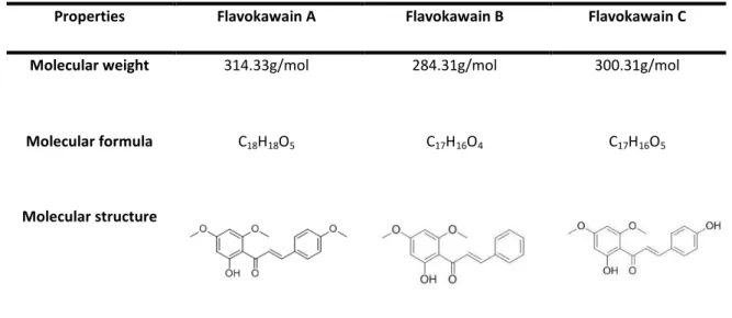
There are 2 major classes of compounds found within the Kava roots extract:
kavalactones and chalcones. Kavalactones, the major components of kava and responsible
for psychoactive effects, include: kawain, methysticin, desmethoxyyangonin and yangonin.
The chalcones are Flavokawain A (FKA), Flavokawain B (FKB), and Flavokawain C (FKC),
representing 0.46%, 0.015% and 0.012% of kava root extracts respectively. These
compounds have similar backbones with different side chains (Singh, 1992; Zi & Simoneau,
2005). FKA contains a methoxy group, FKB only hydrogen and FKC has an extra hydroxyl
group (Table 1) (Singh, 1992; Zi & Simoneau, 2005; Abu et al., 2013).
Table 1: Summary of the molecular properties of Kava-kava chalcones.
Properties Flavokawain A Flavokawain B Flavokawain C
Molecular weight 314.33g/mol 284.31g/mol 300.31g/mol
Molecular formula C18H18O5 C17H16O4 C17H16O5
Molecular structure
Modified from Abu et al., 2013. Images in the table are available at < http://www.albtechnology.com>, accessed on January 10, 2016.
Chalcones are precursor of flavonoids in plants, which have shown to have many
biological activities including antioxidant, anti-inflammatory and anti-cancer action through
multiple mechanisms (Zi & Simoneau, 2005). The basic molecular structure of chalcones
include two aromatic rings linked by an unsaturated three carbon bridge. Because of α,β
hydroxyl groups, thus less likely to react with nucleic acids and therefore are not associated
with mutagenesis as are some other alkylating chemotherapeutics (Zi & Simoneau, 2005;
Mahapatra,2015).
1.1.3 Kava-kava toxicity
Kava drinking have been safely used for thousands of years in the South Pacific region
with no documented side effects, however from 1999 to 2002 herbal hepatotoxicity was
unexpectedly reported in patients worldwide (Showman et al., 2015). Due to these reports, kava was banned in the European Union and Canada, recalled in Australia and included on
the USA Food and Drug Administration (FDA) consumer advisory list (Teschke et al., 2011, Martin et al., 2014; Showman et al., 2015).
Traditionally kava is prepared extracting compounds from its roots using aqueous
solutions, but modern extraction techniques utilizes organic solvents (acetone and ethanol),
reaching significantly higher levels of kavalactones and chalcones in the extract (Table 2)
(Zhou et al., 2010; Martin et al., 2014). Although it was an important concern about liver injury, toxicity cases occurred after the use of kava aqueous solution in the South Pacific
region as well, suggesting that solvents did not play a major role in toxicity (Zhou et al., 2010; Teschke et al., 2011; Abu et al., 2013).
Initially, kavalactones have been proposed to account for kava toxicity, but in vitro
and in vivo experiments did not show significant effects (Singh & Devkota, 2003; Zhou et al., 2010). Other compounds of kava preparations, such as flavokawain B were also pointed as
subject and this affirmation has been refuted for some studies (Teschke et al., 2011; Abu et al., 2013).
Flavokawain B, revealed cytotoxicity against hepatoma cells (HepG2) and normal
immortalized liver cells (L-02). Also, in vivo orally administered FKB (25mg/kg body weight), showed modest signs of hepototoxicity in mice, but in a concentration value 250 times
higher than the usual daily uptake by humans (Zhou et al., 2010; Teschke et al., 2011; Showman et al., 2015). On the other hand, FKB has also demonstrated hepatoprotective effects (Abu et al., 2013; Pinner et al., 2016).
Table 2: Contents of flavokawain B and total kavalactones in Kava roots extracts (mg/g dried weight) by different extraction solvents
Extract solvent FKB (mg/g) Total kavalactones (mg/g)
Water 0.2 46.6
60% acetone 26 474.8
Acetone 33.7 570
95% ethanol 32.3 548.8
Modified from Zhou et al., 2010.
Some concerns were also focused on the possibility that kava raw material could
have been contaminated by mould hepatotoxins. Post haverst storage conditions, the warm
and humid weather of Pacific, could favor mould proliferation. Moreover, mycotoxins, such
as aflatoxins which are potentially toxic to human liver, were encountered in kava
preparations, suggesting that kava hepototoxicity cases might be related to contaminations,
instead of the kava constituents (Li et al., 2008; Teschke et al., 2011; Martin et al., 2014).
In summary, the toxicity related to Kava extract intake is controversial and no
1.1.4 Kava-kava and cancer
A number of countries in the South Pacific have very low cancer incidence compared
to other parts of the world, despite of the high proportion of smokers in these populations.
Considering this, Henderson et al. (1985), has proposed that a dietary protective factor might be involved.
Later, Steiner (2000) observed a close inverse relationship between cancer incidence
and kava consumption. First, he reported an age-standardized cancer incidence for
kava-drinking countries (Vanuatu, Fiji, Western Samoa and Micronesia) to be one fourth to one
third the incidence in non-kava drinking countries (USA) and non-kava drinking Polynesians
(New Zealand). Steiner (2000) also highlighted an unexpected gender ratio of cancer in the
major kava-drinking countries (Vanuatu, Fiji and Western Samoa), where overall cancer
incidence is more frequent in women then in men, which only occur in 10 out of 150 cancer
incidence reporting locations in the world. These reports are very intriguing, since the
majority of kava consumption is performed by men in those regions (Shaver & Sosis, 2014).
Considering the known anticancer properties of chalcones, combined with the
epidemiologic data about Kava, researches have been done in order to determine the
chemopreventive action of kava-kava compounds against many types of cancers (Abu et al., 2013). Among the kava-kava coumpounds, the chalcones but not the kavalactones, showed
cytocidal action against different types of cancer cell lines (Zhou et al., 2010). Between the three flavokawains (FK) encountered in kava extract, FKB showed to be the most potent
Flavokawain A (FKA), one of the chalcones of kava extract, was reported to possess
anti-inflammatory and anti-cancer activities by suppressing the expression of iNOS and
COX-II, through the inhibition of NF-kB pathway in LPS-stimulated macrophage cells (Kwon et al., 2013). Zi and Simoneau (2005) have also shown antitumor effects of FKA against bladder
cancer in vitro and in vivo by involvement of mitochondrial apoptotic pathway. Besides, FKA had antitumor effects against breast cancer cell lines (MCF-7 and MDA-MB231) via intrinsic
apoptotic pathway and cell cycle arrest (Abu et al., 2014). It also inhibited tube formation of endothelial cells in vitro, demonstrating a potential anti-metastatic action (Abu et al., 2014).
Flavokawain C (FKC) has been tested in bladder and colon cancer cell lines for its
anti-cancer activity showing anti-proliferative and apoptotic action as well (Abu et al., 2013; Phang et al., 2016).
1.1.5 Flavokawain B
Flavokawain B, scientifically known as 6’-hydroxy-2,4’-dimethoxychalcone, was found for the first time in the roots of Piper methystcum. Later, it was also reported in other species, such as Aniba riparia, P. triangularis var. palloda and Didymocarpus corchorijolia
(Wollenweber et al., 1981; Kuo et al., 2010; ABU et al., 2013). FKB interacts with several important molecular proteins and signaling pathways, demonstrating anti-inflammatory,
anti-nociceptive and anti-cancer properties (Wu et al., 2002; Feroz et al., 2012; Abu et al.,
2013).
pronounced inhibition of COX-I (77%) and II (16%) enzymes (Wu et al., 2002). Lin et al. (2009) also found that flavokawain B had an impressive effect in inhibiting nitric oxide production,
prostaglandin E2, TNF-α, COX-II and the NF-kB pathway.
The anti-nociceptive activity of flavokawain B was proved by Mohamad et al., (2010), who have demonstrated that intraperitoneal administration of FKB produced markedly
dose-dependent suppression of nociceptive responses, both centrally and peripherally
mediated. Moreover, compared to acetylsalicylic acid, FKB was 68 fold more effective. These
findings might have additional therapeutic implications in the development of a new drug to
treat inflammatory pain (Mohamad et al., 2010; Mohamad et al., 2011).
The anticancer properties of Flavokawain B have been demonstrated in several
cancer cell lines, such as osteosarcoma, oral carcinoma, synovial sarcoma, prostate, colon
and lung cancers, between others. Table 3 summarizes the in vitro half maximal inhibitory concentration (IC50) of Flavokawain A, B and C in selected cell lines according to the available
Table 3: Half maximal inhibitory concentration (IC50) of Flavokawain A, B and C in selected cancer cell lines according to the available publications
Flavokawain Cancer type Cancer cell line IC50 (µM) Treatment (hours) Reference
FKA Bladder T24 16.7 48h Zi & Simoneau, 2005
RT4 20.8 48h Zi & Simoneau, 2005
EJ 17.2 48h Zi & Simoneau, 2005
Liver HepG2 62.38 24h Li et al., 2008
Breast MCF-7 25 72h Abu et al.,2014
MDA-MB231
17.5 72h Abu et al.,2014
Abu et al.,2014
FKB Bladder T24 6.7 48h Zi & Simoneau, 2005
RT4 15.7 48h Zi & Simoneau, 2005
EJ 5.7 48h Zi & Simoneau, 2005
Oral epidermal carcinoma
KB ~70 24h Lin et al., 2012
Breast 4T1 47.5 72h Abu et al., 2015
Cervix Ca Ski 109 48h Abu et al., 2013
Colon carcinoma HCT 116 ~25 24h Kuo et al., 2010
Lung
adenocarcinoma
A549 ~50 24h Kuo et al., 2010
Lung carcinoma H460 18.2 48h An et al., 2012
Prostate cancer LAPC4 32 48h Tang et al., 2010
LANCaP 48.3 48h Tang et al., 2010
PC-3 6.2 48h Tang et al., 2010
DU145 3.9 48h Tang et al., 2010
22RV1 58.4 72h Li et al., 2012
C4-2B 7.7 72h Li et al., 2012
Oral squamous carcinoma
HSC-3 17.2 24h Hseu et al., 2012
Cal-27 26.7 24h Hseu et al., 2012
Melanoma A-2058 18.3 24h Hseu et al., 2012
Synovial sarcoma SYO 10 72h Sakai et al., 2012
HS-SY-II 21 72h Sakai et al., 2012
Adenoid cystic carcinoma
ACC-2 4.69 48h Abu et al., 2013
Uterine
Leiomyosarcoma
SK-LMS-1 ~3.3 72h Eskander et al., 2012
Endometrial adenocarcinoma
ECC-1 ~4.4 72h Eskander et al., 2012
Osteosarcoma 143B ~6.9 72h Ji et al, 2013
OS160 ~19 72h Ji et al, 2013
MG-63 ~13 72h Ji et al, 2013
Liver Saos-2 L-02 HepG2 ~26 35.15 62.38 72h 24h 24h
Ji et al, 2013 Li et al., 2008 Li et al., 2008
FKC Bladder EJ 14.6 48h Abu et al., 2013
T24 10.6 48h Abu et al., 2013
Liver Hep G2 59.48 48h Li et al., 2008
Modified from Abu et al., 2013.
These studies have also determined normal tissue cells viability upon treatment with
cancer cells. Table 4 summarizes the in vitro IC50 of Flavokawain B towards normal cell lines already tested.
Table 4: Half maximal inhibitory concentration (IC50) of Flavokawain B in selected normal cell lines according to the available publications
Tissue type Cell line IC50(µM) Time of treatment (h)
Reference
Human fibroblasts NIH3T3 ~25 24h Kuo et al., 2010
Human fibroblasts HFW 25 – 50 24h Kuo et al., 2010
Prostate epithelial PrECs Not reached (>17.6 µM)
48h Tang et al., 2010
Prostate stromal PrSCs Not reached (>17.6 µM)
48h Tang et al., 2010
Human endometrium fibroblasts
T-HESC Not reached
(>35.,2µM)
72h Eskander et al., 2012
Human gengival fibroblasts
HGF 30 – 40 72h Lin et al, 2012
Murine macrophage RAW 264.7 Not reached (>40 µM)
24h Lin et al, 2009
Liver L-02 35 48h Zhou et al, 2010
The antiproliferative and cell death mechanisms involved in the exposition of
different cancer cells to FKB have shown similar pathways, such as the activation of
apoptosis and cell cycle arrest (Zi & Simoneau, 2005; Li et al., 2008; Kuo et al., 2010; Tang et al., 2010; Zhou et al., 2010; An et al., 2012; Eskander et al., 2012; Hseu et al., 2012; Li et al.,
2012; Lin et al., 2012; Sakai et al., 2012; Ji et al., 2013; Kwon et al., 2013; Abu et al.,2014, Abu et al., 2015). Figure 4, modified from HSEU et al., (2012), illustrates and summarizes the possible mechanisms of FKB against different cancer cells according to the available
Figure 4: Proposed diagram of FKB induced G2/M arrest, apoptosis and Akt/p38 MAPK inactivation of different cancer cells.
Modified from Hseu et al., 2012.
Flavokawain B apoptotic action was demonstrated through typical apoptotic cell
morphology (cell membrane blebbing, shrinkage, nuclear condensation and fragmentation)
and activation of the intrinsic and extrinsic apoptotic pathways with upregulation of caspase
8, 9, 3/7, cleavage of PARP and upregulation of the death receptor 5 (DR5). These
observations and further studies implied that death receptor and mitochondrial-mediated
apoptotic pathways were involved. Altered mitochondrial membrane permeabilization,
release of cytochrome C, upregulation of DR5, Bim, Puma, Bax, Bak and downregulation of
lines (Kuo et al., 2010; Tang et al., 2010; An et al., 2012; Lin et al., 2012). Moreover, it was also observed that flavokawain B induced a G2/M arrest, involving important cyclin/cdk
complexes (Eskander et al., 2012; Lin et al., 2012). The PI3K/Akt signaling pathway is implied in cell proliferation, survival, cell-cycle control, angiogenesis and others. FKB treatment of
human oral carcinoma cells (HSC-3) inhibited the activation of Akt by a decrease on its
phosphorylation (Figure 4) (Hseu et al., 2012).
Further studies in vivo with nude mice have shown that flavokawain B significantly reduced tumor growth in xenograft models of prostate, human squamous carcinoma and
breast cancers (Tang et al., 2010; Li et al., 2012; Lin et al., 2012; Abu et al., 2015).
FKB can also be a promising anti-metastatic drug, since it inhibited the expression of
matrix-metalloproteinase 9 (MMP-9) and urokinase plasminogen activator in oral epidermal
carcinoma cells (KB) and apparently reduced angiogenesis in tumors of a xenograft model
(Lin et al., 2012). Abu et al. (2015) demonstrated in a xenograft model of breast cancer, that the oral treatment with FKB reduced tumor growth and the levels of angiogenesis related
proteins (Abu et al., 2015). Moreover, very recently, Abu et al. (2016), have shown that FKB inhibited cell migration of breast cancer cells, impaired endothelial tube formation in vitro
andregulated several metastasis related proteins, such as VEGF, SNAIL and CXCR4. However,
1.2 Ovarian cancer
1.2.1 Epidemiology
Ovarian cancer is the second most common malignancy of the female genital tract in
developed countries and the third most common in developing countries, where cervical
cancer incidence is still high (Chen & Berek in UpToDate, 2016). It represents the greatest
clinic challenge in gynecology oncology, due to the lack of obvious clinical signs and
symptoms, specific methods for early detection and its high mortality rates (Ramalingam,
2016). For all stages, the 5-year survival rate is 45%, but most of patients are diagnosed with
advanced disease reaching around 30% survival rate in 5 years. (Chien et al., 2007).
The estimated annual incidence of this disease worldwide is over 200,000 cases, with
around 125,000 annual deaths (Siegel et al., 2014). The number of new cases is usually greater in high than in middle- to low-income countries. Age-standardized incidence is 11.7
per 100,000 in the UK, 8.0 per 100,000 in the USA and 5.2 per 100,000 in Brazil (Ramalingam,
2016). According to the Instituto Nacional de Câncer (INCA) database, in Brazil there were
3.283 deaths related to ovarian cancer in 2013 and around 6150 new cases are estimated for
2016 (INCA database <www.inca.gov.br>).
1.2.2 Pathology of epithelial ovarian cancer
The origin and pathogenesis of epithelial ovarian cancer (EOC) have long been
investigated but is still poorly understood. Traditionally, two main hypotheses were
proposed: incessant ovulation and persistent exposure to gonadotropins, both leading to
2016). However, based on the detection of serous tubal intraepithelial carcinomas (STIC) and
its molecular patterns, it has also been proposed that the fallopian tube might be one EOC
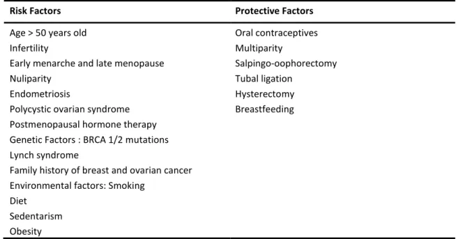
site of origin (Kurman & Shih, 2010; Sorensen et al., 2015). The risk and protective factors that have been related to epithelial ovarian cancer are listed on Table 5 (Sorensen et al.,
2015; Chen & Berek in UpToDate, 2016).
Table 5: Risk and protective factors of ovarian cancer
Risk Factors Protective Factors
Age > 50 years old Infertility
Early menarche and late menopause Nuliparity
Endometriosis
Polycystic ovarian syndrome Postmenopausal hormone therapy Genetic Factors : BRCA 1/2 mutations Lynch syndrome
Family history of breast and ovarian cancer Environmental factors: Smoking
Diet Sedentarism Obesity Oral contraceptives Multiparity Salpingo-oophorectomy Tubal ligation Hysterectomy Breastfeeding
Chen & Berek in UpToDate, 2016. Available at <www.uptodate.com>, accessed on March 26, 2016.
In the past, ovarian carcinoma has been considered to be one single disease;
however studies have shown that it is a heterogeneous pathology that comprises a variety of
tumors with various histopathological and genomic features with different biological
behavior (Bai et al., 2016). The most lethal and the majority of ovarian malignancies (90% percent) are of epithelial origin, the reminder histology types arise from other ovarian cells,
such as germ cell tumors and sex cord-stromal tumors. Epithelial ovarian cancer is
Serous carcinomas are the most frequent category (~50%) and are separated into low and
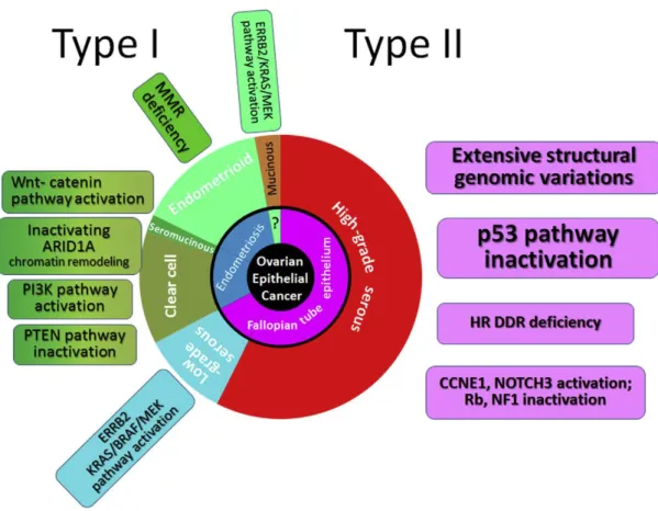
high grade based on the degree of nuclear atypia and mitoses (De Picciotto et al., 2016). Kurman & Shih (2010), have proposed a dualist model, recently revised and expanded
by the same authors (Kurman & Shih, 2016), that categorizes various types of epithelial
ovarian cancer into two groups based on their histology, molecular biology and natural
history (Figure 5) (Kurman & Shih, 2010; Kurman & Shih, 2016).
Type I tumors are clinically indolent and present usually at low stage. They consist of
low-grade serous, endometrioid, clear cell, seromucinous, mucinous carcinomas and
malignant Brenner tumors. These tumors often demonstrate mutations in KRAS, BRAF, PIK3CA, ERBB2 or PTEN (Kurman & Shih, 2016). When confined to the ovary type I tumors have an excellent prognosis, but advanced stage tumors have a poor outcome and usually do
not respond well to chemotherapy (Kim et al., 2012). On the other hand, type II tumors are highly aggressive and almost always are diagnosed in advanced stage (>75%) with
extraovarian disease at the peritoneum and omentum, accompanied by ascites (Kurman &
Shih, 2010). They include high-grade serous, carcinosarcomas and undifferentiated
carcinomas that are frequently associated with mutations in TP53, BRCA1, BRCA2, NF1 and other homologous recombination repair genes. Extended surgery and chemotherapy have improved progression free survival (PFS) and, to a very modest extent, the overall survival,
but most of the patients with type II tumors have recurrences and die because of disease
progression (Kim et al., 2012; Kurman & Shih, 2016).
These findings suggest that the different types of epithelial ovarian cancers develop
represents the dualistic model of ovarian cancers proposed by Kurman & Shih (2016) and its
molecular alterations.
Figure 5: Expanded dualist model of ovarian carcinogenesis and its molecular pathways. Type I carcinomas comprise, low-grade serous, clear cell, endometrioid and mucinous carcinomas. Type II include mainly high-grade serous carcinomas. The inner circle indicates the probable origin of neoplasms and the molecular alterations of each tumor are indicated in the square boxes (Reproduced from Kurman & Shih, 2016).
1.2.3 Treatment and new perspectives
Current management of ovarian cancer in general is based on radical surgery
consisting of total abdominal hysterectomy, bilateral salpingo-oophorectomy, omentectomy,
peritoneal washings and pelvic lymph node sampling, together with chemotherapy.
However, in the last 50 years, there has been only marginal improvement in 5 years overall
Over the years experts have explored different combinations of antitumor drugs in
order to improve the prognosis of ovarian cancer. In 1976, cisplatin reported efficacy in
ovarian cancer an initiated the use of combination chemotherapy. In 1980 paclitaxel,
constituent of the Pacific Yew tree, Taxus brevifolia, showed great activity in EOC, becoming part of the first line therapy. Afterwards, carboplatin, a cisplatin analogue, was reported to
have fewer side effects and replaced cisplatin. The carboplatin-paclitaxel combination is now
the most universal regimen in the treatment of epithelial ovarian cancer (Kim et al., 2012; Della Pepa et al., 2015). Further improvements have been sought in order to increase the efficacy of first-line chemotherapy, such as delivering drugs through the intraperitoneal (IP)
route (Jaaback & Johnson, 2005). Although the IP therapy prolonged survival compared to
the intravenous (IV) administration, there were significantly higher side effects and in many
countries the IV chemotherapy is still preferred (Armstrong et al., 2006).
Despite the activity of first-line chemotherapy, which gives response rates up to 80%,
the majority of patients die of the recurrent resistant disease and it has not substantially
changed overall survival in more than 50 years (Armstrong et al., 2006; Kim et al., 2012; Coward et al., 2015). The strongest independent variable in predicting survival is still residual macroscopic disease after surgery, suggesting that novel regimens of chemotherapy, based
on molecular pathogenesis of ovarian cancer are urgently needed (Kim et al., 2012).
Molecular-targeted therapies, alone or in combination with chemotherapy, have
produced promising results in preclinical and clinical studies (Borley & Brown, 2015; Coward
phosphatidylinositide 3-kinase (PI3K) and Poly (adenosine diphosphate [ADP]-ribose)
polymerase (PARP) inhibitors, besides antiangiogenic drugs.
1.2.3.1 PI3K pathway inhibitors
PI3K-AKT-mTOR is a complex signaling pathway altered in many types of cancers that
coordinates a number of upstream inputs, such as growth factors and tyrosine kinase
receptors. Its stimulation causes activation of a cell surface receptor and phosphorylation of
PI3K (Engelman, 2009). Activated PI3K then phosphorylates lipids on the plasma membrane,
forming the second messenger phosphatidylinositol (3,4,5)-trisphosphate (PIP3). As a result,
the PI3K complex is recruited to the plasma membrane and activates the pyruvate
dehydrogenase kinase 1 (PDK1) and Akt proteins. On the other hand, the phosphatase and
tensin (PTEN) analog protein acts as an endogenous pathway repressor by
de-phosphorylating PIP3 back to PIP2 (Engelman, 2009; Dobbin & Landen, 2013; Cheaib et al., 2015).
Akt is a serine-threonine kinase that is activated through phosphorilation of both
residues: threonine 308 (Thr308) and serine 473 (Ser473) (Vincent et al., 2011). It regulates a large number of downstream targets related to cellular survival and metabolic processes,
such as the transcription of anti-apoptotic genes (X-linked inhibitor of apoptosis protein
[XIAP], Bcl-2 and surviving), oncogenes (p53) and nuclear factor-kB (NF-kB). Akt also promotes cell cycle progression and increases the expression of genes involved in
Activation of the PI3K-AKT-mTOR signaling cascade occurs frequently in epithelial
ovarian cancer and mediates cell-cycle progression, cell survival, migration, invasiveness and
angiogenesis (Wang & Fu, 2015). This pathway is up-regulated in a significant proportion of
ovarian cancers via either direct upstream stimulation (growth factor receptors and their
ligands), indirect activation via cross-talk with the Ras pathway, intrinsically activating
genetic alterations of PI3K/Akt or via loss of function in the tumor suppressor gene PTEN
(Engelman, 2009). Moreover, ovarian cancer cells that either overexpress active Akt/AKT1 or
gene amplification of AKT2 are more resistant to chemotherapy (Bai et al, 2016). Therefore, this pathway is an attractive candidate for therapeutic interventions against EOC and
inhibitors targeting it are in various stages of pre-clinical development with promising
results, such as, Perifosine (Akt inhibitor) and Temsirolimus (mTORC1 inhibitor) (Dobbin &
Landen, 2013; Leary et al., 2013).
Figure 6 illustrates the PI3K-AKT-mTOR signaling pathway alterations with its
implications in ovarian cancer, such as increased expression of anti-apoptotic and angiogenic
Figure 6: The PI3K/Akt/mTOR signaling pathway, frequently up-regulated in ovarian cancer (Reproduced from Leary et al., 2013)
1.2.3.2 PARP inhibitors
PARP enzymes play a vital role in cellular DNA repair, coordinating base-excision
repair pathways. Inhibition of PARP causes accumulation of single-strand brakes, which
causes double-strand breaks during replication. These defects are usually corrected by
homologous recombination proteins, such as BRCA 1 and 2. When BRCA gene is mutated, the inhibition of PARP causes genomic instability and cell death (De Picciotto et al., 2016).
BRCA mutated patients seem to be particularly sensitive to PARP inhibitors alone and in combination with chemotherapy, especially in relapsed platinum-sensitive high grade serous
disease (Scott et al., 2015; De Picciotto et al., 2016; NCCN, 2016). These data implicated in the approval of Olaparib, a PARP-1, -2 and -3 inhibitor, by the FDA, limited to the BRCA1/2
mutation carriers. It was the first biologic agent to treat ovarian cancer based upon
personalized medicine (Wang & Fu, 2015). Many trials are still being done with Olaparib and
other PARP inhibitors in association with different biologic agents in order to enhance
effectiveness. Because of the significant impact on cancer therapy, the BRCA1/2 status screening is now recommended for all patients diagnosed with high grade serous ovarian
cancer (HGSOC) independently of the family history of breast and ovarian cancers (Scott et al., 2015; NCCN, 2016).
1.2.3.3 Antiangiogenic drugs
Cancer has the ability to spread to adjacent or distant organs, what makes it life
threatening. Angiogenesis, one of the hallmarks of cancer, is required for tumor growth
beyond 1-2mm, being essential for tumor invasion and metastasis (Hanahan & Weinberg,
2011; Weis & Cheresh, 2011). Neovascularization requires the recruitment of vasculature,
circulating endothelial cells and signals of pro-angiogenic mediators, such as VEGF (Hanahan
& Weinberg, 2011; Eskander & Tewari, 2014).
Metastatic intra-peritoneal dissemination of ovarian cancer relies on the ability of
floating cells to survive, proliferate and metastasize. Animal models have shown the ability
of intra-peritoneal ovarian cancer cells to attach to avascular areas and form vascular
ovarian tumors overexpress the VEGF ligand and this expression correlates with ascites
formation, poor prognosis and reduced survival (Kim et al., 2012; Colombo et al., 2016). For these reasons, neovascularization is thought to be particularly crucial in the persistence of
ovarian cancer and has been a target of clinical research (Eskander & TewarI, 2014; Wang &
Fu, 2015).
Therapies targeting angiogenesis are currently being investigated, with promising results
against EOC, both as single agents as well as in combination with chemotherapy (Coward et al., 2015). Monoclonal antibodies against VEGF-A or Tie-2 receptor – Bevacizumab and Trebananib – have shown to prolong progression-free survival (PFS) in patients with
recurrent disease (Colombo et al., 2016). Bevacizumab is the most investigated and promising molecular target drugs in ovarian cancer, being able to neutralize all major
isoforms of VEGF, prevent endothelial cell proliferation and vessel formation (Gadducci et al., 2015; Colombo et al., 2016). Phase III clinical trials have already demonstrated that Bevacizumab combined with chemotherapy provides a statistically and clinically significant
improvement in PFS in the primary and recurrent advanced ovarian cancer (Colombo et al., 2016).
Taken together, the described anticancer potential of Flavokawain B combined with the
urgently need of new therapy approaches for ovarian cancer have prompted us to
investigate the FKB action towards ovarian cancer cells and better characterize its
Investigate the potential of FKB as a new additional drug in the treatment of ovarian
cancer.
Examine the cytocidal effect of Flavokawain B against OVCAR-3 cells with different
concentrations and time of drug exposure.
Analyze the effect of Flavokawain B against normal cells (human fibroblasts) and
compare it to its action against cancer cells.
Determine the half-maximal inhibitory concentration (IC50) in vitro of FKB over OVCAR-3 cells and fibroblasts.
Investigate the possible mechanisms of action by which Flavokawain B causes cancer
cells death.
Evaluate the Flavokawain B action over the activation of the Akt pathway.
Better characterize the antiangiogenic potential of Flavokawain B in vitro utilizing Human umbilical endothelial cells (HUVEC) and Human brain microvascular endothelial
cells (HBMEC).
3.1 Drug
Flavokawain B powder was purchased from the LKT Laboratories, Inc. (Product ID:
F4503) and diluted in DMSO at 5mg/mL stock solution.
3.2 Cell Lines
OVCAR-3: Human ovary ephitelial adenocarcinoma cell line was purchased from Banco de células do Rio de Janeiro (BCRJ) – Lote: 0507708. This cell line has been established from the
malignant ascites of a 60 years old caucasian patient with progressive adenocarcinoma of
the ovary, resistant to combination chemotherapy with cyclophosphamide, adriamycin and
cisplatin. It is an aneuploid cell line, with chromosome counts in the sub to near-triploid
range and has TP53 missense mutations described.
HUVEC: Human umbilical vein endothelial cells were purchased from Life Technologies –
GIBCO (Cat# C-003-5C).
HBMEC: Human brain microvascular endothelial cells from ATCC collection (ATCC, USA) were obtained from Dr. Hanna Caldas, laboratory of the Neuro-oncology department at Wake
Forest Baptist Medical Center (North Carolina, USA).
HUMAN FIBROBLASTS: Human skin fibroblasts were established from a skin biopsy of a healthy anonymous donor who voluntarily accepted to contribute with the research. The
volunteer has signed a free and clarified consent term. The procedure has been approved by
3.3 Cell culture conditions and compounds
The OVCAR-3 and HBMEC cells were cultured in RPMI medium containing 10% fetal
bovine serum (FBS); HUVEC cells were cultured in M200 basal media, supplemented with
Large Vessel Endothelial Supplement (LVES) and Fibroblasts were grown in DMEM with 10%
FBS. All culture media used were supplemented with 1% penicillin-streptomycin solution.
Cells were grown at 37oC in 5% CO2 incubator using T75 flasks and 100mm dishes
(SARSTEDT AG & Co, Nümbrecht Germany). All the cell culture procedures were done with
aseptic conditions under a laminar flow hood. The cells were monitored under an inverted
microscope Axiovert 25 (Carl Zeiss, Germany). All the cell culture reagents listed were
purchased from Life Technologies (Carlsbad, USA) or Sigma-Aldrich Co. (St. Louis, CA, USA).
3.4 Antibodies
The antibodies utilized and their respective providers are listed on the following
table:
Table 6: List of antibodies utilized for Western-Blotting
Antibodies Catalog Number Provider
Anti-Actin (C4) #MAB1501 Millipore
Anti -Bcl-2 #2872S Cell Signalling Technology
Anti-Bcl-XL(H-5) #sc-8392 Santa Cruz Biotechnology
Anti- AKT1 #13038S Cell Signalling Technology
Anti-pAKT #75692 Cell Signalling Technology
Anti-BAX (p19) #sc-7480 Santa Cruz Biotechnology
Anti-BAK (G-23) #sc-832 Santa Cruz Biotechnology
Anti-AIF (H300) #sc55519 Santa Cruz Biotechnology
Anti-IgG-Mouse #7076 Cell Signalling Technology
3.5 Primary fibroblasts culture
Primary fibroblast culture was established as described by MARTINS, 2015. A skin
sample of 3mm from the forearm was obtained with a disposable punch, commonly utilized
for dermatology biopsies. The biopsy procedure was performed by a trained doctor after
local antisepsis and under local anesthesia. Immediately after, the skin sample was kept in
DMEM (Dulbecco’s modified Eagle’s medium -High Glucose) supplemented with 10% FBS,
2% penicillin/streptomycin, 1% amphotericin B and 1% sodium pyruvate at 370C until the
moment of culture. All the cell culture reagents listed were purchased from Life
Technologies (Carlsbad, CA, USA) or Sigma-Aldrich Co. (St. Louis, MO, USA).
The skin sample was divided into smaller pieces and transferred to two 100mm cell +
dishes (SARSTEDT AG & Co, Nümbrecht Germany). The fragments were covered with
microscopy coverslips sterilized. It was added 10mL of DMEM high glucose supplemented
with 10% FBS, 1% penicilin/streptomycin and 1% sodium pyruvate at 37oC. The biopsies
were kept at 37oC in 5% CO2 incubator and not manipulated for three days. Cell growth was
monitored using an inverted microscope Axiovert 25 (Carl Zeiss, Germany) and the culture
media was changed twice a week. The coverslips were taken from the dishes after a
significant number of fibroblasts were visualized. The cells were grown until a confluence of
60 to 70% and splitted into new dishes. The experiments were done with the p1 to p4 cell
3.6 Evaluation of cell and nuclear morphology by light and fluoresce microscopy
To evaluate OVCAR-3 cells morphology and viability after treatment with FKB, cells
were analyzed by light and fluorescence microscopy after double staining with
4,6-diamidino-2-phenylindole (DAPI) and Propidium Iodide (PI). DAPI is a fluorescent stain that
strongly binds to cell DNA and can pass through an intact cell membrane. PI is also a
fluorescent agent that binds to nuclei acids, however is cell membrane impermeant, just
being able to stain dead cells after membrane disruption.
Cells were cultivated in coverslips previously coated with Poly-L-lysine and treated
with FKB at 5, 10 and 20µg/mL or DMSO 0.1% as a control. After 24 hours of treatment cells
were double stained with 7µmol/L of Propidium Iodide (PI) and 300 nM DAPI for 30 min. The
cells were washed for 15 min in 2 mL of Phosphate Buffer Saline (PBS) and the coverslips
with the attached cells were carefully adapted to a microscopy perfusion chamber covered
with PBS at 37oC. Cells were then examined by light and fluorescence microscopy (Carl Zeiss,
Germany) with two filters (DAPI fluorescent filter, excitation 340–380 nm, barrier filter 430
nm and a rhodamine filter, excitation 530–560 nm, barrier filter 580 nm). The objectives
were used with oil immersion at a 40 magnification. Cells nuclei showed blue fluorescence of
DAPI and dead cells were indicated by the red fluorescence of PI. The images were acquired
using an Axiovert Zeiss 200 M (Zeiss, Oberkochen, Germany) and for each treatment
condition, three microscopic fields were photographed. Adobe Photoshop 6® software was
3.7 Cell viability MTT assay
The MTT (3-(4,5-dimethylthiazol-2-yl)-2,5-diphenyltetrazolium bromide) is
a colorimetric assay for assessing cell viability. MTT is a yellow tetrazolium salt that is
reduced to purple formazan crystals by the mitochondrial succinate dehydrogenase enzyme
in living cells. Correlation between production of formazan and cell number has been shown
(Denizot & Lang et al., 1986; Vistica et al., 1991). Afterwards, a solubilizing solution (usually dimethyl sulfoxide) is added to dissolve the insoluble purple formazan product into a
colored solution and the absorbance quantified by spectrophotometry.
In the present study, OVCAR-3, human fibroblasts and HUVEC cells were plated at a
density of 7.5 x 10³ cells per well in 96-well culture plates (SARSTEDT AG & Co, Nümbrecht
Germany) in their respective cell medium containing 10% FBS. After 24 hours, the medium
was treated with DMSO 0.1% vehicle control or FKB at different concentrations for 24 and 48
hours. After treatment, MTT reagent was added to the wells at a final concentration of
1mg/mL and incubated at 37o C for 3 hours. After the incubation period, cell media was
carefully taken to preserve the formazan crystals and DMSO was added to dissolve then into
a purple solution. The absorbance was determined at 595nm using Victor™ X4 (Perkin Elmer
Inc., Whaltam, MA, USA) microplate reader. The number of viable cells was evaluated by
uptake and reduction of MTT comparing the treated cells to the control group. All
experiments were performed at least in triplicate on three separate occasions. Data are
3.8 Flow cytometry assay
OVCAR-3 cells at 70 to 80% were harvest and 1x10⁵ cells were seeded in 6well plates
(SARSTEDT AG & Co, Nümbrecht Germany) overnight. The cells were then treated with 0.1%
DMSO or 5, 10 and 20ug/mL of FKB for 24 hours. Afterwards, cells were collected into 2mL
tubes and centrifuged for 5 minutes at 400g. The pellet was then resuspended in 1X Binding
Buffer and stained with Annexin V and 7-aminoactinomycin D (7-AAD) for 15 minutes
according to the manufacturer’s protocol (BD Pharmingen™). All analyses were done
using appropriate scatter gates to exclude cellular debris and aggregated cells. The stained
cells were acquired using FACScanto flow cytometer (BD).
3.9 Western Blotting analysis
Cells were plated at a confluence of 1x10⁶ cells per 100mm plate (SARSTEDT AG & Co,
Nümbrecht Germany) and permitted to adhere overnight at 37oC. After incubation, cells
were treated with DMSO 0.1% as a control or 2.5; 5.0; 7.5 and 10ug/mL of FKB for 24 hours.
After treatment, cells were harvest and lysed in Lysis Buffer containing: Sodium Fluoride 50
mM, Sodium Orthovanadate 1 mM, 1% Triton X100 and protease inhibitors (AEBSF 104 mM;
aprotinina 80 µM; bestatina 4 mM; E-64 110 1,4 mM; leupeptina 2 mM e pepstatina A 1,5
mM).
Protein quantification was performed according the Bradford technique described in
1976 (Bradford, 1976). Between 1,0 to 2,0 µL of protein extract was added to the Bradford
(Sigma-Aldrich Co., St. Louis, MO, USA) reagent using 96well-plates (SARSTEDT AG & Co,
X4) at 595nm. For each quantification procedure, a standard curve with serum albumin (0.2
to 2.0 µg) was utilized.
Volumes of clarified protein lysates containing the same amount of protein (30ug)
were electrophoretically resolved on denaturing SDS-polyacrylamide gel 12%, transferred to
nitrocellulose membranes, blocked with serum albumin solution for 1 hour and probed with
primary antibodies overnight. Proteins were revealed using horseradish
peroxidase-conjugated anti-mouse or anti-rabbit antibodies, incubated with ECL Plus kit (GE, Fairfield,
CA, USA) and visualized by the enhanced chemiluminescense detection system ImageQuant
400 (GE, Fairfield, CA, USA). The desitometric analysis of the bands was performed using the
ImageJ software (National Institutes of Health, USA).
3.10 Live cell microscopy
Time lapse microscopy was utilized to observe tube formation assay of HBMEC cells.
1x10⁵ cells were seeded in a 100mm dish coated with Matrigel® and 15ng/mL of VEGF was
added to the cell media. DMSO 0.1% was added to the control dish and 5µg/mL of FKB was
added to the treated dish. Cells were placed inside a transparent incubator at 37oC and 5%
CO2 for 8 hours. Multiple microscope images sequences were recorded using a FV1200
microscope. The images were then viewed at a greater speed and a time-lapse movie was
produced. This experiment was performed in Dr. Hanna Caldas laboratory at the
3.11 Tube formation assay
Tube formation assay was performed using the Angiogenesis Starter Kit® (Life
Technologies, GIBCO) and according to the manufacturer protocol. A 24well-plate
(SARSTEDT AG & Co, Nümbrecht Germany) was coated with 200µl of Geltrex® Matrix
solution and incubated at 37oC for 30 minutes to allow the matrix to solidify. Gently 5x10⁴
HUVEC cells were seeded into each well with 0.1% DMSO or 1, 2.5 and 5.0 ug/mL of FKB and
incubated for 18 hours at 37oC in a humidified atmosphere of 5% CO2. At the end of the
incubation period, cells were photographed with a digital camera attached to a
stereomicroscope (SMZ 1500 Nikon) with 10X magnification. HUVECs in control group
formed tube-like structure, which were defined as endothelial cord formations that
connected at both ends. The anti-angiogenic activities were accessed by manual counting of
the branch points in which at least three tubes joined and total number of tubes using
ImageJ software (National Institutes of Health, USA). The average numbers of branches and
tubes were calculated from at least 6 randomly photographed fields. Three independent
experiments were performed in duplicates for each condition.
3.12 Wound Healing assay
The in vitro wound healing assay was performed to access cell migration. Confluent HUVEC monolayers were grown on 6 well plates (SARSTEDT AG & Co, Nümbrecht Germany).
The monolayer cells were wounded by scratching with 200µl pipette tip and then washed
with warm PBS to remove the non-adherent cells. M200 complete media together with 0.1%