Sevinç Mutlu
Dissertation presented to obtain the Ph.D degree in Biology/Neuroscience Instituto de Tecnologia Química e Biológica António Xavier | Universidade Nova de Lisboa
Oeiras, December, 2016
Anatomical and functional
mapping of striatal circuits
controlling licking
TABLE OF CONTENTS
Acknowledgements..………3 Summary..………....5 Resumo.………...9 Abbreviation List ………..13 CHAPTER 1 | INTRODUCTION 15CHAPTER 2 | DIFFERENT STRIATAL DOMAINS PROJECT ONTO SPECIFIC AREAS OF DOWNSTREAM TARGETS 39
Summary………....41
Introduction………....42
Results..………..…………...44
Discussion……….…….51
Materials and methods……...……….57
Acknowledgements………....61
References. ………62
CHAPTER 3| THE ROLE OF VENTROLATERAL STRIATUM IN CONTROLLING LICKING 67
Summary………69
Introduction………70
Results………73
Discussion………..………..119
Materials and methods………..127
Acknowledgements………..131
ACKNOWLEDGEMENTS
In neuroscience, different levels of specific functional questions are generally studied separately. The anatomy of a circuit and its function has been generally studied separately. However, it is important to bring together different levels of knowledge to fully understand a functional question. Thanks to my amazing collaborators, I was able to study a functional question at anatomical, functional and
behavioral level.
I would like to thank my supervisor Dr. Rui M. Costa for accepting me into his team, for giving me the opportunity to learn and perform great research, and for his supervision during my PhD. I feel lucky that I had the opportunity to meet him, learn about neuroscience and the scientific process from him and his team.
To my colleagues in the Neurobiology of Action Laboratory, for teaching me amazing hands on neuroscience techniques, for great discussions, and for the friendly environment.
Especially to Dr. Fatuel Tecuapetla, for teaching me the basics of behavioral training and optogenetics, and the basics of basal ganglia research, Dr. Catia Feliciano for teaching me molecular cloning techniques, Dr. Eran Lottem for great discussions and his enthusiasm, Dr. Joaquim Silva for his everyday collaboration and fruitful
discussions during my PhD., Dr. Ana Machado, Dr. Megan Carey, Dr. Eran Lottem and Dr. Zach Mainen for collaborating with us and bringing our research into a more interesting, exciting level together, and to Ivo Marcelo and Dr. Jonathan Tang for their valuable feedback on the thesis.
To our support staff Ricardo Riberio, Carlos Mao de Ferro, Artur Silva, Filipe Carvalho, Ana Cunha, Sergio Casimiro and Susana Dias for their help.
To my thesis committee Luisa Vasconcelos and Joe Paton, and to my thesis examners Eric Burgueire, Ana Joao Rodrigues and Maria Armenia Carrondo for their time evaluating our work and their valuable comments.
Last but not least, I would like to thank to Fabian and my family for their patience and support during my PhD.
SUMMARY
The basal ganglia receive information about sensory-motor state, internal state, the recent history of actions and their outcomes. They integrate information in order to select the optimal action in the right sensory environment, to receive or avoid the predicted outcome based on the recent history. Eventhough models, such as reinforcement learning, reward prediction error, direct and indirect pathway antagonism, linking cognitive-behavioral phenomenon with neural data are widely agreed upon, they are not sufficient to explain a vast amount of experimental data. Therefore the roles of basal ganglia structures in action selection are yet to be understood.
Striatum is considered to be the main basal ganglia structure that receives input from the whole cortex, many thalamic nuclei and midbrain dopaminergic cells and integrates these inputs and projects onto basal ganglia output structures. Therefore, striatum could be the key structure involved in optimal action selection by integrating information from different brain structures, together with dopaminergic input and where the decision for optimal action is made. Therefore understanding the role of striatum in action selection could be the key step in understanding basal ganglia functioning. Different striatal populations were shown to project onto different regions of GPe and SNr. However, the rules of these projections were not described in detail. Therefore, we first mapped projection patterns of striatonigral and striatopallidal pathways onto their output nuclei. We observed that striatonigral projections kept their striatal
mediolateral position, but inverted their dorsoventral position onto SNr. Striatopallidal projections directly translated their striatal
position onto GPe. Dorsomedial and dorsolateral striatum were shown to be involved in goal directed and habitual behaviors,
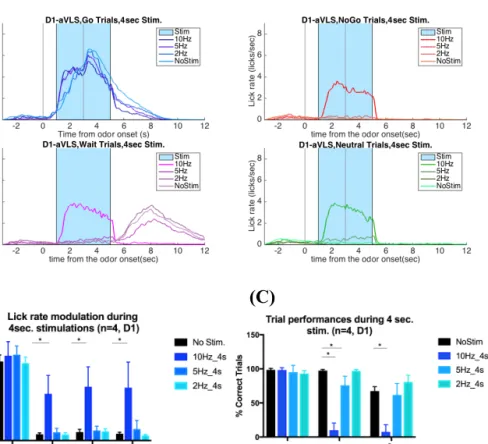
respectively. We observed that dorsomedial and dorsolateral striatum project onto different parts of target regions, GPe and SNr, in line with studies suggesting that they could be components of different basal ganglia loops. We also showed that direct pathway ventrolateral striatal cells projects on the “core” region of SNr, while indirect pathway ventrolateral striatal cells projects on ventrolateral GPe. Core region of SNr was suggested to be involved in orofacial motor control. We showed that ventrolateral striatum receives input from orofacial areas of motor cortices, and is involved in the control of orofacial movements in different conditions. We developed a head fixed-olfactory guided operant task and investigated the role of ventrolateral striatum striatonigral and striatopallidal populations in orofacial motor control. We trained mice to respond to different olfactory cues by licking to receive water reward, not licking to avoid punishment, and withholding licking to receive a delayed water reward. Striatonigral ventrolateral striatum stimulations induced licking, and suggested a context-dependent involvement in control of licking. Indirect pathway ventrolateral striatum stimulations stopped licking for all conditions. Population calcium imaging of striatonigral and striatopallidal pathway aVLS cells suggested that both pathways were active during initiation of instrumental licking, and
striatopallidal pathway was also active during different stages of instrumental licking. These results support the previous observations that activity of both pathways might be involved in initiation of
instrumental actions, and also suggest that these pathways are involved differently in different aspects of instrumental actions. In summary, in this thesis, we mapped inputs to and outputs from different striatal domains, and uncovered a striatal circuit in
ventrolateral striatum that specifically controls licking, which could serve as a novel model to accurately study the role of basal ganglia structures in action selection and performance.
RESUMO
Os gânglios da base recebem a informação sobre o estado sensitivo e motor, o estado interno e a história recente das ações e os seus resultados. São os gânglios da base que integram esta informação por forma a selecionar a ação mais apropriada num dado ambiente sensorial, permitindo ou evitando um determinado resultado com base na história recente de ações. Os mecanismos através dos quais é feita essa seleção de ação complexa são ainda desconhecidos. O estriado é a principal estrutura de entrada de informação dos gânglios da base, onde a entrada de informação cortical, talâmica e dopaminérgica é integrada. Assim, o estriado pode ser visto como a estrutura-chave para a compreensão do mecanismo através do qual os gânglios da base estão envolvidos na seleção de uma ação. Começámos por mapear os padrões de projeção das vias nigral e estriado-palidal até aos núcleos de saída. Observámos que as projeções estriado-nigrais mantiveram a sua posição medio-lateral relativa, mas inverteram sua posição dorso-ventral no SNr. Além disso, verificámos que as projeções estriado-palidais traduziram diretamente sua posição striatal no GPe.
Observámos ainda que, o estriado dorso-medial e dorso-lateral, que se sabe estarem envolvidos, respectivamente, em comportamentos dirigidos e hábitos, projetavam para diferentes partes das regiões alvo, GPe e SNr, sugerindo que estes constituíam componentes de diferentes ‘loops’ dos gânglios da base.
Mostrámos ainda que as células ventro-laterais da via direta projetam para a região central do SNr, enquanto que as células ventro-laterais indiretas projetam sobre o GPe ventrolateral.
Em seguida revelámos que o estriado ventrolateral anterior recebe entrada de áreas orofaciais dos córtices motores e está envolvido no controlo de movimentos orofaciais em ratinhos e treinados e sem serem treinados. Para investigar o papel dos neurónios estriado-nigrais e estriado-palidais do estriado ventrolateral anterior no controle orofacial desenvolvemos uma tarefa operante de cabeça fixa, dependente do olfacto. Treinámos os ratinhos para lamberem de modo a receberem uma recompensa de água, a pararem de lamber para evitarem uma punição e a deixarem de lamber para receberem uma recompensa de água mais tarde.
Estimulações da via estriado-nigral do estriado ventro-lateral anterior induziram os animais a lamberem, de forma dependente do contexto. As estimulações da via indireta levaram os animais a pararem de lamber durante todas as condições. ‘Calcium imaging’ da população de células VLS estriado-nigrais e estriado-palidais sugeriu que ambas as vias estavam ativas durante o início da lambidela, e que a via estriatopalidal também estava ativa durante a execução das lambidelas. Esses resultados corroboram as observações de que a atividade de ambas as vias é necessária para a iniciação de ações instrumentais, mas sugerem um papel diferencial para essas vias na execução de ações instrumentais.
Em resumo, nesta tese, mapeámos entradas e saídas de diferentes domínios estriatais e descobrimos um circuito estriatal no estriado ventrolateral anterior que controla especificamente os movimentos orofaciais e lambidelas tanto expontâneas como instrumentais.
ABREVIATION LIST
MSN Medium spiny neurons
aVLS Anterior ventrolateral striatum
aDLS Anterior dorsolateral striatum
aNacc Anterior Nucleus Accumbens
DMS Dorsomedial striatum
DLS Dorsolateral striatum
VLS Ventrolateral striatum
aDMS Anterior dorsomedial striatum
mDMS Mid dorsomedial striatum
pDMS Posterior dorsomedial striatum
aDLS Anaterior dorsolateral striatum
mDLS Mid dorsolateral striatum
pDLS Posterior dorsolateral striatum
aVLS Anterior ventrolateral striatum
mVLS Mid ventrolateral striatum
pVLS Posterior ventrolateral striatum
GPe Globus Pallidus external segment
aGPe Anterior Globus Pallidus external
segment
mGPe Mid Globus Pallidus external segment
pGPe Posterior Globus Pallidus external
segment
GPi Globus Pallidus internal segment
SNr Substantia Nigra Reticulata
SNc Substantia Nigra Compacta
PPN Peduncular pontine nucleus
SC Superior Colliculus
LTP Long term potentiation
LTD Long term depression
PcRT Parvicellular reticular nucleus
IRT Intermediate reticular nucleus
Gi Gigantocellular reticular nucleus
INTRODUCTION
Basal ganglia diseases such as Parkinson’s disease, Huntington’s disease, Hemibalism, obsessive-compulsive spectrum disorders and many others, disrupt one’s ability to transform decision to actions. In Parkinson’s, Huntington’s and Hemibalism this disruption appears in the form of difficulties in controlling unwanted movements. Patients with obsessive-compulsive spectrum disorders on the other hand, seem to overly perform actions independent of their consequences. Therefore one’s ability to evaluate consequences of actions seems to be disrupted. Therefore basal ganglia are thought to be the set of subcortical structures that together are the key involved in the transformation of decisions to actions (Smith et al., 2014). In everyday life, we either perform actions that are exploratory (spontaneous), or actions that we do in order to receive or avoid their expected outcomes. We repeat some of these actions so many times that their outcomes become predictable and we reduce attention to their execution. However, some actions that we repeat less frequently require more attention, show variability in their execution, and they can be disrupted easily by unexpected sensory events. The basal ganglia are a set of subcortical nuclei that are thought to be composed of the critical circuits involved in action selection, action-outcome associations, and stimulus response associations.
The basal ganglia receive information about sensory-motor state, internal state, recent history of actions and their outcomes. They integrate this information in order to select the next action in the right sensory environment to receive or avoid the predicted outcome, based
on the recent history. The mechanisms via which such complex action selection might be implemented are not clear.
The striatum is the largest basal ganglia nucleus that receives input from most of the cortex, many different thalamic nuclei, amygdala, hippocampus, dorsal raphe and peduncular pontine nucleus (Graybiel, 1998, Silberberg et al., 2015). The main modulator of striatal activity is dopamine (Surmeier et al., 2007, Gerfen et al., 2011). The sources of the striatal dopamine are SNc and VTA dopaminergic cells projecting onto striatum, spanning the whole structure (Gerfen et al., 2011). Around 90% of the cells in striatum are spiny GABAergic projection neurons that express either D1 or D2 dopamine receptors (and rarely both receptors) (Gerfen et al., 1990, Gerfen, 1992, Gerfen et al., 2011). Although cells of these two populations are similar in soma size and spacial distribution in striatum, they express different dopamine receptors and project onto different structures (Gerfen, 1992, Silberberg et al., 2015). D1 dopamine receptor expressing cells project to internal segment of Globus Pallidus (GPi) and Substantia Nigra (SN) and are therefore called striatonigral pathway. D2 dopamine receptor expressing cells, project to GPe and are called striatopallidal pathway (Gerfen et al., 1990, Gerfen, 1992). D1 receptor depolarizes and D2 receptor hyperpolarizes the MSN’s in response to dopamine agonist binding (Gerfen et al., 1990). Although both MSN types show LTP and LTD, D1 receptor activation
promotes expression of LTP and D2 receptor activation promotes expression of LDT (Shen, et al., 2008).
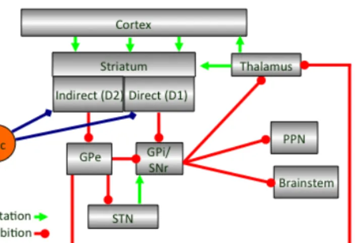
Figure 1.1| Main input and output connections of basal ganglia structures.
Disinhibition within basal ganglia
Disinhibition is thought to be the main mechanism via which basal ganglia allows movement to be executed. SNr activity was shown to decrease during movement (Chevalier et al., 1985, Deniau et al., 1985, Chevalier et al., 1990). Striatonigral cells project onto SNr directly, inhibiting it, while striatopallidal cells disinhibit SNr (Chevalier et al., 1985, Deniau et al., 1985, Chevalier et al., 1990). Striatopallidal cells project onto GPe, which projects onto either SNr or STN, and both of these structures disinhibit SNr in response to striatopallidal pathway activation (Chevalier et al., 1985, Deniau et al., 1985, Chevalier et al., 1990, Gerfen et al., 1990). Striatonigral and striatopallidal pathways are thought to work antagonistically to
control movement; the striatonigral pathway facilitates movement and the striatopallidal pathway suppresses it (Albin, et al., 1989,
Disinhibition of SNr is thought to increase the inhibition onto premotor regions, increasing the threshold for movement, while decrease of disinhibition on SNr would have the opposite effect and reduce the threshold for movement (Chevalier et al., 1985, Deniau et al., 1985, Albin et al., 1989, Chevalier et al., 1990). While the main effect of striatal stimulation on SNr was suggested to be inhibitory, 20-25 % of SNr cells projecting to SC and thalamus showed excitation (Chevalier et al., 1985, Deniau et al., 1985). Therefore, when animals are immobile, striatum is silent and STN might be keeping SNr under high level of inhibition, and when animals are moving striatal direct and indirect pathways modulate SNr activity to allow movement to be performed (Chevalier et al., 1985, Deniau et al., 1985).
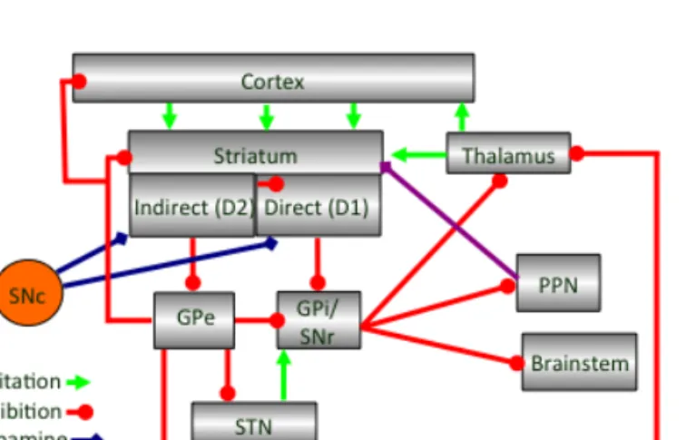
However, the number of circuits involved in disinhibition of SNr to allow movement to be performed are much complex than simple striatonigral pathway mediated inhibition, and striatopallidal mediated disinhibition model. Multiple new functional connections were described between basal ganglia nuclei that contribute to either inhibition or disinhibition of SNr; intrastriatal connectivity mostly from striatopallidal onto striatonigral cells (Tecuapetla et al, 2009), intrastriatal inhibitory neurons inhibiting striatopallidal and
striatonigral cells (reviewed in Smith et al., 1998, Kreitzer, 2009), arkypallidal cells projecting from GPe back to striatum inhibiting it (Mallet et al., 2012, Mallet et al., 2016), GABAergic GPe projections directly to cortex (Saunders et al., 2015), and cholinergic Projections to striatum (Dautan et al., 2014). Therefore, intra-basal ganglia circuits seems to be more complex than initially proposed, with
implications for new roles of direct and indirect pathway in movement and action selection.
Still, models of inhibition and disinhibition have been useful to understand the basal ganglia disorders, where imbalance between direct and indirect pathways may play a role (Albin, et al., 1989, Alexander, et al., 1990). In Parkinson’s disease loss of dopamine cells, was reported to cause spine loss and increased firing rates in high affinity D2 dopamine receptor expressing population (Day et al., 2006, Mallet et al., 2006). In early and middle stages of Huntington’s disease degeneration of striatopallidal population was more
prominent than degeneration of striatonigral population (Reiner et al., 1988). It is possible that striatonigral and striatopallidal pathway cells are affected differently by perturbations in disease states.
Figure 1.2| Updated scheme of main input and output connections of basal ganglia.
D2 receptor expressing striatopallidal population has higher affinity for dopamine, is more excitable than striatonigral population and could inhibit striatonigral activity via intra-striatal lateral inhibition (Tecuapetla et al, 2009), projects back to striatum (Mallet et al., 2012), frontal cortex (Saunders et al., 2015), and thalamus which projects back to striatum or cortex (Gerfen et al., 1990, Mastro et al. 2014, Gittis et al., 2014). On the other hand striatonigral cells project directly onto SNr to inhibit it and allow movement (Gerfen et al., 1990, Chevalier et al., 1990). Considering the complexity of the circuitry and the divergence of indirect pathway output it is possible that indirect pathways might be involved in multiple different aspects of actions selection showing richer functional heterogeneity
compared to direct pathway (Tecuapetla, et al., 2016). Recent optogenetic manipulations suggested that striatonigral pathway activation promoted locomotion while striatopallidal pathway activation stopped it (Kravitz et al., 2010). However, population calcium imaging and recordings from optogeneticly identified striatonigral and striatopallidal populations suggested that both populations were simultaneously active preceding initiation of an instrumental action (Jin et al., 2010, Cui, et al, 2013, Jin, et al., 2014). Both striatonigral and striatopallidal populations were also simultaneously active preceding initiation of spontaneous locomotion (not associated with a particular outcome) (Tecuapetla et al., 2014). However, the cells of both pathways that showed activity preceding initiation were suggested to be sub-populations and both pathways showed heterogeneous activity during performance of an instrumental action (Jin et al., 2014). Subpopulations from both pathways showed
execution related inhibited, sustained (more striatonigral cells showed sustained activity and more striatopallidal cells showed cells of inhibited activity) or stop related activity (Jin et al., 2014). Following these findings, optogenetic manipulations of both pathway
populations before initiation and during execution showed that balanced activity of both pathways were necessary for proper initiation and execution of instrumental actions and striatonigral pathway might be facilitating action initiation and performance and indirect pathway might be inhibiting competing actions (Mink, 1996, Hikosaka et al., 2000, Tecuapetla et al., 2016).
Corticostriatal projection patterns in striatum
Similar to primates, rodent motor cortical projections onto striatum showed somatotopic organization (Nambu et al., 2011, Ebrahimi et al., 1992, Hintiryan et al., 2016). Corticostriatal input in both primates and rodents was shown to make specific patterns. Limbic cortex and amygdala input was constrained into immunohistochemically identifiable patchy regions called “patches” (Goldman-Rakic, 1982, Gerfen et al., 1987). MSNs into patches projected to dopamine cells (Goldman-Rakic, 1982, Gerfen et al., 1987). Sensory-motor cortex input was occupying the regions around the patches, called “matrix”, and MSNs in the matrix region projected to SNr (Goldman-Rakic, 1982, Gerfen et al., 1987). In primates sensory-motor cortical input to striatum, representing the same body part, diverges into partially connected “set of zones” called matrisomes which then project onto a small group of spatially constrained neurons on GPe (Graybiel et al., 1994, Flaherty et al., 1991). Matrisomes, subregions of matrix that receive similar sensory-motor input, might be multiple regions of
integration of information related to the specific body part (Graybiel et al., 1994, Flaherty et al., 1991). In rodents, divergence and
convergence of cortical input onto striatum have been addressed (Mailly et al., 2013, Hintiryan et al., 2016, Heilbronner et al., 2016). Even though, projections from mouse cortex onto striatum has been described in detail and showed somatotopic organization, and
different levels of convergence and divergence, it has not been shown matrisome-like patterns and if “divergence-reconvergence” of
specific sensory-motor input exist rodents (Graybiel et al., 1994, Flaherty et al., 1991, Hintiryan et al., 2016, Heilbronner et al., 2016). Therefore, in rodents, less is known about how striatum processes cortical information to allow action selection.
In chapter 3, we labeled primary and secondary orofacial motor cortex and showed that their input was restricted to ventrolateral striatum. Primary and sensory motor cortex projected onto the same striatal region with primary orofacial motor cortex projecting more laterally and secondary orofacial motor cortex projecting more
medially. Forelimb region of secondary motor cortex projected dorsal to secondary orofacial motor cortex input. All these projection
patterns were similar to observations reported for primate putamen (Nambu, 2011).
Striatal activity during spontaneous sensory-motor events (that are not associated with particular outcomes)
In striatum, neurons were shown to respond to sensory stimuli of both single modality and of multimodal nature (Brown et al., 1996, Nagy et al., 2005, Nagy et al., 2006, Schultz et al., 2009). Visual, auditory and somatosensory receptive fields appear to be extremely large and
they did not show somatotopic organization in striatum (Brown et al., 1996, Nagy et al., 2005, Nagy et al., 2006, Schultz et al., 2009). The sites of sensory integration in striatum of rodents have not been studied as much as the motor outputs of the striatal populations (Reig, et al., 2014). However, it appears that somoatotopic organization model might not explain the integration of sensory information in striatum. Even though sensory feedback is required for proper execution of movement, somatotopic organization model seems to partially explain the motor output of striatum, but sensory integration in striatum might be happening via different mechanisms (Reig, et al., 2014).
Electrophysiological recordings from primate putamen showed increase in activity related to movement of tongue, arm and leg (DeLong, 1972). In mouse striatum, both striatonigral and
striatopallidal pathway cells showed increase in activity preceding angular velocity of contralateral turns (Tecuapetla et al., 2014). Strong turn related activity in dorsolateral striatum was also observed during early stages of training in a T-maze task (Jog et al., 1999). Single unit recordings in dorsolateral striatum of mice showed correlations with movement of specific body parts such that more cells in dorsal part of dorsolateral striatum fired during forelimb, hind limb, trunk and whisker movements and more cells in ventrolateral striatum fired during orofacial movements such as, licking, tongue reaching and jaw movements (Carelli, et al., 1991, Mittler, et al., 1994). This somatotopy observed in mouse striatum was also observed in primate putamen (Nambu, 2011).
In adult mice, “natural actions” such as grooming, locomotion and consummatory-orofacial movements are some of the movements that require little experience for their accurate execution (Colonnese et al., 1996, Aldridge, et al., 1998, Jin et a., 2010). During postnatal
development, or even during embryonic development, these action sequences are performed frequently and crystalized into “neutral actions” with specific “action syntaxes” that show little variability in their performances (Lashley, 1951, Colonnese et al., 1996, Aldridge, et al., 1998). In rats, even though some phases of the grooming syntactic chain are observed as early as E20-E21, the stereotyped “grooming action syntax” is only observed on the second-third postnatal week (Colonnese et al., 1996, Berridge et al., 1992). This grooming action syntax development coincided with the
developmental window for striatal maturation, and striatal (DLS) lesions caused chronic deficits in the grooming action syntax (Fentress, 1992, Berridge et al., 1992). These observations suggest that striatum might be necessary to create “action syntax”, i.e. the serial order of action (Lashley, 1951, Berridge et al., 1992). The same group also showed that SNr cells responded with higher rates to the same grooming phases depending on if they were performed within the grooming synthetic chain or independent of the chain (Meyer-Luehmann, et al., 2002).
In rats, adult pattern of locomotion was also observed at the end of second postnatal week (Vinay et al., 2002). Even though locomotion is a much simpler action sequence compared to grooming, activity of basal ganglia structures preceded both actions sequences (Meyer-Luehmann, et al., 2002, Tecuapetla et al., 2014). SNr cells started
increasing their firing rate before grooming syntax chain initiation and they slowly decay during chain (Meyer-Luehmann, et al., 2002). Striatal cells of both pathways also increased their activity before initiation of contralateral turning and decayed fastly (Cui et al., 2013, Tecuapetla et al., 2014). Therefore, it is possible that these “neutral action-sequences” are encoded differently in basal ganglia compared to novel action sequences (Jin, et al., 2010).
However, it is also possible that movement related responses at different stages of instrumental conditioning are encoded differently in different parts of the striatum. It was previously suggested that, in humans, in early stages of instrumental conditioning anterior striatum showed correlations with movement and in late stages posterior striatum showed movement related activity (Jueptner, et al., 1997, Graybiel, 1998). Similar shift of activity during acquisition and consolidation of an instrumental action were observed between dorsomedial (associative) and dorsolateral (sensory-motor) striatum (Miyachi, et al., 1997, Miyachi, et al., 2002, Yin et al., 2009, Jin et al., 2010).
Single unit recordings from striatum showed that striatal cells increased firing during different spontaneous behaviors (motor events) with some units start increasing firing before the event, and some decay slower and some faster (Carelli, et al., 1991, Mittler, et al., 1994, Venkatraman, et al., 2010). In addition to limb movement related increase in firing rate in striatum, increase in mean firing rate during spontaneous active state (locomotion) compared to quiescent phase, in all basal ganglia structures (striatum, GPe, SNr and STN)
was observed in rats during spontaneous movements (Shi, et al., 2004)
The studies discussed above suggest that increases in striatal activity of both pathways before spontaneous and instrumental actions might be necessary for their initiation. Tecuapetla et al., 2016 also suggested that balanced activity of both pathways is necessary for their proper initiation and execution. However, the role of striatonigral and striatopallidal populations in initiation and execution of instrumental actions is still not clear.
Most of these studies were performed in DLS. However, one of the challenges in interpretation of DLS activity comes from its
heterogeneous input from motor cortex, therefore heterogeneous motor functions. DLS activity has been implicated in the control of locomotion and different limb movements (Carelli, et al., 1991, Mittler, et al., 1994, Venkatraman, et al., 2010, Hintiryan et al., 2016).
The ventrolateral striatum has been suggested to be involved in orofacial motor control and receive input mostly from orofacial motor cortex (von Krosigk, et. al., 1992, Mittler, et al., 1994, Hintiryan et al., 2016). Therefore it suggests a less heterogeneous motor function compared to DLS, and a novel model for understanding the role of striatonigral and striatopallidal pathways in initiation and execution of instrumental actions, action selection.
Specific movements such as saccades, mastication, vocalization, swallowing, and locomotion are thought to be generated by specific neural networks in brainstem and spinal cord, called central rhythm
generators (CPG) (Hikosaka et al., 1983, Scott et al., 2003, Dusterhoft et al., 2000, Grillner et al., 2003, Amirali et al., 2001). It was
previously suggested that basal ganglia might be involved in action selection via two (path)ways; modulating thalamocortcial networks therefore modulating its own functioning, and modulating brainstem motor networks therefore modulating motor output directly (Hikosaka et al., 2000).
Coordinated orofacial movements were suggested to be controlled via specific brainstem circuits (CPG’s) such as PcRT, IRt, Gi (Travers, et al., 1997, von Krosigk, et. al., 1992, Stanek, et al., 2014). Therefore, studying the role of ventrolateral striatum in orofacial motor control could also help us understand the circuit mechanisms via which basal ganglia acts on specific CPG controlled actions to allow proper action selection and motor control.
REFERENCES
Albin, R. L., Young, A. B. & Penney, J. B. (1989) The functional anatomy of basal ganglia disorders. Trends Neurosci. 12, 366–375
Aldridge, J.W., Berridge, K.C.(1998) Coding of serial order by neostriatal neurons: A “Neutral action” approach to movement sequence. J. Neurosci. 18(7):2777-2787
Alexander, G.E., Crutcher, M.D. (1990) Functional architecture of basal ganglia circuits: neural substrates of parallel processing. Trends Neurosci. 13: 266-271
Amirali, A., Tsai, G., Schrader, N., Weisz, D., Sanders, I. (2001) Mapping of brainstem neuronal circuitry active during swallowing. Ann. Otol. Rhinol. Laryngol. 110:502-513
Bergman, H., Graybiel, A.M., Kimura, M., Plenz, D., Seung, H.S., Surmier, D.J., Wickens, J.R. (2004) Microcircuits: The interface between neurons and global brain function, Report of the 93rd Dahlem Workshop, Berlin, April 25-30 2004: 165-190
Berridge, K.C., Whishaw, I.Q. (1992) Cortex, striatum and cerebellum: Control of a serial order in a grooming sequence. Exp Brain Res 90: 275-290
Bolam, J.P., Smith, Y. (1992) The striatum and the globus pallidus send convergent synaptic inputs onto single cells in the entopeduncular nucleus of the rat: a double anterograde labeling study combined with
postembedding immunocytochemistry for GABA. J. Comp. Neurol. 321: 456-476
Brown, E.E., Hand, P.J., Divac, I. (1996) Representation of a single vibrissa in the rat neostriatum: peaks of energy metabolism reveal a distributed functional module. Neuroscience 75: 717-728
Carelli, R., M., West, M., O. (1991) Representation of the body by single neurons in the dorsolateral striatum of the awake, unrestrained rat. J. Comp. Neurology 309:231-249
Chevalier, G., Vacher, S., Deniau, J.M., Desban, M. (1985) Disinhibition as a basic process in the expression of striatal functions. I. The striato-nigral influence on tecto-spinal/tecto-diencephalic neurons. Brain Res. 334(2):215-26
Chevalier, G., Deniau, J., M. (1990) Disinhibition as a basic process in the expression of striatal functions. TINS, 13(7): 277-280
Colonnese, M., Stallman, E. L., Berridge, K. C. (1996) Ontogeny of action syntax in altricial and precocial rodents: Grooming sequences of rat and guinea pig pups. Behavior, 133:1165-1196
Cui, G., Jun, S. B., Jin, X., Pham, M.D., Vogel, S.S., Lovinger, D.M., Costa, R.M. (2013) Concurrent activation of striatal direct and indirect pathways during action initiation. Nature 494(7436): 238-42.
Day, M., Wang, Z., Ding, J., An, X., Ingham, C., A., … Arbuthnott, G., W., & Surmeier, D., J. (2006) Selective elimination of glutamatergic synapses on striatopallidal neurons in Parkinson’s disease models. Nat. Neurosci. 9:251-259
Dautan, D., Huerta-Ocampo, I., Witten, I.B., Deisseroth, K., Bolam, J.P., Gerdjikov, T., Mena-Segovia, J. (2014) A major external source of
cholinergic innervation of the striatum and nucleus accumbens originates in the brainstem. J.Neurosci. 34(13): 4509-18
DeLong, M.R. (1978) Activity of basal ganglia neurons during movement. Brain Research 40: 127-135
Deniau, J., M., Chevalier, G. (1985) Disinhibition as a basic process in the expression of striatal functions. . II. The striato-nigral influence on
thalamocortical cells of the ventromedial thalamic nucleus. Brain Res. 334(2):227-33
Dusterhoft, F., Hausler, U., Jurgens, U. (2000) On the speech for the vocal pattern generator. A single unit recording study. Neuroreport 11:231-234 Ebrahimi, A., Pochet, R., Roger, M. (1992) Topographical organization of the projections from physiologically identified areas of the motor cortex to the striatum in the rat. Neuroscience Research 14: 39-60
Fentress, J.C. (1992) Emergence of pattern in the development of mammalian movement sequences. J. Neurobiol. 23(10): 1529-56
Flaherty, A.W., and Graybiel, A.M. (1991). Corticostriatal transformations in the primate somatosensory system. Projections from physiologically mapped body-part representations. J. Neurophysiol. 66, 1249–1263. Gerfen , C.R. (1985) Neostriatal mosaic: I. Compartmental organization of projections from the striatum to the substantia nigra in the rat. J. Comp. Neurol. 236(4): 454-76
Gerfen , C.R., Herkenham, M., Thibault, J. (1987) Neostriatal mosaic: II. Patch- and matrix-directed mesostriatal dopaminergic and
non-dopaminergic systems. J. Neurosci. 7(12): 3915-3934
Gerfen, C., R., Engber, T., M., Mahan, L., C., Susel, Z., Chase, T., N., Monsma, F., J., Sibley, D., R. (1990) D1 and D2 dopamine receptor regulated gene expression of striatonigral and striatopallidal neurons. Science 250 (4986):1429-1432
Gerfen, C. R. (1992) Neostriatal mosaic: multiple levels of compartmental organization. TINS 15(4): 133-139
Gerfen, C. R., Surmier, D. J., (2011), Modulation of striatal projection system by dopamine. Annu Rev Neurosci (34): 441-466
Gittis, A.,H. , Berke, J.D., Bevan, M.D., Chan, C.S., Mallet, N., Morrow, M.M., Schmidt, R. (2014) New roles of the external globus pallidus in basal ganglia circuit and behavior. J. Neurosci. 34 (46): 15178-15183
Goldman-Rakic, P.S. (1982) Cytoarchitectonic heterogeneity of the primate neostriatum: Subdivisions into island and matrix cellular components. J. Comp. Neurol. 205: 398-413
Graybiel, A.M., Aosaki, T., Flaherty, A. W., Kimura, M. (1994) Basal ganglia and adaptive motor control. Science, 265:1826-1831
Graybiel, A. M. (1998) The basal ganglia and chunking of action repertoires. Neurobiol. Learn. Mem. 70(1/2): 119-136
Grillner, S. (2003) The motor infrastructure: from ion channels to neural networks. Nat. Rev. Neurosci. 4: 573-586
Helibronner, S.R., Rodrigues-Romaguera, J., Quirk, G.J., Groenewegen, H.J., Heber, S.N. (2016) circuit based corticostriatal homologies between rat and primate. Biol. Psychiatry 80(7): 509-521
Hikosaka, O., Wurtz, R.H. (1983) Visual and occulomotor functions of monkey substantia nigra pars reticulate. Vol.II. Visual response related to fixation of gaze. J. Neurophysiol. 49: 1254-1267
Hikosaka, O., Takikawa, Y., Kawagoe, R. (2000) The role of basal ganglia in the control of purposive saccadic eye movements. Physiol. Rev. 80:953-978
Hintiryan, H., Foster, N.F., Bowman, I., Bay, M., Song, M.Y., Guo, L., Yamashita, S., Bienkowski, M. S., Zingg, B., Zhu, M., Yang, X.W., Shih, J.C., Toga, A.W., Dong, H.W. (2016) The mouse cortico-striatal
projectome. Nat. Neurosci. 19(8): 1100-14
Jueptner, M., Stephan, K.M., Frith, C.D., Brooks, D.J., Frackowiak, R.S.J., Passingham, R.E. (1997a) Anatomy of motor learning. 1.Frontal cortex and attention to action. J. Neurophys. 77(3): 1313-24
Jin, X., Costa, R.M. (2010) Start/stop signal emerge in nigrostriatal circuit during sequence learning. Nature 466(7305): 457-462
Jin, X., Tecuapetla, F., Costa, R.M. (2014) Basal ganglia subcircuits
distinctively encode the parsing and concatenation of action sequences. Nat. Neurosci. 17(3): 423-30
Jog, M.S., Kubota, Y., Connolly, C.I., Hillegaart, V., Graybiel, A.M. (1999) Building neural representations of habits. Science 286(5445): 1745-9 Kravitz, A.V., Freeze, B.S., Parker, P.R., Kay, K., Thwin, M.T., Deisseroth, K., Kreitzer, A.C. (2010) Regulation of parkinsonian motor behaviors by optogenetic control of basal ganglia circuitry. Nature 466(7306): 622-6 Kreitzer, A. (2009) Physiology and pharmacology of striatal neurons. Annu.Rev. Neurosci. 2009. 32:127-47
Lashley, K.S. (1951) The problem of serial order in behavior. In Jeffress, L.A. (ed.), Cerebral Mechanisms in Behavior. Wiley, New York, pp. 112-146
Mailly, P., Aliane, V., Groenewegen, H.J., Heber, S.N., Deniau, J.M. (2013) The rat prefrontostriatal system analyzed in 3D: Evidence for multiple interacting functional units. J. Neurosci. 33(13):5718-27
Mallet, N., Ballion, B., Le Moine, C., Gonon, F. (2006) Cortical input and GABA interneurons imbalance projection neurons in the striatum of Parkinsonian rats. Journal of Neurosicence 26(14): 3875-3884
Mallet, N., Micklem, B. R., Henny, P., Brown M.T., Williams, C., Bolam, J.P., Nakamura, K.J., Magill, P.J. (2012) Dichotomous organization of the external globus pallidus. Neuron 74(6): 1075-86
Mallet, N., Schmidt, R., Leventhal, D., Chen, F., Amer, N., Boraud, T., Berke, J.D. (2016) Arkypallidal cells send a stop signal to striatum. Neuron 89(2): 308-16
Meyer-Luehmann, M., Thonbson, J., Berridge, K.C., Aldridge, J.W. (2002) Substantia nigra pars reticulata neurons code initiation of a serial pattern: implications for natural action sequences and sequential disorders. Eur. Journ. Neurosci. 16: 1599-1608
Miyachi, S., Hikosaka, O., Miyashita, K., Karadi, Z., Rand, M.K. (1997) Differential roles of monkey striatum in learning of sequential hand movements. Exp. Brain Res. 115: 1-5
Miyachi, S., Hikosaka, O., Lu, X. (2002) Differential activation of monkey striatal neurons in the early and late stages of procedural learning. Exp. Brain Res. 146: 122-126
Mink, J.W. (1996) The basal ganglia: Focused selection and inhibition of competing motor programs. Prog. Neurobiol. 50: 381:425DeLong, M.R. (1990) Primate models of movement disorders of basal ganglia origin. Trends Neurosci. 13:281-285
Mittler, T., Cho, J., Peoples, L., L., West, M., O. (1994) Representation of the body in the lateral striatum of freely moving rats: single neurons related to licking. Exp Brain Res 98: 163-167
Nambu, A. (2011) Somatotopic organization of primate basal ganglia. Front Neuroanat. 5:26
Nagy, A., Paroczy, Z., Norita, M., Benedek, G. (2005). Multisensory responses and receptive field properties of neurons in the substantia nigra and in the caudate nucleus. Eur. J. Neurosci. 22, 419–424.
Nagy, A., Eordegh, G., Paroczy, Z., Markus, Z., Benedek, G. (2006). Multisensory integration in the basal ganglia. Eur. J. Neurosci. 24, 917–924. Reig, R., Silberberg, G. (2014) Multisensory integration in mouse striatum. Neuron 83: 1200-1212
Reiner, A., Albin, R.,L., Anderson, K.,D., D’Amato, C., D., Penney J., B., & Young, A., B. (1988) Differential loss of striatal projection neurons in Huntington disease. Proc Natl Acad Sci USA 85(15): 5733-5737
Saunders, A., Oldenburg, I.A., Berezovski, V.K., Johnson, C.A., Kingery, N.D., Elliot, H.L., Xie, T., Gerfen, C.R., Sabatini, B.L. (2015) A direct GABAergic input from the basal ganglia to frontal cortex. Nature 521, 85-89
Schultz, J.M., Redgrave, P., Mehring, C., Aertsen, A., Clements, K.M., Wickens, J.R., Reynolds, J.N. (2009) Short latency activation of striatal spiny neurons via subcortical visual pathways. J. Neurosci. 29: 633-6347 Scott, G., Westberg, K.G., Vrentzos, N., Kolta, A., Lund, J.P. (2003) Effects of lidocaine and NMDA injections into the medial pontobulbar reticular formation on mastication evoked by cortical stimulation in anesthetized rabbits. Eur. J. Neurosci. 17:2156-2162
Shen, W., Flajolet, M., Greengard, P., Surmeier, D.J., (2008) Dichotomous dopaminergic control of striatal synaptic plasticity. Science 321:848-851 Shi, L.H., Luo, F., Woodward, D.J., Chang, J.Y. (2004) Neural responses in multiple basal ganglia regions during spontaneous and treadmill locomotion task in rat. Exp. Brain. Res. 157: 303-314
Silberberg, G., Bolam, J. P. (2015) Local afferent synaptic pathways in the striatal microcircuitry. Curr. Op. Neur. 33: 182-187
Smith, Y., Bolam, J.P., (1989) Neurons of the substantia nigra reticulata receives a dense GABA containing input from the globus pallidus in the rat. Brain Research, 493:160-167
Smith, Y., Bevan, M.D., Shink, E., Bolam, J.P. (1998) Microcircuitry of the direct and indirect pathways of the basal ganglia. Neuroscience 86(2): 353-387
Stanek, E., Cheng, S., Takatoh, J., Han, B. X., Wang, F. (2014) Monosynaptic premotor circuit tracing reveals neural substates for oro-motor coordination. Elife 2014; 3:e02511
Surmeier, D.J., Ding, J., Day, M., Wang, Z., Shen, W. (2007) D1 and D2 dopamine receptor modulation of striatal glutamatergic signaling in striatal medium spiny neurons. TINS 30(5): 228-235
Tecuapetla, F., Koos, T., Tepper, J.M., Kabbani, N., Yeckel, M.F. (2009) Differential dopaminergic modulation of neostriatal synaptic connections of striatopallidal axon collaterals. J.Neurosci. 29: 8977-8990
Tecuapetla, F., Matias, S., Dugue, G. P., Mainen, Z.F., Costa, R.M. (2014) Balanced activity in basal ganglia projection pathways is critical for contraversive movements. Nat. Commun. Jul 8; 5:4315.
Tecuapetla, F., Jin, X., Lima, S.Q., Costa, R.M. (2016) Complementary contributions of striatal projection pathways to action initiation and execution. Cell 166(3): 703-15
Travers, J.B., Dinardo, L.A., Karimnamazi, H. (1997) Motor and premotor mechanisms of licking. Neurosci. Biobehav. Rev. 21(5): 631-47
Venkatraman, S., Jin, X., Costa, R.M., Carmena, J.M. (2010) Investigating neural correlates of freely behaving rodents using inertial sensors. J. Neurophys. 104(1): 569-575.
Vinay, L., Brocard, F., Clarac, F., Norreel, J.C., Pearlstein, E., Pflieger, J.F. (2002) Development of posture and locomotion: An interplay of
endogenously generated activities and neurotropic actions by descending pathways. Brain Res Brain Res Rev 40 (1-3): 118-129
von Krosigk, M., Smith, Y., Bolam, J. P.& Smith, A. D. (1992) Synaptic organization of gabaergic inputs from the striatum and the globus pallidus onto neurons in the substantia nigra and retrorubral field which project to the medullary reticular formation. Neuroscience 50(3): 531-549
Yin, H.H., Knowlton, B.J. (2004) Contributions of striatal subregions to place and response learning. Learn. Mem. 11(4): 459-462
Yin, H.H., Mulcare, S.P., Hilaro, M.R., Clouse, E., Holloway, T., Davis, M.I., Hansson, A.C., Lovinger, D.M., Costa, D.M. (2009) Dynamic
reorganization of striatal circuits during the acquisition and consolidation of a skill. Nat. Neurosci. 12(3): 333-41
CHAPTER 2| DIFFERENT STRIATAL
DOMAINS PROJECT ONTO SPECIFIC
DIFFERENT STRIATAL DOMAINS PROJECT ONTO SPECIFIC AREAS OF THE DOWNSTREAM TARGETS SUMMARY
Striatonigral and striatopallidal pathway cells comprise >90% of striatal cells. They express different dopamine receptors and project onto different target structures. Striatonigral pathway cells project onto SNr while striatopallidal pathway cells project onto GPe. It has been shown that different striatal populations project onto different parts of SNr and GPe. However, a systematic study showing the projection patterns of these populations throughout the structures, and comparing projections of different, simultaneously labeled
populations was missing.
We used transgenic lines to label striatonigral and striatopallidal pathway cells specifically. We simultaneously labeled two domains of each pathway populations using two different fluorescent proteins, EYFP and tdTomato. We mapped projection patterns of 9 different striatonigral and striatopallidal populations onto SNr and GPe. We showed that DMS, DLS, VLS populations project onto different regions of SNr and GPe, creating parallel pathways. The intra-striatal position of striatopallidal pathway cells was directly translated onto GPe by their projections, on both dorsoventral and mediolateral axis. However, striatonigral pathway projections made complex patterns in SNr. They inverted their intra-striatal cell body position on
dorsoventral axis, and translated it directly on the mediolateral axis. Striatonigral pathway projections seemed to wrap around SNr making complex patterns that require 3D reconstruction for their
INTRODUCTION
Striatonigral and striatopallidal MSNs are homogeneously intermingled in the mouse striatum, except perhaps for the most posterior part of the striatum, analogous to caudate tail in primates, which appears to be populated mostly by striatonigral cells and to contain fewer striatopallidal cells (Gangarossa et al., 2013).
The distributions of corticostriatal projections from different cortical regions permitted the division of the dorsal striatum into three domains that receive functionally distinct inputs; the dorsomedial (DMS), dorsolateral (DLS) and ventrolateral striatum (VLS) (Ebrahimi et al., 1992). This classification has also been used to define developmental patterns of striatal circuits (Bayer et al., 1982). The development of striatal domains follows two gradients; from posterior to anterior and from ventrolateral to dorsomedial. According to this pattern, the ventrolateral striatal cells were born earliest,
dorsolateral striatal cells were born after and dorsomedial striatal cells were born the latest (Bayer et al., 1982).
In rodents, the VLS receives input from orofacial and head motor cortex, while the DLS receives input from forelimb, whisker and trunk motor cortex, and the most medial part of DLS receives input from hind limb and trunk motor cortex (Deniau et al., 1996, Ebrahimi et al., 1992, Hintiryan et al., 2016). Throughout the mouse striatum, only a small dorsomedial region does not receive motor input, but receives input from visual areas and more associative cortical areas (Hintiryan et al., 2016)
The combination of retrograde and anterograde labeling of different striatal populations showed that different striatal regions project onto
different parts of SNr (Gerfen et al., 1985, Deniau et al., 1992, Deniau et al., 1996). However, in these studies the regions were not divided into these three domains; DMS, DLS and VLS. In some studies, the cortical input was labeled together with their SNr output to relate the postsynaptic SNr region with the presynaptic cortical region (Deniau et al., 1996). However, to our knowledge, a detailed mesoscopic mapping of direct and indirect pathway projections onto SNr and GPe from genetically defined subpopulations of striatonigral and
striatopallidal cells is missing, in the mouse.
Based on the input maps and developmental patterns we defined DMS, DLS and VLS as functionally different dorsal striatum domains (Deniau et al., 1996, Ebrahimi, et al., 1992, Hintiryan, et al., 2016). We produced DIO-EYFP-WPRE and AAV2.2-EF1a-tdTomato-WPRE viruses to simultaneously express different
fluorescent proteins in different dorsal striatal domains. We used D1-Cre (FK150-D1-Cre) and D2-D1-Cre (Adora-D1-Cre) mouse lines to target striatonigral or stiatopallidal subpopulations. 250-300 nl of each virus was injected and 3-4 weeks expression time was allowed.
We also considered the anterior-posterior axes and therefore we mapped the output of 9 striatal domains: anterior-dorsomedial (aDMS), mid-dorsomedial (mDMS) and posterior-dorsomedial (pDMS), anterior-dorsolateral (aDLS), mid-dorsolateral (mDLS) and posterior-dorsolateral (pDLS), and anterior-ventrolateral (aVLS), mid-ventrolateral (mVLS) and posterior-ventrolateral (pVLS)
domains. We labeled two different domains simultaneously using two different fluorescent proteins, and compared their projection patterns onto target regions. The data below shows the projections of
striatonigral and striatopallidal subpopulations onto the specific regions of GPe and SNr, and will hopefully permit soon the 3D reconstruction of these pathways. Therefore, for the moment, the discussions on the projection patterns observed below are based on qualitative observations.
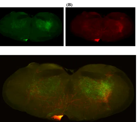
Figure 2.1 | Diagram showing three
domains of dorsal striatum; dorsomedial striatum (DMS), dorsolateral striatum (DLS), ventrolateral striatum (VLS). (The arrow indicates their developmental order.)
RESULTS
We produced viruses, with the same promoters, and that only differ in the fluorescent proteins they express. Therefore, we cloned tdTomato fluorescent protein into a pAAV-EF1a-DIO-EYFP-WPRE construct replacing EYFP, and used AAV2.2-EF1a-DIO-EYFP-WPRE and pAAV-EF1a-DIO-tdTomato-WPRE for Cre dependent expression of EYFP and tdTomato in different striatal domains. We used D1-Cre (FK150-Cre) and D2-Cre (Adora2a-Cre) transgenic mouse lines. We injected 250-300nl virus in each domain and waited for 3-4 weeks expression time.
We optimized coordinates for each transgenic line separately. Nine different domains were labeled: aDMS, mDMS, pDMS, aDLS, mDLS, pDLS, aVLS, mVLS, and pVLS. Sequential images of whole brain slices were acquired at 10X magnification.
Projections of striatonigral and striatopallidal neurons from different domains targeted different downstream areas
Striatonigral projections of different striatal domains seemed to have different patterns of projection onto SNr. Relative mediolateral position of cell bodies in striatum seemed to be conserved by their projections onto the target structures, i.e., medial striatal domains targeted medial parts of SNr while lateral striatal domains projected onto the lateral parts of SNr. However, on the dorsoventral axis, striatal position was inverted by their projections, i.e., more dorsal striatal populations projected more ventrally and ventral striatal populations projected more dorsally onto SNr. Striatonigral
projections kept their relative mediolateral position and never crossed each other to reach their target region in SNr. Therefore, they made parallel pathways projecting onto SNr.
Unlike striatonigral cells, striatopallidal populations projected onto GPe directly translating their striatal cell body position, on both mediolateral and dorsoventral axis. Striatopallidal populations also kept their mediolateral relative position on GPe and never crossed each other to reach their target region on GPe. Therefore,
striatopallidal pathway projections also made parallel pathways projecting onto GPe.
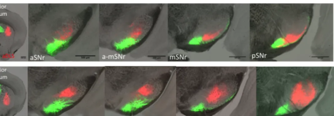
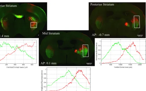
Striatonigral projections on aSNr and pSNr showed differences
aDMS, aDLS, mDMS and mDLS striatonigral projections targeted larger, relatively dorsal regions of SNr, closer to SNc. However, on posterior-SNr (pSNr) they stayed on the most ventrolateral site, away from SNc. Interestingly, aVLS and mVLS projections targeted dorsolateral sites on anterior-SNr (aSNr) and kept targeting dorsolateral sites of pSNr, but they increased the targeted area on pSNr compared to aSNr. pDMS, pDLS, and pVLS striatonigral domains projected on small regions and did not seem to change the size of their target area throughout SNr. Therefore, it is likely that on aSNr all aDMS, aDLS, mDMS, mDLS, aVLS and mVLS might be occupying similar regions. However, on pSNr, aVLS and mVLS projections seemed to be occupying impressively larger areas compared to aDMS, aDLS, mDMS and mDLS projections.
Example images from injection sites, from anterior-SNr (aSNr), mid-SNr (mmid-SNr), and posterior-mid-SNr (pmid-SNr) are presented in Figure 2.1, Figure 2.2, and Figure 2.3. For each image, we separated two fluorescent protein channels and used Otsu’s method to convert images to binary (Otsu, 1979). We took each binary channel of the same image as vectors and calculated their correlation coefficient. We calculated the level of spatial overlap, independent of the intensity of the signal of two labeled populations using Pearson’s correlation coefficient. We used Pearson’s correlation coefficient as a measure of spatial overlap of projections.
Example images in Figure 2.1, Figure 2.2 and Figure 2.3 suggested that projections from different striatonigral domains might show different levels of overlap at different anterior posterior levels of SNr.
Figure 2.2 | Example images of striatonigral pathway aDLS and
aDMS-aVLS double labeled populations showed different projection patterns onto SNr. Non-overlapping aDMS-aDLS populations partially overlapped on aSNr and mSNr but not on pSNr (aDMS-aDLS: r_striatum=-0.01, r_aSNr= 0.1, r_mSNr= 0.2, r_pSNr= -0.006, Pearson’s correlation coefficient, n=2). Non-overlapping aDMS-aVLS populations targeted non-overlapping SNr regions (aDMS-aDMS-aVLS:
r_striatum=-0.01, r_aSNr= -0.02, r_mSNr= -0.02, r_pSNr= -0.02, Pearson’s correlation coefficient, n=2).
Figure 2.3 | Example images of direct pathway mDMS-mDLS and mDMS-mVLS
populations showed different projection patterns on SNr. Targeted area of projections was different on the aSNr, mSNr and pSNr. Non-overlapping mDMS-mDLS and mDMS-mVLS populations targeted non-overlapping SNr regions (mDMS-mDLS: r_striatum=-0.01, r_aSNr= -0.009, r_mSNr= -0.009, r_pSNr= 0.03,
mDMS-mVLS: r_striatum=-0.005, r_aSNr= -0.02, r_mSNr= -0.02, r_pSNr= -0.04, Pearson’s correlation coefficient, n=2).
aDMS striatonigral populations projected on the most ventrolateral SNr. mDMS projections targeted the same region however they also targeted the thin ventrolateral layer of SNr that was suggested to be occupied by mostly superior colliculus (SC) projecting cells
(Grofova, et al., 1989). pDMS projected only to the thin ventrolateral layer of SNr.
Figure 2.4 | Example images of direct pathway pDMS-pDLS and pDMS-pVLS
populations showing different projection patterns on SNr. Targeted area of projections was different on the aSNr, mSNr and pSNr. Non-overlapping pDMS-pDLS populations partially overlapped on aSNr, mSNr, and pSNr (r_striatum=-0.01, r_aSNr= 0.4, r_mSNr= 0.4, r_pSNr= 0.4, 2D-Pearson’s correlation
coefficient, n=2) pDMS-pVLS populations targeted non-overlapping SNr regions (pDMS-pVLS: r_striatum=-0.01, r_aSNr= 0.03, r_mSNr= -0.03, r_pSNr= -0.01, Pearson’s correlation coefficient, n=2).
DLS striatonigral projections generally targeted the SNr regions between the targets of DMS and VLS projections. mDLS projections targeted similar but more ventral regions on SNr compared to aDLS projections. pDLS projections targeted the most ventral (thin layer) regions around the projections of aDLS and mDLS.
VLS projections generally occupied similar regions to DLS
projections on aSNr, but extended their projection area on pSNr, on the dorsolateral site of pSNr. aVLS and mVLS projected on similar regions with mVLS targeting more ventral compared to aVLS projections. pVLS projections targeted similar but more dorsolateral regions on SNr compared to aVLS and mVLS projections.
pDMS and pDLS seemed to project only on the thin ventrolateral layer along the anterior posterior axis of SNr, which was reported to be occupied by cells projecting to SC (Grofova, et al., 1989).
Therefore, it is possible that pDMS and pDLS striatonigral populations are mostly targeting SC circuits.
It was previously suggested that striatonigral projection patterns resemble the corticostriatal projection patterns (Gerfen, 1985). Our results supported this observation. Similar to corticostriatal
projections targeting the whole anterior-posterior axis of striatum, each labeled striatonigral population targeted the whole anterior-posterior axis of SNr.
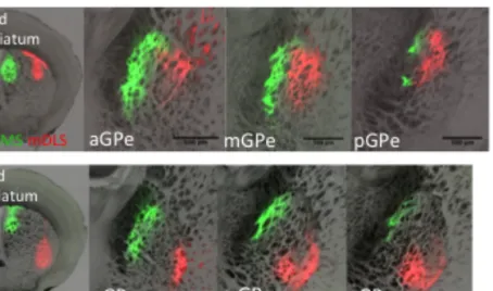
Striatopallidal projections of different populations showed similar patterns on aGPe and mGPe
We also observed that on the mediolateral axis, DMS and DLS projections of striatopallidal cells directly translate their striatal cell body position onto GPe, creating vertical bands, similar to
striatopallidal projection patterns described before (Wilson, et al., 1982, Hazrati, et al., 1992, Sadek et al., 2007).
Example images in Figure 2.4 and Figure 2.5 suggested that non-overlapping striatopallidal populations targeted non-non-overlapping GPe
regions. DMS, DLS and VLS populations projected on different regions on GPe. Therefore, as suggested above, it is possible that striatopallidal projections are more segregated compared to striatonigral projections.
Figure 2.5 | Example images of striatopallidal pathway aDLS and
aDMS-aVLS populations showed different projection patterns on GPe. Non-overlapping aDMS-aDLS and aDMS-aVLS populations targeted non-overlapping GPe regions (aDMSaDLS: r_striatum=0.01, r_GPe_1= 0.02, r_GPe_2= 0.02, r_GPe_3= -0.01, aDMS-aVLS: r_striatum=--0.01, r_GPe_1= -0.02, r_GPe_2= -0.03, r_GPe_3= -0.01, Pearson’s correlation coefficient, n=3).
Both aDMS and mDMS projected onto GPe occupying the most medial region, creating the most medial band onto GPe. aDLS and mDLS occupied the most lateral region of GPe creating the most lateral band. However aVLS and mVLS projections occupied the most ventrolateral region of GPe, sometimes creating a V-shape, but not a vertical band.
Figure 2.6 | Example images of indirect pathway mDLS and
mDMS-mVLS populations showed different projection patterns on GPe. Non-overlapping mDMS-mDLS and mDMS-mVLS populations targeted non-overlapping GPe regions (mDMS-mDLS: r_striatum=-0.01, r_GPe_1= -0.03, r_GPe_2= -0.01, r_GPe_3= 0.01, mDMSmVLS: r_striatum=0.01, r_GPe_1= 0.03, r_GPe_2= -0.04, r_GPe_3= -0.01, Pearson’s correlation coefficient, n=3)
Interestingly all aDMS, mDMS, aDLS, mDLS, aVLS, and mVLS targeted strongly onto aGPe and mGPe but sent weak projections onto pGPe. Even though it was suggested that the most posterior striatum showed low expression of D2 receptors, and fewer striatopallidal neurons, it will be important to map the projections of the most posterior striatopallidal populations (Gangarossa et al., 2013).
DISCUSSION
Anatomical mapping of circuits is important not only to understand the structural organization of the brain, but also for it functional understanding. Anatomical data can sometimes lead to functional
projections onto their target regions would be to create a projectome, and report projection patterns of each population in detail and leave the functional interpretations to the readers. However, it is very hard to test specific hypothesis on connectome or projectome data reported thus far, since it has been hard to reach the data and analyze it for specific questions. Therefore, another approach is to start with
functional questions and functional domains, analyze anatomical data for specific functional domains, and report the answers to specific questions.
Accordingly, we started with defining three striatal functional domains, DMS, DLS, and VLS, based on previous anatomical and functional data, and compared their projection patterns onto their target regions. We used AAVs that express EYFP and tdTomato and simultaneously labeled two subpopulations of either striatonigral or striatopallidal populations using D1-Cre (FK150-Cre) and D2-Cre (Adora2a-Cre) mouse lines. We labeled 9 different subpopulations described before by keeping DMS populations as reference and labeling either DLS or VLS on the same anterior-posterior axis. We acquired whole brain anatomical data. This technique allowed us to compare projection patterns within and between brains. The data collected can hopefully help identifying projection patterns that might be plausible candidates to explain functional data (see example in chapter 3).
Using this approach, we showed that both striatonigral and
striatopallidal population projections created parallel pathways. Both pathway projections followed similar organization on the
mediolateral axis, by conserving their relative mediolateral striatal position onto the target structures, but they also followed different organizational rules.
In one hand, striatonigral projections inverted their cell body position on the dorsal-ventral axes, suggested larger degrees of overlap, and crated complex shapes in SNr. On the other hand, striatopallidal projections translated their striatal position directly onto GP creating vertical bands and V-shaped bands.
In primates, it was suggested that GPi receives most of the limb and trunk input while SNr receives mostly orofacial and occulomotor input (Nambu, 2011). In the mouse SNr, VLS populations in general, target larger regions that DMS and DLS populations. Therefore, it is still possible that, in the mouse, a larger population of SNr is involved in orofacial motor and occulomotor control, compared to other motor functions.
It was previously suggested that basal ganglia is involved in action selection via two (path)ways, modulating thalamocortcial networks, and modulating brainstem motor networks (Hikosaka et al., 2000). It was also suggested that anterior two thirds of SNr projects to
thalamus and SC, and the posterior one third of SNr projects to thalamus and brainstem (Grofova, et al., 1982, Deniau, et al., 1996). All the striatonigral populations that were labeled projected to the whole anterior-posterior axis of SNr. DMS and DLS striatonigral populations of anterior, mid and posterior striatum projected on larger areas on aSNr and to smaller areas on pSNr. However, unlike DMS and DLS populations, VLS striatonigral populations projected on larger areas on pSNr compared to aSNr.
Therefore, aSNr, via its stronger thalamic projections, might be providing information feeding back to the thalamo-cortico basal-ganglia loops. pSNr, via its stronger projections to brainstem, might be directly modulating the premotor regions, motor output. It is likely that DMS, DLS and VLS target thalamo-cortico-basal-ganglia loops similarly, but VLS sends stronger motor output compared to DMS and DLS.
Our labeling techniques do not discriminate between patch and matrix compartments of striatum, but it was previously suggested that
anterior striatum receives more input from limbic cortex regions and have more patches compared to the other striatal regions (Gerfen et al., 1987, Graybiel, 1998). It was suggested that anterior striatum was involved execution of instrumental actions in early stages of learning while posterior striatal was involved in execution of instrumental actions in late stages of learning (Jueptnter et al., 1997a, Yin et al., 2004, Yin et al., 2009). If we take this at facevalue, together with the fact that pSNr is the motor output region of SNr, then it would be expected that the posterior striatum would be projecting weaker to aSNr and stronger to pSNr (Grofova, et al., 1982). Our first observations suggested that pDMS, pDLS and pVLS populations might indeed be projecting into smaller regions on aSNr, than aDMS, aDLS and aVLS populations. However, the same comparison on pSNr requires more analysis. Therefore, it is likely that posterior striatum and pSNr might be involved in execution in late stages of learning, while anterior populations might be involved in early stages of instrumental learning (Jueptnter et al., 1997a, Hikosaka, et al., 2000, Yin et al., 2004, Yin et al., 2009).
It was also suggested that striatonigral projections create layers around SNr, form “onion-like” structures, and the dendritic fields of SNr projection cells were distributed within these layers of
converging inputs (Faul et al., 1978, Grofova et al.,1982, Deniau et al., 1996). To understand the organization of such a complex structure coronal, or sagittal images of SNr would not be sufficient. Therefore, 3D reconstruction of projections would be necessary to understand the organization of different striatopallidal population projections onto SNr. We are currently pursuing these efforts.
Similar to primates’ putamen projections, in the mouse, we showed that somatotopic organization on the striatum was directly projected onto GPe via striatopallidal projections (Nambu, 2011).
DMS projected strongly onto aGPe and mGPe. DMS projections seemed to occupy larger region than DLS or VLS projections onto aGPe, and its projections seemed weaker on the pGPe.
It was previously suggested that two types of cells showed different intrinsic projection patterns in GPe (Stanek et al., 2007). The first group was located within the 100um thick, outer layer of GPe that was on the striatal border and was occupied by cells that arborized within the same layer and send collaterals to the inner layer of GPe (Stanek et al., 2007). Second group was arborized only within the larger inner layer of GPe (Stanek et al., 2007). We did not observe differences in the striatopallidal projection patterns between the inner and the outer layer of GPe. It is likely that these two structures did not differ in their striatal input but only differ in their within-GPe
It was previously suggested that PV+ cells were located more laterally on GPe and cells expressing Lhx6 were located more medially (Mastro, et al., 2014). These cells were also shown to project onto different targets; PV+ cells projected stronger onto Pf, SNr and STN, while Lhx6 expressing cells projected stronger onto DLS, SNc and Rt (Mastro, et al., 2014). We observed that medial part of GPe received only DMS input and lateral region received DLS and VLS input. DMS input might be transmitted to target structures by Lhx6 population such as DLS, SNc and Rt, whereas, DLS and VLS input might be transmitted to Pf, SNr and STN via PV+ cells, distributing information from different domains of striatopallidal pathway to different circuits.
Even though specific regions of SNr and GPe received specific input from striatum, dendritic fields of SNr and GPe cells span large areas, with striatal input targeting distal dendrites of both GPe and SNr cells (Grofova et al., 1982, Smith et al, 1998, Stanek et al., 2007, Bolam et al., 2000). Therefore, even though these observations do not clarify the advantages of having parallel projecting striatonigral and
striatopallidal pathways, circuit mapping shows that striatal cells that receive specific motor input project to regions of SNr which in turn projects to downstream motor regions involved in same specific movements (Deniau et al., 1996, Grofova et al., 1989). Therefore, large dendritic fields of SNr and GPe cells might allow them to integrate different contextual input to gate motor information to the pre-motor output centers, while also allowing specific actions to be performed in different contexts, in a cue guided manner or self initiated.
It would be interesting to compare the dendritic regions of GPe and SNr targeted by functionally similar striatal domains compared to functionally different striatal domains. These differences might help us better understand the information processing on basal ganglia output cells.
In summary, corticostriatal projections, striatonigral and
striatopallidal projections showed complex projection patterns, and the complexity increased by large dendritic fields of SNr and GPe cells. All these circuit complexity might allow movement patterns to be learned and executed in different contexts, in response to different stimuli.
In order to study the complex anatomical patterns together with their motor functions on initiation and execution of specific movements, on the rest of the thesis we focused on the anterior-ventrolateral striatum and in addition to its striatonigral and striatopallidal projections, we investigated the role of this region in naïve and instrumental orofacial actions.
MATERIAL AND METHODS Animals
All procedures were reviewed and performed in accordance with the Champalimaud Center of the Unknown Ethics Committee guidelines and approved by the Portuguese Veterinary General Board (Direccao Geral de Veterinaria, approval 0421/000/000/2014). GENSAT BAC transgenic lines D1-Cre (FK150) and D2- Cre (Adora2a) are used to specifically target striatonigral or striatopallidal cells. Animals between 3-6 months of age, that were housed in normal light cycle