U
NIVERSIDADE DEL
ISBOAF
ACULDADE DEC
IÊNCIASD
EPARTAMENTO DEB
IOLOGIAV
EGETALG
ENOTYPIC
A
NALYSIS OF THE
L
EISHMANIA
INFANTUM
R
ESISTANCE TO
C
ONVENTIONAL
D
RUGS
AND
N
EW
C
HEMICALLY
S
YNTHESIZED
C
OMPOUNDS
David Jorge Santos Mateus
D
ISSERTAÇÃOM
ESTRADO EM
M
ICROBIOLOGIA
A
PLICADA
U
NIVERSIDADE DEL
ISBOAF
ACULDADE DEC
IÊNCIASD
EPARTAMENTO DEB
IOLOGIAV
EGETALG
ENOTYPIC
A
NALYSIS OF THE
L
EISHMANIA
INFANTUM
R
ESISTANCE TO
C
ONVENTIONAL
D
RUGS
AND
N
EW
C
HEMICALLY
S
YNTHESIZED
C
OMPOUNDS
David Jorge Santos Mateus
D
ISSERTAÇÃOM
ESTRADO EM
M
ICROBIOLOGIA
A
PLICADA
Dissertação orientada por Prof. Dr. Gabriela Santos-Gomes (IHMT-UNL)
e Prof. Dr. Francisco Dionísio (FCUL)
G
ENOTYPIC
A
NALYSIS OF THE
L
EISHMANIA
INFANTUM
R
ESISTANCE TO
C
ONVENTIONAL
D
RUGS AND
N
EW
C
HEMICALLY
S
YNTHESIZED
C
OMPOUNDS
David Jorge Santos Mateus
2014
This thesis was fully performed at Center for Malaria and Other
Tropical Diseases of Institute of Hygiene and Tropical Medicine, New
University of Lisbon under the direct supervision of Prof. Dr. Gabriela
Santos-Gomes.
Prof. Dr. Francisco Dionísio was the internal designated supervisor in
the scope of the Master in Applied Microbiology of the Faculty of
Sciences of the University of Lisbon.
C
ONTENTS
ACKNOWLEDGEMENTS ... 1 LIST OF ABBREVIATIONS ... 2 RESUMO ... 4 ABSTRACT ... 8 I.INTRODUCTION ... 9 1. Leishmaniasis ... 9 1.1. Epidemiology ... 9 1.1.1. In humans ... 9 1.1.2. In dogs ... 10 1.1.3. Geographic distribution ... 11 1.2. Life cycle ... 111.3. Parasite/vertebrate host interaction ... 12
1.4. Diagnostic ... 14
1.5. Control ... 15
1.6. Treatment ... 15
2. Gene amplification in Leishmania ... 17
2.1. Gene amplification ... 17
2.2. Circular amplicons ... 18
2.3. Mechanism ... 19
2.4. Response to drug pressure ... 20
3. Drug resistance in Leishmania ... 21
3.1. Pentavalent antimonials ... 21
3.2. Miltefosine ... 22
3.3. Amphotericin B ... 24
3.4. Strategies to combat drug resistance ... 24
3.5. New drugs ... 25
4. Dinitroanilines derivatives ... 26
II.OBJECTIVES ... 27
III.MATERIALS AND METHODS ... 28
1. In vivo infection and treatment ... 28
1.1. Animals and parasites ... 28
1.1.1. Animals ... 28
1.2. Infection ... 28
1.3. Treatment ... 29
1.4. Parasitic load determination ... 29
2. In vitro resistant parasites ... 30
2.1. Parasites ... 30
2.2. Drug-resistant promastigotes ... 30
2.2.1. Drug dilutions and plating ... 30
2.2.2. Evaluation of resistant promastigotes ... 31
2.2.3. Sample collection ... 31
3. Primer selection and plasmid cloning ... 31
3.1. Primer selection and optimization ... 31
3.2. Plasmid cloning ... 33
4. Quantification of gene number by real-time PCR ... 34
4.1. DNA extraction ... 34 4.2. Real-time PCR ... 34 5. Statistical analysis ... 35 IV.RESULTS ... 36 1. In vivo experiments ... 36 1.1. Parasitic load ... 36 1.2. Gene amplification ... 36 2. In vitro experiments ... 37
2.1. Effective drug concentration ... 37
2.2. Gene amplification ... 37
V.DISCUSSION ... 40
1. Antileishmanial activity of the drugs ... 40
2. Gene amplification and mechanisms of drug action ... 40
3. General conclusions and future perspectives ... 43
A
CKNOWLEDGEMENTS
Foremost, I would like to express my sincere gratitude to my supervisor Prof. Dr. Gabriela Santos-Gomes. Without her knowledge, support, patience and guidance this dissertation would not have been possible. I am eternally grateful for her willingness to take me into her research team, for the support on the good and bad moments and for being the best supervisor imaginable. She was not only a mentor but also became a good friend.
I would like to thank Prof. Dr. Francisco Dionísio for accepting to be my internal supervisor in this project, for his insightful comments and advices. My thanks to Prof. Dr. Eugénia Cruz and Dr. Manuela Carvalheiro for giving us the new antileishmanial formulations necessary for this research project.
My sincere appreciation to Armanda Rodrigues for the practical orientation given when I first arrived in the laboratory and for lending me “half a brain” once in a while from then on. My thanks also goes for my fellow colleagues Ana Bolas, Cristina Branco, Mafalda Claro, Maria Pereira, Mariana Fernandes and Joana Cavaco Silva. Without all of you and your friendship our laboratory is not the same. I am glad you are always there to cheer up my days.
A special thanks to all my dearest friends, especially to André Soares, Claudio Monterosso, Diogo Pereira, Filipa Marta, Inês Gato, João Robalo, Joana Vale and Miriam Silva. Thank you for being always there for me, for giving me motivation and, most of all, for making my world a better place.
At last, but by no means the least, I would like to give my most heartfelt thanks and appreciation to my mother. Thank you for creating me and for never stop supporting me throughout my life.
This work was funded by the Portuguese Foundation for Science and Technology (FCT) and the European Union (FEDER) through the projects PTDC/CVT/098290/2008 and PTDC/CVT/113121/2010.
L
IST OF
A
BBREVIATIONS
ACR2 – arsenate reductase 2 AmB – amphotericin B
AQP1 – aquaglyceroporin bp – base pairs
CanL – canine leishmaniasis CL – cutaneous leishmaniasis CoA – coenzyme A
DMSO – dimethyl sulfoxide DNA – deoxyribonucleic acid dNTP – deoxynucleotide
DOTS – directly observed treatment, short-course EC100 – effective concentration to kill 100% of parasites
EDTA – ethylenediaminetetraacetic acid EU – European Union
FBS – fetal bovine serum
FCUL – Faculty of Sciences of the University of Lisbon FW – forward
GSH – glutathione
GSH1 – gamma-glutamylcysteine synthase GSH1 – gamma-glutamylcysteine synthase gene HIV – human immunodeficiency virus
IHMT – Institute of Hygiene and Tropical Medicine IP – intraperitoneally
IPTG – isopropyl β-D-1-thiogalactopyranoside LB – lysogeny broth
LDA – limiting dilution assay m/v – mass/volume concentration MDR1 – P-glycoprotein MDR1 MDR1 – P-glycoprotein MDR1 gene MGA – meglumine antimoniate MILT – miltefosine
ML – mucocutaneous leishmaniasis MRPA – ABC-thiol transporter MRPA MRPA – ABC-thiol transporter MRPA gene MT – putative miltefosine transporter protein
ORZ – oryzalin
PBS – phosphate buffered saline PCR – polymerase chain reaction PEG – polyethylene glycol
POL1 – DNA polymerase alpha catalytic subunit gene ppg – per gram of tissue
PTR1 – pteridine reductase PTR1 – pteridine reductase gene RNA – ribonucleic acid
ROS – reactive oxygen species rpm – rotations per minute RV – reverse
SbIII – trivalent antimonial
SbV – pentavalent antimonial
SCHN – Schneider medium
SOC – super optimal broth with catabolite repression STB – sodium stibogluconate T(SH)2 – trypanothione TAE – tris-acetate-EDTA TAN – annealing temperature TDR1 – thiol-dependent reductase 1 TFL – trifluralin
TFL-A – trifluralin analogue TFL-A3 – trifluralin analogue 3 TFL-A6 – trifluralin analogue 6 TR – trypanothione reductase TS2 – trypanothione disulfide
TSS – transformation and storage solution U – units
UK – United Kingdom
UNL – New University of Lisbon USA – United States of America v/v – volume/volume concentration VL – visceral leishmaniasis
WHO – World Health Organization WT – wild-type
R
ESUMO
A leishmaniose, doença parasitária com manifestações clínicas diversas, é considerada uma das mais importantes patologias parasitárias. É causada por cerca de 20 espécies de protozoários intracelulares do género Leishmania sendo, na maioria dos casos, uma zoonose de mamíferos selvagens ou domésticos que atinge acidentalmente o Homem. Este parasita é transmitido aos hospedeiros vertebrados pela picada de flebotomídeos do género Phlebotomus no Velho Mundo e Lutzomyia no Novo Mundo. O ciclo de vida deste inclui dois estadios com duas formas morfológicas distintas: forma amastigota no interior dos macrófagos do hospedeiro vertebrado e forma promastigota no intestino do insecto vector. Com base nas suas manifestações clínicas a leishmaniose pode ser classificada em três tipos principais: leishmaniose cutânea, leishmaniose mucocutânea e leishmaniose visceral. Leishmania infantum é o agente responsável pela leishmaniose visceral zoonótica, manifestação severa e mortal da doença, que se distribui pela região da bacia do Mediterrâneo incluindo Portugal.
Há mais de 70 anos que os antimoniais pentavalentes (SbV), como estibogluconato
de sódio (STB) e antimoniato de meglumina (MGA), têm sido usados como tratamento de primeira linha em todas as manifestações clínicas da doença. Estes fármacos, apesar de serem bastante eficazes no combate à leishmaniose, apresentam toxicidade elevada, sendo responsáveis por efeitos secundários graves. Nos últimos anos, a sensibilidade do parasita a estes fármacos tem sofrido alterações problemáticas em algumas regiões endémicas para Leishmania. Novos fármacos mais eficazes e/ou menos tóxicos têm vindo a ser usados em resposta à diferenciação de estirpes do parasita com menor sensibilidade aos antimoniais ou em situações clinicas de maior gravidade. Porém, o elevado custo torna impossível a utilização regular destes fármacos nos países mais afectados, uma vez que esta doença está frequentemente associada a países de baixa renda. Apesar da elevada quantidade de fármacos anti-Leishmania actualmente em uso, nenhum deles conjuga elevada eficiência, baixa toxicidade e custo acessível às populações afectadas. No seu conjunto, estes factores fazem da investigação de novos fármacos anti-Leishmania uma prioridade. Actualmente a investigação de novos fármacos está muito focada em compostos existentes com actividade terapêutica dirigida a outras doenças, compostos de origem natural ou formulações de herbicidas. Entre estes últimos compostos encontram-se os derivados de dinitroanilinas, nomeadamente a orizalina (ORZ) e compostos análogos da trifluralina (TFL), tendo o seu potencial no tratamento de leishmaniose sido demonstrado.
A diminuição da sensibilidade do parasita aos fármacos em uso foi relacionada com a habilidade de amplificar selectivamente o número de cópias de alguns genes em
resposta ao contacto com as formulações. A amplificação génica é considerada o principal mecanismo de resistência a fármacos dentro do género Leishmania. O pequeno genoma e a reduzida quantidade de genes fazem deste parasita um alvo ideal para o estudo deste fenómeno. A ausência de controlo da transcrição é também um dos factores de sucesso da amplificação génica como mecanismo adaptativo, tendo sido demonstrado que o DNA amplificado pode atingir 10% do DNA total do parasita. Alterações na permeabilidade da membrana, permitindo uma inferior acumulação do fármaco dentro da célula, redução na importação do fármaco, inactivação do fármaco ou sequestro em compartimentos intracelulares são os mecanismos usados por Leishmania para sobreviver ao contacto com fármacos anti-Leishmania. O aumento do número de cópias dos genes das proteínas responsáveis por estes mecanismos está directamente relacionado com o aumento do respectivo nível de expressão mas também com o aumento da probabilidade de ocorrência de mutações pontuais em algumas destas cópias que poderão vir a ser vantajosas para a sobrevivência do parasita quando em contacto com fármacos.
Neste estudo foi avaliado o potencial anti-Leishmania em modelo animal de um fármaco anti-Leishmania clássico, o estibogluconato de sódio, da dinitoanilina orizalina e de novos compostos sintéticos análogos da trifluralina, TFL-A3 e TFL-A6, que demonstraram elevada actividade anti-Leishmania e baixa toxicidade in vitro. Em modelo animal o tratamento não conduz à eliminação completa dos parasitas, tendo-se colocado em hipótese que os parasitas sobreviventes serão menos sensíveis ou resistentes aos fármacos usados. Neste estudo os parasitas que sobreviveram ao tratamento foram quantificados e recolhidos para extracção de DNA e o número de cópias de genes seleccionados foi quantificado por PCR em tempo real. Estudos anteriores efectuados em amostras recolhidas de pacientes de áreas endémicas de leishmaniose demonstraram que a menor susceptibilidade do parasita aos fármacos está relacionada com a amplificação dos seguintes genes: os genes para os transportadores de membrana MRPA (MRPA) e MDR1 (MDR1), o gene para a enzima gama-glutamil-cisteína sintetase (GSH1), envolvida na síntese de glutationa e tripanotiona, e o gene para a enzima pteridina redutase (PTR1), que intervém na cascata metabólica do folato. A quantificação absoluta do número de cópias de cada gene por parasita foi possível recorrendo a um gene de cópia única no genoma de Leishmania, o gene para a subunidade catalítica alfa da DNA polimerase. Foi também analisada a amplificação génica dos quatro genes seleccionados na forma promastigota do parasita (in vitro). Promastigotas de L. infantum foram tornados resistentes a antimoniato de meglumina, miltefosina, TFL-A3 e TFL-A6, e o número de genes quantificado por PCR em tempo real.
Foi demonstrado que a actividade anti-Leishmania dos compostos TFL-A6 e orizalina é bastante promissora, sendo muito semelhante à actividade do estibogluconato de sódio. TFL-A3 apresentou actividade anti-Leishmania mais reduzida. Os parasitas que in vivo sobreviveram ao tratamento com estibogluconato de sódio apresentaram aumento significativo do número de cópias dos genes MDR1 e PTR1, sugerindo amplificação da capacidade de expulsão deste fármaco do interior do parasita e alterações na via metabólica do folato, reduzindo a quantidade de intermediários de espécies reactivas de oxigénio e azoto. Apesar de existirem duvidas devido ao facto das amostras serem provenientes de doentes que poderiam ter estado sujeitos a fármacos adicionais, a amplificação de MDR1 tinha sido demonstrada em estudos anteriores. O presente estudo vem mostrar que a amplificação de MDR1 ocorre de facto em parasitas que estiveram unicamente em contacto com estibogluconato de sódio. Os parasitas provenientes de murganhos tratados com TFL-A3 apenas apresentaram aumento do número de cópias do gene MDR1. Os parasitas isolados a partir do grupo tratado com TFL-A6 apresentaram amplificação dos genes MDR1, MRPA e PTR1. Esta amplificação indica que o mecanismo de resistência está associado ao efluxo do fármaco pelo parasita, ao sequestro do fármaco para compartimentos intracelulares do parasita e a alterações na quantidade de espécies reactivas de oxigénio e azoto que se formam. Nos parasitas que estiveram em contacto com a orizalina os genes GSH1, MDR1 e PTR1 apresentam aumento do número de cópias. No caso da orizalina o mecanismo usado parece estar relacionado com o aumento de efluxo do composto, alterações na quantidade de espécies reactivas de oxigénio e azoto e alterações no balanço do potencial reductor no interior do parasita. Nas experiências in vitro os promastigotas resistentes a antimoniato de meglumina, miltefosina, TFL-A3 ou TFL-A6 demonstraram resultados distintos dos encontrados nos parasitas resultantes dos murganhos que foram sujeitos a tratamento. Estas diferenças podem ser explicadas pelo modo como diferentes formas morfológicas do parasita reagem quando contactam com o fármaco. Nas experiências in vivo a forma amastigota encontrava-se internalizada pelos macrófagos do hospedeiro, retida no interior dos fagolisossomas, enquanto nas experiências in vitro a forma promastigota encontrava-se livre no meio de cultura. Os diferentes ambientes (amastigota – intracelular, rodeado por duas membranas, pH ácido, 37ºC, sujeito a acções do sistema imunitário do hospedeiro; promastigota – livre, meio de cultura, pH neutro, 24ºC) induzem a diferenciação pelo parasita de diferentes mecanismos de adaptação e sobrevivência. Os promastigotas resistentes a antimoniato de meglumina demonstraram um aumento significativo do número de cópias de GSH1 e redução do número de cópias de MDR1. Os parasitas resistentes a miltefosina não apresentaram qualquer alteração significativa no número dos genes estudados. Os
parasitas resistentes a TFL-A3 apresentaram amplificação do gene GSH1 e os resistentes a TFL-A6 demonstraram um aumento significativo do número dos genes GSH1, MDR1 e PTR1.
Com este estudo é possível concluir que uma elevada actividade anti-Leishmania e uma baixa toxicidade pode não ser suficiente para um composto ser considerado uma boa alternativa aos fármacos actualmente usados. Resistência a TFL-A6 parece dever-se a indução de mecanismos dever-semelhantes aos responsáveis pela resistência aos fármacos actualmente em uso. O desenvolvimento de menor sensibilidade ou resistência pode conduzir à rápida diminuição da eficiência anti-Leishmania deste composto ou mesmo à ineficácia no combate a estirpes já resistentes aos fármacos em uso. Todos estes factores devem ser tidos em consideração no desenvolvimento de novos compostos anti-Leishmania, prevenindo o aparecimento de estirpes menos susceptíveis mas também como forma de minimizar o investimento em fármacos que à partida não deveriam ser considerados como alternativa para tratamento da leishmaniose.
Palavras-chave: Leishmaniose, Leishmania infantum, resistência, novos fármacos, amplificação génica.
A
BSTRACT
Leishmaniasis, caused by intracellular protozoa of the genus Leishmania, is considered one of the most important human parasitic diseases in the world. Leishmania infantum is the agent responsible for zoonotic visceral leishmaniasis, the most severe and fatal form of the disease, in the Mediterranean region, including Portugal. Recently, the variation of parasite sensitivity to several drugs became a problem in some endemic areas for Leishmania. It was observed that the ability of Leishmania to selectively increase the number of gene copies is the main adaptation of these parasites to drug pressure, therefore responsible for resistance. Furthermore, all the currently used antileishmanial drugs do not conjugate high efficiency, low toxicity and an affordable cost. Therefore the development of new therapeutic compounds for leishmaniasis still is a priority. The potential of trifluralin (TFL) for the treatment of leishmaniasis was recently demonstrated and two analogues, TFL-A3 and TFL-A6, which presented higher antileishmanial activity and less cytotoxicity than TFL were analyzed in the present study. Another drug, oryzalin (ORZ), and conventional antileishmanial drugs (sodium stibogluconate, meglumine antimoniate and miltefosine) were also assessed. In vivo and in vitro studies were performed to confirm their antileishmanial activity and to access their potential to induce drug resistance in L. infantum. The genes for ABC-thiol transporter (MRPA), P-glycoprotein (MDR1), the enzyme gamma-glutamylcysteine synthase (GSH1) and pteridine reductase 1 (PTR1), already associated with drug resistance in previous studies, were selected and quantified by real-time PCR. The results revealed that TFL-A6 and ORZ have an antileishmanial potential similar to the conventional antileishmanial drugs. However, it was demonstrated that a relatively short period of treatment (10 days) is enough to induce significant gene amplification in the parasites that survived in vivo to the treatment. In vitro experiments demonstrated that the level of gene amplification in the promastigote form is different than in the intracellular amastigote form of the parasite when exposed to the same drugs. This study accentuates the need for understanding the mechanisms and evaluate the appearance of resistances when designing and investigating new antileishmanial drugs.
Keywords: Leishmaniasis, Leishmania infantum, drug resistance, new drugs, gene amplification.
I.
I
NTRODUCTIONI.
I
NTRODUCTION
1. Leishmaniasis
1.1. Epidemiology
Leishmaniasis is a parasitic disease caused by intracellular protozoa belonging to the family Trypanosomatidae Doflein, 1901 and the genus Leishmania Ross, 1903 [Shaw, 1994; Croft et al., 2006]. More than 20 species of Leishmania were identified as responsible for the disease in humans [Schallig & Oskam, 2002]. The parasite is transmitted to the vertebrate host by sandflies of the genus Phlebotomus Rondani & Berté, 1843 in the Old World and Lutzomyia França, 1924 in the New World [Desjeux, 2001].
Leishmania infections are in most cases zoonosis from wild or domestic mammals, and accidentally the parasite infects humans, although in East Africa and Indian subcontinent anthroponotic form of the disease can be found. The main domestic animal reservoirs are the dogs, while the sylvatic ones are small rodents and larger mammals, like foxes, wolves and jackals.
1.1.1. In humans
Leishmaniasis is considered one of the most important human parasitic diseases in the world, both in mortality and morbidity. According to the World Health Organization (WHO) there is an estimate of 2 million new cases every year, 350 million people are at risk of being infected, around 12 million people are already infected and 60 000 die every year. Increase of incidence and severity of this disease is due to infected human and dog migration, global warming altering the distribution of the vector, co-infection with immunosuppressive diseases and poverty [Desjeux, 2001; Neuber, 2008; Alvar et al., 2012].
Leishmaniasis different clinical manifestations can be classified into three types: cutaneous leishmaniasis (CL), mucocutaneous leishmaniasis (ML) and visceral leishmaniasis (VL).
Cutaneous leishmaniasis is the most common form of the disease. The clinical manifestations can go from a single self-healing skin lesion to multiple localized or diffused lesions leading to a chronic state [Sádlová, 1999]. The clinical outcome depends mostly on the species of the parasite, but it is also influenced by the species of the vector
I.
I
NTRODUCTIONand the innate or acquired resistance of the human host [Herwaldt, 1999]. Although the majority of the cases lead to spontaneous healing and the skin lesions disappear in less than a year, around 15% of the cases lead to relapses and 2 to 40% of the cases of CL evolve into the mucocutaneous form of the disease. The species responsible for this form of leishmaniasis are: Leishmania major, L. tropica, L. aethiopica, L. infantum, L. shawi, L. mexicana, L. amazonensis, L. braziliensis, L. peruviana, L. panamiensis, L. guyanensis, L. lainsoni and L. naiffi [Murray et al., 2005; Neuber, 2008].
Mucocutaneous leishmaniasis consists in a progression of the cutaneous form, it happens when the infection reaches the mucosal regions. Can affect and destroy the tissues of the oronasal and pharyngeal cavities, and can lead to severe and painful facial mutilations. This form can happen as soon as one week after the appearance of the first skin lesion as well as many years after the cutaneous manifestations, showing that the parasite persists in the tissues for a long time. The species responsible for this form of leishmaniasis are: L. braziliensis, L. panamensis and L. guyanensis. There are also reports of mucocutaneous leishmaniasis caused by L. donovani, L. major and L. infantum in individuals with immunosuppressive diseases [Desjeux, 1996].
Visceral leishmaniasis is the most severe and fatal form of the disease, comprising a broad range of clinical manifestations. The parasite invades and multiplies in the organs of mononuclear phagocyte system such as the spleen, liver and lymph nodes and the symptoms are characterized by prolonged and irregular fever, splenomegaly, lymphadenopathy, hepatomegaly, pancytopenia, progressive anemia, weight loss and hypergamma-globulinemia with hypoalbuminemia. In many cases the infection does not take an acute or chronic course, remaining asymptomatic or subclinical and can even lead to a self-healing scenario [Sahni, 2012]. The species responsible for this form of leishmaniasis are: L. donovani and L. infantum [Berman, 1997].
1.1.2. In dogs
Canine leishmaniasis (CanL) is a chronic and systemic disease with a broad spectrum of non-specific clinical manifestations caused by L. infantum in domestic dogs (Canis lupus familiaris). The most frequent manifestations are skin lesions, however, the animals can show other clinical signs unrelated to cutaneous lesions as their main symptoms such as renal disease or any other symptoms characteristic of human VL. Since infection with the parasite does not always mean clinical illness there is a high prevalence of subclinical and asymptomatic infected animals in the endemic regions. In addition to the fact that the domestic dogs share the same habitat, frequently contact
I.
I
NTRODUCTIONand often travel with humans, this makes dog the most important reservoir for L. infantum. Other species of Leishmania were also found infecting dogs (L. braziliensis, L. panamensis and L. peruviana) and even though this species do not cause disease in these animals it may be that dogs act as reservoirs for this species as well [Reithinger et al., 2003; Solano-Gallego et al., 2011; Palatnik-de-Sousa, 2012].
1.1.3. Geographic distribution
Leishmania occurs mostly in tropical and sub-tropical areas and is present in all continents, except Antarctica. Until the last decade, it was thought that Oceania and Southeast Asia were Leishmania free but recent findings prove otherwise [Rose et al., 2004; Conlan et al., 2011]. Endemic regions can be found in South and Central America, North and East Africa, Middle-East, Indian sub-continent, Central and Eastern Asia and South Europe, putting together a total of 98 countries considered endemic for Leishmania. More than 90% of all VL cases occur in just six countries: India, Bangladesh, Sudan, South Sudan, Brazil and Ethiopia. The prevalence of CL is more widely spread, with 70 to 75% occurring in ten countries around the globe: Afghanistan, Algeria, Colombia, Brazil, Iran, Syria, Ethiopia, North Sudan, Costa Rica and Peru [Alvar et al., 2012].
In Portugal as in the Mediterranean Basin countries, the species responsible for the majority of leishmaniasis cases is L. infantum, both in humans and animals. There are reports of leishmaniasis across the entire continental territory of Portugal, but three areas stand out as the most important endemic regions: Metropolitan area of Lisbon, Alto Douro and Algarve. The prevalence of CanL can go up to 20% in these endemic areas and between 2000 and 2009 there were 173 new cases reported of VL, mostly in children and immunocompromised individuals [Santos-Gomes et al., 1998; Campino & Maia, 2010].
1.2. Life cycle
Leishmania is a dimorphic parasite: promastigote form (Figure 1) occurs in the invertebrate host (sandfly) and the vertebrate host has the amastigote form (Figure 2). The promastigote, found in the midgut of the insect vector, has an elongated shape and a well-developed anterior flagellum. It is possible to characterize two stages of promastigote development: the procyclic stage and the metacyclic stage. Procyclic
I.
I
NTRODUCTIONpromastigotes are not infectious, are in continuous division and have a shorter flagellum. On the other hand, metacyclic promasti-gotes are highly infectious, are long and narrow and have a large flagellum [Killick-Kendrick, 1990; Ashford, 2000]. Amastigotes have a round shape and no external flagellum. Being an obligatory intracellular form it can only survi-ve and multiply inside parasito-phorous vacuoles of phagocytic cells.
Parasites are transmitted to the vertebrate host when the infected female sandfly takes a blood meal, inoculating the promastigotes in the vertebrate’s dermis. Parasites are then phagocytized by macrophages and once inside the cell they transform into amastigotes. Amastigotes multiply inside phagocytic cells that are later ingested by another sandfly when it takes a blood meal. Ingested cells are digested in the sandfly gut and the free amastigotes reach the midgut where they
transform into promastigotes. Promastigotes multiply and later migrate into the proboscis of the sandfly, from where they will be introduced into the vertebrate host dermis when a blood meal occurs
[Cohen-Freue et al., 2007;
Dostálová & Volf, 2012] (Figure 3).
1.3. Parasite/vertebrate host
interaction
In the vertebrate, destruction of invading microorganisms is essen-tially executed by two types of
Figure 1. Schematic representation of a Leishmania promastigote. a – flagellum; b – axoneme; c – basal corpuscle; d –
mitochondria; e – nucleus; f – subpellicular microtubules; g – glycosome; h – lipid inclusions; i – acidocalcisome; j – endoplasmic reticulum; k – multivesicular tubule; l – nucleolus; m – kinetoplast; n – Golgi complex. Adapted from Teixeira et al. (2013).
Figure 2. Schematic representation of a Leishmania amastigote. a – basal corpuscle; b – lipid inclusions; c –
subpellicular microtubules; d – mitochondria; e – megasome; f – glycosome; g – endoplasmic reticulum; h – nucleus; i – nucleolus; j – acidocalcisome; k – kinetoplast; l – Golgi complex; m – flagellar pocket; n – axoneme; o – flagellum. Adapted from Teixeira et al. (2013).
I.
I
NTRODUCTIONcells, the macrophages and the neutrophils. When inoculated in the vertebrate dermis, Leishmania promastigotes are phagocytized by macrophages (Figure 4). Within these cells, the parasites stay inside parasitophorous vacuoles that fuse with lysosomes
Figure 3. Life cycle of Leishmania in the mammalian host and insect vector. 1 – Female sandfly takes a blood
meal and inoculates the promastigotes; 2 – Promastigotes are phagocytized by macrophages; 3 – Transformation into amastigotes inside the parasitophorous vacuole; 4 – Amastigote replication; 5 – Ingestion of infected macrophages or free amastigotes by the female sandfly when taking a blood meal; 6 – Transformation into promastigotes in the sandfly midgut; 7 – Promastigotes replication and transformation into metacyclic promastigotes. Adapted from Teixeira et al. (2013).
I.
I
NTRODUCTIONcontaining proteolytic enzymes, forming phagolysosomes [Bogdan & Röllinghoff, 1998]. Within phagolysosomes production of reactive oxygen and reactive nitrogen molecules occur in order to destroy the
invader organism [Slauch, 2011]. Leishmania has the ability to slow down the fusion of these vacuoles assuring the successful transfor-mation into amastigotes. Amasti-gotes present high enzymatic activity and using catalase, superoxide dismutase and glutathione peroxidase are able to eliminate the products of
oxidative burst [Meshnick & Eaton, 1981; Murray, 1981]. The lipophosphoglycan of Leishmania is also a potent inhibitor of the proteins responsible for activation of oxidative burst as well as capable of interfering with the production of nitric oxide inside the phagolysosomes [Bogdan & Röllinghoff, 1998].
1.4. Diagnostic
Because of the wide spectrum of clinical manifestations this disease represents a diagnostic challenge, requiring confirmatory tests to decide if patients should be treated. These tests require a high sensitivity and specificity due to the leishmaniasis clinical severity and to the fact that drugs currently used for the treatment have high toxicity and can be very expensive. Microscopy still is the standard technique for leishmaniasis diagnosis in endemic areas [Reithinger & Dujardin, 2007]. Giemsa-stained lesion biopsy smears or aspirates from the lymph node, bone marrow or spleen are used with high specificity to identify the presence of the parasite. Laboratory cultures of the biopsies and aspirates are also used to diagnose leishmaniasis. However, microscopy and culture diagnosis tend to have low sensitivity and be highly variable [Herwaldt, 1999]. Several immunological techniques are also used as complementary to direct parasitological detection. Tests based on indirect fluorescence antibody, enzyme-linked immunosorbent assay and western blot have shown high diagnostic accuracy. Two serological tests were specifically developed for VL, the direct agglutination test and the rK39-based immunochromatographic test. Both tests showed values higher than 90% for both sensitivity and specificity [Chappuis et al., 2007]. One test was developed for CL, the
Figure 4. Scanning electron microscopy micrograph of a
Leishmania promastigote being phagocytized by a macrophage.
I.
I
NTRODUCTIONMontenegro skin test where an intradermal injection of dead promastigotes is given to the patient and examined 48 hours later to see if a delayed-type hypersensitivity has formed. This test has a very high sensitivity and specificity however does not distinguish between past and present infections. The most reliable approach in leishmaniasis diagnosis at this moment is the molecular approach based on polymerase chain reaction (PCR) assays. PCR has the highest sensitivity and specificity (up to 100%) of all the diagnostic methods for leishmaniasis, can diagnose independently of parasite species and have been shown to be better than direct parasitological test specially on samples with low parasite loads or samples from less intrusive sources [Reithinger & Dujardin, 2007].
1.5. Control
Control strategies focus on two major areas: vector control and reservoir control. For vector control the most widely strategy used is the spraying of households with insecticide and the use of insecticide treated curtains and bed nets. However this strategy was only proved to be effective in small cluster areas and the results cannot be extrapolated elsewhere [Davies et al., 2003]. For reservoir control there are several strategies. The culling of dogs have been widely used in Brazil and China. But while in China the indiscriminate culling of dogs had positive results, recent studies showed that has not been effective in Brazil, where only serological positive dogs were euthanized. The use of insecticide treatment of dogs and insecticide dog collars has shown good results in controlling parasite transmission to humans. The treatment of reservoirs has also shown good results in reducing transmission [Piscopo & Azzopardi, 2006]. With the recent introduction of vaccines for CanL (in Europe and Brazil) a decrease in parasite transmission is expected, but is still too soon to reach conclusions.
1.6. Treatment
The first-line treatment for every type of leishmaniasis still remains the pentavalent antimonials (SbV) in the forms of sodium stibogluconate (STB) and meglumine
antimoniate (MGA). In many endemic areas these drugs have been used in the combat of Leishmania infection for more than 70 years. In the last two decades cheaper generic forms with equivalent results of the branded products emerged and its use was even more widespread. Despite having good efficacy these drugs are also toxic and
I.
I
NTRODUCTIONresponsible for life-threatening adverse side effects, including cardiac arrhythmia and acute pancreatitis [Chappuis et al., 2007]. STB (100 mg/ml of SbV) and MGA (85 mg/ml
of SbV) are given intravenously and intramuscularly, usually at a dose of 20 mg of
SbV/kg/day for up to 28 days, with a recommended maximum dose of 850 mg/day.
Shorter periods of treatment have been proposed with equal efficiency (minimum of 10 days) [Wortmann et al., 2002] but this may increase the predisposition to drug-resistance even more [Piscopo & Azzopardi, 2006]. In some areas, especially in India, the failure rate for treatment with SbV is more than 50% due to drug-resistance [Sundar, 2001],
largely because of inappropriate use of antimony treatment. Because of the increasing drug-resistance and the adverse side-effects, research on new drugs was continued and lead to second-line drugs, amphotericin B (AmB) and miltefosine (MILT), most commonly used nowadays to fight resistant infections or the most severe cases of the disease.
AmB is mostly used in resistant cases of leishmaniasis where the SbV treatment fails,
but its high toxicity and prolonged period of treatment (given intravenously for up to 60 days) are a limiting factor despite the high cure rate (97%). An alternative is the liposomal form of AmB which is equally effective but less toxic. However it is still prohibitively expensive, especially in poor countries where the resistances to SbV are more significant
and this drug is needed most [Piscopo & Azzopardi, 2006]. In Southern Europe the use of this drug as a first line-treatment to replace SbV have increased, possibly because the
access to the drug is economically less restrictive and because of the growing resistance to SbV augmented by the use of MGA to treat dogs with leishmaniasis [Gradoni et al.,
2003].
MILT is the first effective drug against Leishmania (94% cure rate) to be taken orally. MILT is a simple, very stable, relatively safe and highly efficient molecule with the capacity not only to directly kill Leishmania parasites but also to enhance both T cell and macrophage activation and the production of reactive nitrogen and oxygen intermediates by these cells [Soto et al., 2007]. These properties and the potential to be distributed in resource-poor areas make this drug an excellent candidate for a generalized first-line drug. However concerns about compliance with treatment and resistance have already been expressed [Murray et al., 2005].
Other compounds have been proven efficient antileishmanial drugs, particularly drugs directed for CL treatment. Pentamidine, paromycin, fluconazole and ketoconazole are examples of drugs already used in the treatment of CL, but as the more commonly used first-line drugs there are life-threatening adverse side effects and the potential for the emergence of drug-resistance. Drugs as allopurinol and imiquimod are also being used in the treatment of leishmaniasis in combination with SbV in cases of drug resistance or
I.
I
NTRODUCTION2. Gene amplification in Leishmania
2.1. Gene amplification
When a slow accumulation of RNA and proteins is not enough for the normal functioning of the cells, multiple copies of genes can be found, allowing a faster expression than the one achieved from a single gene copy. Multiple gene copies can be found in most cells, although in some cases specific genes, or sets of genes, are amplified in specific types of cells. Developmentally regulated gene amplification is common. However, gene amplification not related to cell development has been described as an abnormal process found in both prokaryotes and eukaryotes. Gene amplification can be found within chromosomes or in extrachromosomal elements. Within chromosomes is found in homogeneously staining regions, following the same pattern of replication and segregation as the rest of the chromosome. The extrachromosomal elements can appear as either linear or circular amplicons, with direct (head-to-tail) or inverted (head-to-head) repeats. These amplicons usually lack centromeres and telomeres and in the absence of a selective pressure tend to be lost [Stark & Wahl, 1984]. Gene amplification has been studied and related to several adaptations, ranging from the transport of nutrients within the cells to adaptive modifications in domesticated animals, and also plays a major role in adaptation to antibiotics and drugs in several types of cells. This adaptation to drugs via gene amplification was reported and widely studied in mammal tumor cells [Kondrashov, 2012].
Leishmania infantum, like other species of Leishmania, have a small genome (approximately 32100000 bp) with a small amount of genes (8241 coding genes), providing an easy target for gene amplification studies [Rogers et al., 2011]. Also, the unusual lack of transcriptional control of Leishmania, where expression is regulated almost exclusively by post-transcriptional mechanisms [Martínez-Calvillo et al., 2003; Martínez-Calvillo et al., 2010] is an advantage for success of gene amplification. Studies have shown that amplified DNA can reach as much as 10% of the total DNA in Leishmania, as extrachromosomal circular or linear amplicons. Subchromosomal amplifications were also reported in Leishmania, consisting of relatively small chromosomes (250000 bp or less) occurring in multiple copies (up to 40 per cell) and only present in some but not all isolates from numerous Leishmania species. Initially thought that this was the result of some kind of horizontal transmission, current data suggest that is the result of extrachromosomal amplification where the amplicons acquired at least one new telomere during their formation, presumably by the action of
I.
I
NTRODUCTIONleishmanial telomerase. The greatest number of copies of these amplicons found in some isolates suggests evasion of the mechanisms that normally limit the number of chromosomes. Since no biological role for this subchromosomal amplicons have been demonstrated so far it is believed that they may provide subtle growth advantages, particularly during adaptation to in vitro culture [Beverley, 1991].
2.2. Circular amplicons
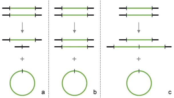
Opposite to tumor cells where there is little or no need of homologous sequences for the rearrangement of chromosomal sequences to produce circular amplicons, in Leishmania homologous sequences are needed for the generation of amplicons [Grondin et al., 1996]. Also, unlike mammalian amplifications, Leishmania amplifications appear to be homogenous and possess the minimum number of DNA rearrangements necessary to generate an amplicon from the chromosomal gene; one for the direct amplification (Figure 5a) and two for the inverted amplification (Figure 5b). They usually range from 30000 bp to
200000 bp. Secondary rearrangements of amplified DNA were considered rare events in Leishmania but studies showed that gene amplification is more dyna-mic than anticipated. It is now suggested that gene rearrangements leading to gene amplification are widespread and that circular amplicons are generated from linear amplicons [Grondin et al., 1998]. There are three types of
amplifi-cation described in Leishmania. Deletion amplifiamplifi-cation (Figure 6a), where the copy of wild-type chromosomal locus is deleted during the generation of the extrachromosomal amplicon, resulting in a heterozygous deletion line. Conservative amplification (Figure 6b), where the generation of the amplicon produces no alterations in chromosomal structure or ploidy. And, duplicative amplification (Figure 6c), where several additional Figure 5. Schematic representation of DNA structures of direct amplification (a) and inverted amplification (b) in Leishmania. The blue
boxes represent repetitive DNA sequences whose orientations are indicated by black arrows. Green arrows indicate the orientation of the gene being amplified (green line).
I.
I
NTRODUCTIONcopies are found inserted into the locus in addition to the amplified amplicon. As stated before, this extrachromosomal amplicons can be either a result of direct amplification or inverted amplification [Beverley, 1991].
2.3. Mechanism
Leishmania amplicons, whose structure have been characterized, seem to rule out gene amplification models that imply alteration of the parental chromosome structure (like sister chromatid exchange or recombination in the absence of re-replication). Extrachromosomal amplicons are likely to be generated from extra copies of the chromosomal locus formed by re-replication during the cell cycle. The onionskin model (Figure 7) seems to explain the initial structure of the re-replication. This hypothetical
structure has this name
because it resembles the skin layers of an onion when multiple rounds of replication initiate in the same origin within a single replication bubble in a given replication cycle. The flexibility of this model allows to virtually
Figure 6. Schematic representation of deletion (a), conservative (b) and duplicative (c) amplification. The black
lines represent flanking chromosomal DNA, the green lines represent DNA segments that give rise to extrachromosomal circular amplicons in addition to being either deleted, conserved or duplicated in the chromosome.
Figure 7. Schematic representation of the onionskin model.
Overreplication gives rise to a layer structure. The black lines represent genomic DNA and the blue areas correspond to repetitive DNA sequences flanking the DNA fragments (green) that will be amplified.
I.
I
NTRODUCTIONobtain all types of amplification. Unaltered DNA strands can maintain the normal chromosome structure after the collapse of the onionskin, and the copies generated are released as extrachromosomal amplicons, explaining this way the conservative amplification. Rearrangements in the chromosome strand can lead to deletion plus amplicon formation, explaining the deletion amplification. Rearrangements between the chromosomal strand and the replicated segments can lead to duplicative amplification within the chromosome, with the release of the extra copies generated as extrachromosomal amplicons. It also explains the generation of linear amplicons whenever the released copies acquire telomeres by the action of a leishmanial telomerase. Since the formation of amplicons in Leishmania is associated with homologous repetitive sequences it is supposed that these sequences give rise to hotspots for DNA rearrangement and amplification. Genes flanked by direct repeats undergo direct amplification (Figure 5a) while genes flanked by inverted repeats undergo inverted amplification (Figure 5b). The final step of the mechanism consists in the ability to increase and maintain the copy number of the circular amplicons. A selective pressure can ensure that a sufficient number of copies of a specific gene are maintained in the parasites. However, little is known about the mechanism for autonomous replication of the amplicons, only that no specific sequences seem to be required for replication [Beverley, 1991; Papadopoulou et al., 1994].
2.4. Response to drug pressure
Leishmania differs from other closely related protozoan parasites in the reaction to drug pressure. In Trypanosoma brucei gene amplification is extremely rare and in parasites of genus Plasmodium the gene amplification is mainly chromosomal, therefore in these parasites the increase of gene copy number is very reduced, so other mechanisms of overexpression of multidrug resistance genes play an important role. On the other hand, it was observed that the ability of Leishmania to selectively increase the number of gene copies is responsible for resistance. This was possible due to the relatively low genomic complexity of Leishmania. Gene amplification, mostly as extrachromosomal amplicons, is now considered the main adaptation response of these parasites to drug pressure [Beverley, 1991; Papadopoulou et al., 1998]. Changes in membrane permeability, allowing a reduction of intracellular drug accumulation, or a decrease in drug intake and inactivation of the drug by metabolism or sequestration are the mechanisms used by Leishmania to survive when in contact with antileishmanial compounds. The increase of gene copy number will not only directly increase the level
I.
I
NTRODUCTIONof expression but also augment the probability of point mutations to appear, which may confer advantage to the parasite when in contact with antileishmanial drugs [Croft et al., 2006].
3. Drug resistance in Leishmania
3.1. Pentavalent antimonials
Pentavalent antimonials (SbV) are considered pro-drugs. In order to acquire
antileishmanial activity these compounds require biological reduction to the trivalent form (SbIII). Although the site of reduction remains unclear data suggests that it happens inside
the parasite as well as within the macrophage. The pentavalent form is known to stimulate the infected macrophages, increasing the oxidative/nitrosative stress on the intracellular parasites. SbIII acts directly on the parasite, deregulating its redox-balance.
SbV is taken up by the parasite by an unidentified transporter and SbIII enters via
aquaglyceroporin (AQP1). Reduction of SbV to SbIII inside the parasite can be a thiol
dependent non-enzymatical spontaneous reduction or enzymatically catalyzed by thiol-dependent reductase 1 (TDR1) or arsenate reductase 2 (ACR2). Maintenance of redox balance inside the parasite is performed by trypanothione (T(SH)2) and trypanothione
reductase (TR) which reduces trypanothione disulfides (TS2) to T(SH)2, keeping the
redox potential low. T(SH)2 is synthesized by condensation of glutathione and
spermidine, where gamma-glutamylcysteine synthase (GSH1) is the key enzyme for glutathione (GSH) synthesis. SbIII inhibits TR and can form conjugates with T(SH)
2 and
glutathione, leading to an increase in redox potential (Figure 8) [Wyllie et al., 2004; Decuypere et al., 2012].
In order to acquire resistance to antimonials it was shown that Leishmania can use different strategies. A decrease in the reduction of SbV to SbIII, by reducing the levels of
TDR1 and ACR2 inside the parasite leads to increase resistance. However, it is not effective in protecting from external SbIII. In strains exhibiting decreased susceptibility to
SbIII a decrease in AQP1 expression was noted, although no difference in gene copy
number was found. Decreasing the influx of SbIII is probably the first barrier of the
parasite to counteract the action of antimonials. An increase in the synthesis of glutathione and trypanothione from cysteine was also reported in resistant strains. This helps to restore thiol redox potential perturbed by the accumulation of TS2, caused by
the inhibition of TR activity by SbIII. It also increases the spontaneous formation of SbIII
I.
I
NTRODUCTIONsequestered by ABC-thiol transporter MRPA (MRPA) into intracellular organelles or pumped out by an uncharacterized transporter (Figure 8). Previous studies also shown that an increase in MRPA alone cannot confer resistance to SbIII in Leishmania and other
factors are needed. GSH1 seems to play a major role in the resistance conferred by MRPA, and although an increase of GSH1 substantially increases the resistance when conjugated with an increase of MRPA, it was shown that normal levels of GSH1 are enough for the MRPA-related resistance to occur [Callahan et al., 1994; Grondin et al., 1997; Haimeur et al., 2000; Croft et al., 2006; Ashutosh et al., 2007; Jeddi et al., 2011].
3.2. Miltefosine
Miltefosine (MILT) also known as hexadecylphosphocholine was only recently introduced as an antileishmanial drug but even before this introduction concerns about the emergence of resistance to MILT were already present. Data from a phase IV Figure 8. Overview of Leishmania pathways involved in response and resistance to pentavalent antimonials
(SbV). ACR2 – Arsenate reductase 2; AQP1 – aquaglyceroporin; GSH – Glutathione; GSH1 –
Gamma-glutamylcysteine synthase; MRPA – ABC-thiol transporter MRPA; ROI – Reactive oxygen intermediates; RNI – Reactive nitrogen intermediates; SbIII – Trivalent antimonials; SbIII/thiols – Conjugates of SbIII with T(SH)2 and/or GSH;
TDR1 – Thiol-dependent reductase 1; TR – Trypanothione reductase; T(SH)2 – Trypanothione; T(S)2 – Trypanothione
I.
I
NTRODUCTIONtreatment trial show a relapse rate twice as high when compared to first-line treatment. The multitude of proposed mechanisms of action for MILT and the contradictory data from several studies may indicate that more than one molecular site of action is used. These mechanisms seem to be related to alterations in the lipid metabolism, mediation of apoptosis-like cell death, mitochondrial dysfunction and immunomodulation. A perturbation of ether-phospholipid metabolism related to inhibition of glycosomal alkyl-specific acyl-CoA acyltransferase was found in Leishmania when treated with MILT. Also, a decrease in phosphatidylcholine and an increase in phosphotidylethanolamine was observed. This suggests that MILT is responsible for the alteration of the composition of the parasite membrane. Apoptosis-like cell death was also reported in parasites exposed to MILT. This usually happens after exposure to reactive oxygen species (ROS) and lead to DNA fragmentation, nuclear condensation, loss of cell volume and consequently cell death. Therefore, MILT may be responsible for disrupting the intracellular redox balance of the parasite that allows it to tolerate the ROS inside the phagolysosome. The involvement of mitochondrial dysfunction was also investigated and the inhibition of cytochrome-c oxidase was observed. This inhibition was responsible for a substantial reduction in the mitochondrial membrane potential. As an additional contributory factor MILT was shown to have immunomodulatory proprieties, being able to enhance interferon-gamma receptors in infected macrophages and thereby increase T helper cell type 1 response, necessary to fight the parasite infection [Croft et al., 2006; Soto et al., 2007; Dorlo et al., 2012].
Emergence of drug resistance to MILT in vivo has not yet been described, although a strain of L. infantum with decreased susceptibility was recently isolated from a non-responsive HIV/VL patient. In vitro studies demonstrated that it is easy to induce resistance to MILT in Leishmania. These studies concluded that a defect in drug internalization and increased drug efflux from the parasite were the possible mechanisms of resistance. Two Leishmania lipid translocases, putative miltefosine transporter protein (MT) and MT non-catalytic subunit protein ROS3 play an important role in maintaining the phospholipid asymmetry of the parasite membrane. Their low expression was proven to be directly related to resistance to MILT, since a lower amount of these proteins leads to a decrease in the internalization of the drug into the parasite. Overexpression of P-glycoprotein MDR1 (MDR1) and ABC subfamily G-like transporters ABCG6 and ABCG4 was also implicated in MILT resistance, being responsible for a decrease in drug accumulation in the parasite. Several single point mutations in the genes coding these proteins were also observed in vitro [Coelho et al., 2012; Dorlo et al., 2012; Luque-Ortega et al., 2012].
I.
I
NTRODUCTION3.3. Amphotericin B
Amphotericin B (AmB) is a polyene antibiotic that binds preferentially to ergosterol, the major sterol of Leishmania. AmB binding to ergosterol causes the formation of transmembrane AmB channels which alter the permeability to cations, water and glucose and affect membrane-bound enzymes. These alterations eventually lead to the parasite death. Studies have also shown that AmB binds to the cholesterol present in the macrophages, decreasing the ability of the parasite to enter these cells and modulates macrophage activity by inducing the production of proinflammatory cytokines and ROS [Chia & McManus, 1990; Mozaffarian et al., 1997].
Apart from relapses of VL patients co-infected with HIV, no other signs of resistance were found so far for AmB. These relapses were also proven not to be a cause of resistance of the parasite to AmB, thus all the data available arrive from in vitro experiments. AmB-resistant Leishmania showed significant changes in membrane sterol composition, where most of the ergosterol was replaced by a precursor, cholesta-5,7,24-trien-3β-ol. This was a result of a loss of function of sterol-methyltransferases responsible for ergosterol biosynthesis whose genes most likely suffered deletion amplification [Croft et al., 2006; Paila et al., 2010].
3.4. Strategies to combat drug resistance
There are several strategies to combat drug resistance that only now are starting to be taken into consideration, mostly because of the increase and spreading of resistance to SbV.
The variation of parasite sensitivity to several drugs is a problem in areas endemic for more than one species of Leishmania. This can be solved by improvement of the diagnostic methods, especially non-invasive tests with high sensitivity and specificity for precocious detection of antileishmanial antigen and able to distinguish different species of Leishmania. A precocious, robust and accurate diagnosis would improve the treatment, allowing a correct choice of drug, the right dosage and determine the period of treatment. This would also aid to minimize the possible failure of treatment and consequently reduce the probability of the emergence of drug resistance.
The monitoring of the therapy also plays an important role. One of the causes for the appearance of drug-resistant Leishmania is believed to be the reduced level of compliance with treatment, since disease symptoms disappear before the full clinical cure. A better strategy in this field is needed, perhaps the implementation of control
I.
I
NTRODUCTIONprograms like the ones already in practice by the World Health Organization for tuberculosis. These programs rely on the directly observed treatment, short-course (DOTS) strategy. The DOTS strategy combines government commitment, case detection, standardized treatment, direct observation of the patience and compliance with treatment by healthcare workers, regular drug supply and standardized recordings and, a reporting system that allows assessment of the treatment results.
The distribution and cost of the drugs are also another important factor. The elevated cost of the more efficient and safe drugs and their unavailability in several public health institutions (especially in countries of low income) is something that needs to be changed. Either by researching on new, highly effective and cheaper drugs or by implementation of programs by governments or private institutions able to distribute the right drugs among those that need them.
Another way to prevent the emergence of drug resistance is to monitor the susceptibility of the parasite strains to the common drugs. This could be done by create and implement a routine test in endemic areas with high risk of resistance emergence. This would allow a better choice of treatment and higher efficacy rate in the elimination of the disease. Also, the use of combination therapies would be facilitated. Given that the possibility of resistance development to a single therapeutic agent is high, the chance of developing simultaneous resistance to two compounds with different targets would lower the probability of resistance emergence by a great amount.
Furthermore, the most relevant strategy is probably the research of new drugs and new targets. An adequate amount of drugs with different targets and no cross-resistance is the most efficient way to avoid the appearance of drug resistance [Sundar, 2001; Sundar & Rai, 2002; Croft et al., 2006; Jain, 2010; Antinori et al., 2012].
3.5. New drugs
In spite of all the currently used antileishmanial drugs, none of them conjugates high efficiency, low toxicity and an affordable cost. So it is still a priority the development of new therapeutic compounds for leishmaniasis.
Current research on new antileishmanial drugs is especially focused on already existing compounds used for the treatment of other diseases, compounds from natural sources, like plants, and even formulations already in use as herbicides. Several compounds of diverse molecular structure with potential antileishmanial activity have been isolated. Among these compounds, the most notable are dinitroanilines, alkaloids, particularly indoles, naphtylisoquinolines, bisbenzylisoquinolines, benzoquinolizidines,
I.
I
NTRODUCTIONtriterpenes, steroids, saponins, sesquiterpenes, diterpenes and flavonoids, mainly isoflavones and chalcones [Marques et al., 2008; Passero et al., 2013].
4. Dinitroanilines derivatives
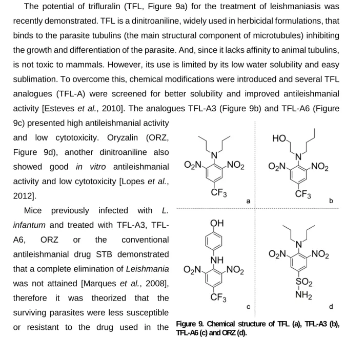
The potential of trifluralin (TFL, Figure 9a) for the treatment of leishmaniasis was recently demonstrated. TFL is a dinitroaniline, widely used in herbicidal formulations, that binds to the parasite tubulins (the main structural component of microtubules) inhibiting the growth and differentiation of the parasite. And, since it lacks affinity to animal tubulins, is not toxic to mammals. However, its use is limited by its low water solubility and easy sublimation. To overcome this, chemical modifications were introduced and several TFL analogues (TFL-A) were screened for better solubility and improved antileishmanial activity [Esteves et al., 2010]. The analogues TFL-A3 (Figure 9b) and TFL-A6 (Figure 9c) presented high antileishmanial activity
and low cytotoxicity. Oryzalin (ORZ, Figure 9d), another dinitroaniline also showed good in vitro antileishmanial activity and low cytotoxicity [Lopes et al., 2012].
Mice previously infected with L. infantum and treated with A3, TFL-A6, ORZ or the conventional antileishmanial drug STB demonstrated that a complete elimination of Leishmania was not attained [Marques et al., 2008], therefore it was theorized that the surviving parasites were less susceptible or resistant to the drug used in the treatment.
Figure 9. Chemical structure of TFL (a), TFL-A3 (b), TFL-A6 (c) and ORZ (d).
II.
O
BJECTIVESII.
O
BJECTIVES
Since it was theorized that parasites that survived exposure to conventional and new antileishmanial drugs in animal models were less susceptible or resistant to the drug used in the treatment, the main objective of this study was to analyze the level of gene amplification of resistance-related genes in these L. infantum strains and evaluate the resistance achieved by the parasite to the different drugs used.
To evaluate the antileishmanial activity of a commonly used antileishmanial drug (STB) and new chemically synthesized compounds (ORZ, TFL-A3 and TFL-A6) and their potential to generate drug-resistant parasite strains, previously infected mice were treated with these compounds, and the parasites that survived to the treatment quantified and collected for DNA extraction and real-time PCR analysis.
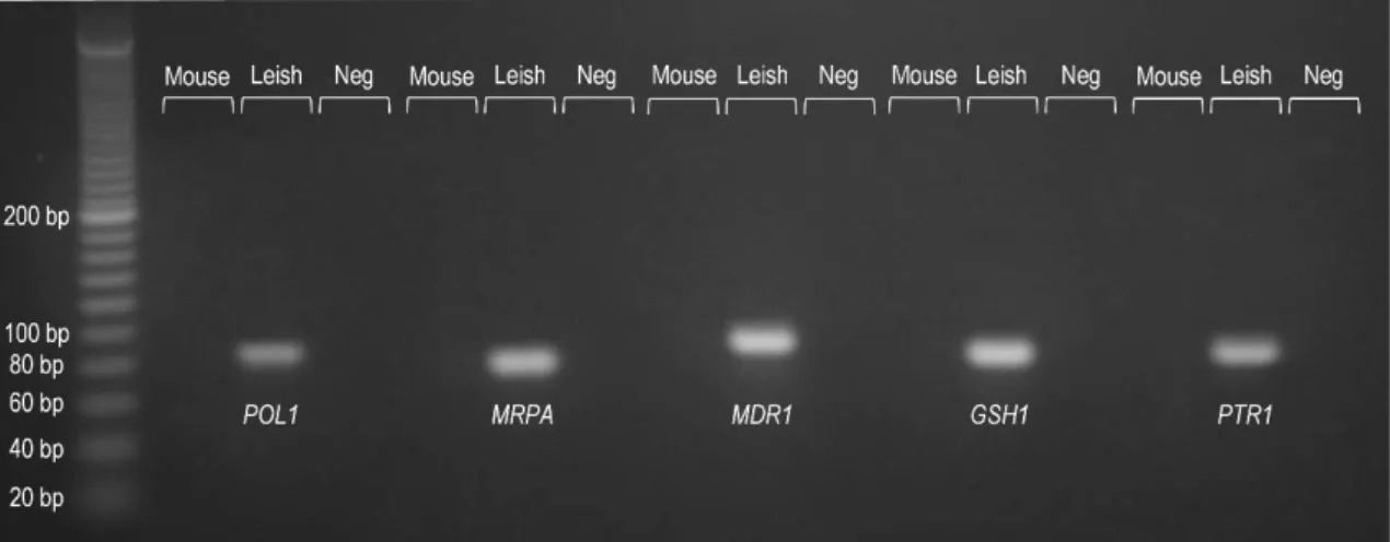
Previous studies linked the amplification of four specific genes to drug resistance in clinical strains of L. infantum: the genes for energy depended transporters MRPA (MRPA, RefSeq XM_001465669.1) and MDR1 (MDR1, RefSeq XM_001468445.1), the gene for the enzyme GSH1 (GSH1, RefSeq XM_001464941.1) involved in glutathione and trypanothione synthesis and, the gene for pteridine reductase 1 (PTR1, RefSeq XM_001465671.1) an enzyme involved in the folate metabolic pathways [Mary et al., 2010]. Therefore, these four genes (MRPA, MDR1, GSH1 and PTR1) were selected as targets for gene amplification analysis.
To analyze the gene amplification in an in vitro scenario, L. infantum promastigotes were made resistant to several antileishmanial compounds (MGA, MILT, TFL-A3 and TLF-A6) and the level of gene amplification accessed by real-time PCR. For absolute quantification by real-time PCR to be possible, the number of copies of each gene were normalized relatively to the number of copies of the single copy invariant gene for DNA polymerase alpha catalytic subunit (POL1, RefSeq XM_001464606.1) for every sample analyzed.
By investigating the differences in gene copy number of the selected genes in the different resistant strains it is also possible to clarify the mechanisms responsible for the parasite survival when exposed to antileishmanial compounds.
III.
M
ATERIALS ANDM
ETHODSIII.
M
ATERIALS AND
M
ETHODS
1. In vivo infection and treatment
1.1. Animals and parasites
1.1.1. Animals
A total of 65 female BALB/c Mus musculus mice were used in three independent experiments. All the animals were infected with L. infantum. For the first two experiments 20 mice were randomly divided into four groups of five mice each. One untreated control group and each of the other three groups treated with a different drug: STB, TFL-A3 and TFL-A6. In the third experiment 25 mice were randomly divided into five groups of 5 mice each. One untreated control group and each of the remaining four groups were treated with a different drug: STB, TFL-A3, TFL-A6 and ORZ. The animals were purchased and maintained in the IHMT animal facility according to the EU requirements (86/609/CEE) and Portuguese law (DR DL129/92 and Portaria 1005/92).
1.1.2. Parasites
L. infantum zymodeme MON-1 (MHOM/PT/89/IMT151) was used to infect BALB/c mice in these experiments. L. infantum MON-1 was maintained in the laboratory by successive passages in BALB/c mice. The spleens of infected mice were extracted and homogenized with a tissue disaggregator with 50 µm separator screen (Medicons, Syntec International, Ireland) to isolate a suspension of single cell amastigotes. This cell suspension was added to Schneider medium (SCHN, Sigma-Aldrich, Germany) supplemented with 10% (v/v) of heat-inactivated (30 minutes at 56ºC) fetal bovine serum (FBS, Sigma-Aldrich), penicillin-streptomycin (Biochrom, Germany) at 100 U/ml and 100 µg/ml respectively (complete SCHN medium), and incubated at 24ºC. In axenic cultures the intracellular amastigotes change into free metacyclic promastigotes.
1.2. Infection
Concentration (promastigotes/ml) from cultures of metacyclic promastigotes was calculated by optical microscopy using a Neubauer Chamber. The culture was centrifuged at 1800xg for 10 minutes and resuspended in the appropriate amount of
III.
M
ATERIALS ANDM
ETHODSsterile saline solution (0.90% m/v of NaCl) in order to achieve a concentration of 1x10⁷
promastigotes/ml. Each mouse was intraperitoneally (IP) inoculated with 100 µl of this solution.
1.3. Treatment
Forty-five days post infection mice were randomly sorted into groups of 5 mice each. The control group was IP injected with 200 µl of trehalose-citrate buffer (10 mM sodium citrate, 135 mM NaCl, 29 mM trehalose, pH 5.5) once a day for 10 days. Groups treated with TFL-A3 and TFL-A6 (dissolved in trehalose-citrate buffer with 5% Tween 80) were IP injected with a dose of 25 mg TFL-A/kg/day for 10 consecutive days. The group treated with ORZ (dissolved in trehalose-citrate buffer with 5% Tween 80) was intravenously injected for 10 consecutive days with a dose of 25 mg ORZ/kg/day. Fifty days post infection, the mice treated with STB were injected subcutaneously with 15 mg STB/kg/day for 5 consecutive days.
1.4. Parasitic load determination
Three days after the conclusion of the treatment mice were sacrificed and their spleen aseptically removed and weighted. The spleens were homogenized individually in 4 ml of complete SCHN medium using Medicons. Viable parasite loads were estimated by limiting dilution assay (LDA) in 96 well plates. An additional 1:2 dilution of the cell suspension was made and 200 µl of this cell suspension was placed in the first well of each row of 96 well plates and fourfold serial dilutions were made until the last column of the plate. For each homogenized cell suspension, four rows of a 96 well plate were used in order to have four replicates. The LDA plates were sealed and incubated during 15 days at 24ºC. After incubation each well was examined by optical microscopy and labelled as positive if promastigotes were present or negative if no parasites were detected. The highest dilution for which the well contained promastigotes was used to calculate the number of parasites per gram of tissue (ppg) as follows:
ppg=
�Reciprocal titer of highest positive dilution ×Volume of first well × Dilution factor Volume of cell suspension� Weight of homogenized tissue (g)