R E S E A R C H
Open Access
Cardiorespiratory alterations in rodents
experimentally envenomed with
Hadruroides lunatus
scorpion venom
Fernanda Costal-Oliveira
1, Clara Guerra-Duarte
1, Maira Souza Oliveira
2, Karen Larissa Pereira de Castro
1,
Leticia Lopes-de-Sousa
1, Aline Lara
3, Enéas Ricardo de Morais Gomes
3, Cesar Bonilla
4, Sílvia Guatimosim
3,
Marília Martins Melo
2and Carlos Chávez-Olórtegui
1*Abstract
Background:Hadruroides lunatusis the most abundant scorpion species in the Peruvian central coast, where most of the accidents involving humans are registered. In spite of its prevalence, there are only very few studies onH. lunatusenvenomation. The aim of the present study was to analyze the cardiorespiratory alterations caused byH. lunatusenvenomation in rodents.
Methods:Wistar rats injected withH. lunatusscorpion venom were submitted to electrocardiography. After euthanasia, rat lungs were collected and histopathologically analyzed. Mouse cardiomyocytes were used to perform immunofluorescence and calcium transient assays. Data were analyzed by ANOVA or Student’s t-test. The significance level was set atp< 0.05.
Results:It was observed thatH. lunatusvenom increased heart rate and caused arrhythmia, thereby impairing the heart functioning. Lungs of envenomed animals showed significant alterations, such as diffuse hemorrhage. In addition, immunofluorescence showed thatH. lunatusvenom was capable of binding to cardiomyocytes. Furthermore, mouse ventricular cardiomyocytes incubated withH. lunatusvenom showed a significant decrease in calcium transient, confirming thatH. lunatusvenom exerts a toxic effect on heart.
Conclusion:Our results showed that H. lunatusvenom is capable of inducing cardiorespiratory alterations, a typical systemic effect of scorpionism, stressing the importance of medical monitoring in envenomation cases.
Keywords:Hadruroides lunatusvenom, Cardiorespiratory alterations, Electrocardiography, Immunofluorescence, Calcium transient
Background
The genus Hadruroides comprises 22 species of
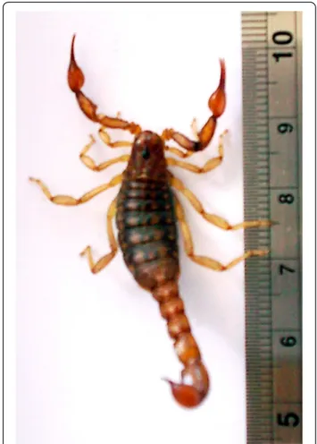
scor-pions, distributed throughout Ecuador, Peru, northern Chile and islands around Galapagos, occupying habi-tats predominantly of arid climate [1]. Scorpions from the speciesHadruroides lunatusare abundant in Peru-vian central coast, particularly around the city of Lima, in rocky areas with “lomas” formations. This species comprises small to medium-sized scorpions that have
brownish coloration, with dorsal lighter spots (Fig. 1). H. lunatusdiffers from other Hadruroidesscorpions by the curved morphology of the pedipalp fixed finger, creating a gap when fingers are closed, and by the rect-angular shape of the spots on the tergites [2].
H. lunatus is the most medically relevant species in Peru [3]. During 2009, the Health Ministry of Peru [4] reported 41 cases of human accidents caused by this scoprion in Lima. Although these stings are not consid-ered lethal, intense pain, edema, ulceration and necrosis are among the reported symptoms and signs.H. lunatus venom has not been extensively studied and there are not sufficient case reports describing human envenom-ation by this species. However, considering our previous * Correspondence:olortegi@icb.ufmg.br
1Department of Biochemistry and Immunology, Institute of Biological
Sciences, Federal University of Minas Gerais, Belo Horizonte CP: 486 CEP: 31270-901, MG, Brazil
Full list of author information is available at the end of the article
works [5, 6], we hypothesize that this scorpion has po-tential to cause significant damage to their victims.
In a first attempt to characterize the effects of this venom, our group reported initial data of H. lunatus experimental envenomation in rodents [5, 6]. Although H. lunatus scorpion venom (Hlsv) was classified as
moderately toxic when compared with Tityus spp.
venoms, symptoms such as excitability, agitation, sali-vation, eye secretions, convulsions, leg paralysis, as well as serological, biochemical and enzymatic alter-ations were detected in envenomed animals. These symptoms closely resemble those produced by the venom of scorpions pertaining to the Buthidae family, which con-tains the most medically relevant species that possess, in some cases, neurotoxins in their venoms [7, 8]. These molecules are peptides that act on ion channels and result in great release of neurotransmitters, seriously affecting hemodynamic and cardiorespiratory systems [9, 10].
These previous works suggestedHlsvmay have cardio-toxic effects, since its activity was associated with high serum levels of creatine kinase (CK) and its isoenzyme MB (CK–MB) [5]. The aim of this study was to confirm this possible cardiotoxic activity ofHlsv.
Methods
Animals and venom
Twelve male Wistar rats (weighing 100–150 g) and
four male C57BL/6 (18–22 g) mice were maintained at the animal facility of the Institute of Biological Sci-ences, Federal University of Minas Gerais (UFMG), Belo Horizonte, MG, Brazil, and received water and food under controlled environmental conditions. The experimental protocols were approved by the Ethics Committee on the Use of Laboratory Animals of UFMG (CETEA-UFMG protocol 092/11).
H. lunatus scorpions were collected in the region of Atocongo (Lima, Peru) and maintained in the National Institute of Health (INS), in Lima, Peru. Scorpions were
kept in plastic boxes with water ad libitum and fed
weekly with cockroaches. Venom was obtained by tel-son electrical stimulation (12 V) [11]. The venom was diluted in Milli-Q water and stored at−20 °C until use. Protein concentration was measured by the Lowry method [12].
SDS-PAGE
Different amounts (5, 10 and 20 μg) of Hlsv were di-luted in sample buffer under reducing conditions and separated in 15 % SDS-PAGE gel, according to Laemmli [13]. The gel ran at 200 V and was stained with silver.
Electrocardiography (ECG)
Rats were anaesthetized using 2.5 % isoflurane with a Metalvet Plus anesthetic inhaler (Metalvet, Brazil) and placed in supine position. Pre-anesthetic medication (morphine 2.5 mg/kg and diazepam 2.5 mg/kg) was ad-ministrated via intramuscular injections [14]. Electrodes were attached to forelimbs and hindlimbs. Control group (n= 6) received 0.4 mL of Milli-Q water via subcutane-ous (SC) injection, whilst Hlsv treated group (n= 6) re-ceived Milli-Q water containing 750 μg of Hlsv. This dose was chosen as the amount of venom corresponding to one third of the LD50 stablished for this venom [5]. Computer ECG (ECG-PC TEB, Brazil) tracings were taken prior to the experiment (T0) and variables were analyzed each 5 min throughout the examination, com-prising seven time points (T0, T5, T10, T15, T20, T25, T30). Heart rate (HR), heart rhythm, wave measurement and intervals were evaluated. As the ECG software gives the RR interval in millisecond (ms), the heart rate (beats per minute) was calculated by dividing 60,000/RR inter-val (ms), as 1 min corresponds to 60,000 milliseconds. ECG was recorded at speed of 50 mm/s, sensitivity of 2 N and lead II was considered for analysis.
Histopathological examination
Rats were euthanized by hypovolemia under anesthesia and submitted to necropsy. Lungs were removed, fixed
in 10 % buffered formalin and embedded in paraffin [15]. Histological sections (4 mm) were stained with hematoxylin and eosin, and analyzed in optical microscope.
Immunofluorescence and calcium transient measurements
Cardiac ventricular myocytes were isolated from C57BL/6 mice by standard enzymatic solution, as previously de-scribed [16]. Briefly, animals were euthanized; hearts quickly removed and perfused using a customized Langendorff apparatus with a solution containing type II collagenase (Worthington, USA). After isolation, cells were maintained in Dulbecco’s modified Eagle’s medium
(DMEM –Sigma, USA) containing 10 % of fetal bovine
serum until use.
For immunofluorescence assay, 500 μL of a solution
containing freshly isolated cardiomyocytes (in DMEM containing 10 % fetal bovine serum) was incubated with
0.05 or 2 μg/mL of Hlsv at room temperature under
agitation for 30 min, followed by incubation with 200 μg/mL of rabbit IgG anti-Hlsv for 30 min. Cells were centrifuged, the supernatant removed, and cell pellet was resuspended in medium containing anti-rabbit IgG conjugated to Alexa Fluor 488 (Invitrogen, USA) for 30 min. After centrifugation, the supernatant
was removed and 500 μL of fresh medium was added.
Images were acquired with a Zeiss LSM 510META con-focal microscope (Zeiss Jena, Germany) and analyzed with ImageJ software (NIH, USA). Control cells were incubated only with anti-rabbit IgG conjugated to Alexa Fluor 488 or with anti-Hlsv IgG plus IgG conju-gated to Alexa Fluor 488.
To measure the intracellular calcium (Ca2+) transi-ent, cardiomyocytes were incubated with a calcium sensitive fluorescent probe (fluo 4 AM–5μmol/L) for
30 min, and then with 0.05 μg/mL of Hlsv for 5–
20 min. Calcium transient amplitude was examined in field-stimulated cells at 1 Hz, with a square pulse of 5 ms and 30 V. After application of eight electric pulses, a line-scan imaging was performed in the longitudinal axis of the cells with an acquisition frequency of 1.54 ms, using Zeiss 510 Meta confocal microscope. Thirty-four cells were analyzed in the control group and 32 inHlsvgroup.
Statistical analysis
All variables were submitted to normality and homosce-dasticity analyses and then analysis of variance (ANOVA). Parametric variables were studied by Student-Newman-Keuls (SNK) posttest and non-parametric variables were evaluated by either Kruskal-Wallis or Friedman posttests. Regression analysis was accessed for HR throughout all time points. Significance was considered for 5 % (p< 0.05). Analyses were done in R (2.11 version) software program.
Student’st-test was used to access significance of variabil-ity among groups in calcium transient analysis.
Results
SDS-PAGE
To evaluate venom content, electrophoresis of different amounts ofHlsv(5, 10 and 20 μg) under reducing con-ditions was performed. The venom profile showed, in addition to a high content of low molecular weight compounds compatible with neurotoxins, a consider-able amount of proteins in the range between 12 and 14 kDa. Another group of proteins were also visualized above 30 kDa (Fig. 2).
Electrocardiographic analysis
At T0, all animals from both groups showed similar ECG tracings with no visible alterations. As usual, RR interval distance was considered to calculate the HR (Table 1). The mean HR on Hlsvgroup significantly in-creased from T0 to T5. The RR interval from an animal
fromHlsvgroup went from 133 ms (HR of 451 bpm) at
T0 to 97 ms (HR of 619 bpm) at T5. It means that after 5 min of envenomation, animals presented early signs of poisoning, demonstrated by the significant increase of HR (mean of 424 to 463 bpm), being detected a HR of 619 bpm in one of the animals. At this time (T5), no dif-ference was observed on the control group. At T10, the HR of Hlsv group decreased to a value similar to T0,
remaining normal up to T30. Within Hlsv group, HR
values fitted cubic regression (R2= 74.36 %; p= 0.0486)
Fig. 2SDS-PAGE 15 % ofHlsv.In lane 1, the low molecular weight marker. In the other lanes, 20, 10 and 5μg ofHlsvunder reducing
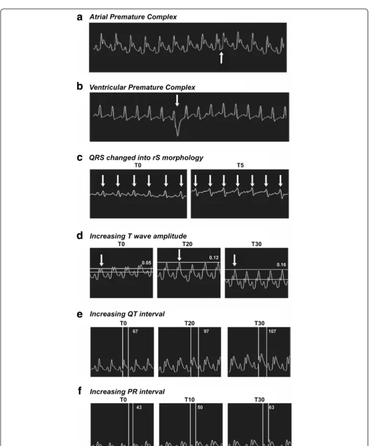
and T5 indicated the highest HR value (p= 0.0380), be-ing others similar to T0 (Table 1). Within control group, HR values fitted simple linear regression (R2= 93.04 %; p= 0.0004) and no differences were detected in all times. Arrhythmias were detected only onHlsvgroup at T15. Alterations such as atrial premature complex (APC) (Fig. 3a) and ventricular premature complex (VPC) (Fig. 3b) were detected in two different animals.
An individual fromHlsv group showed rS wave at T5
(Fig. 3c). None of these alterations were observed in control group. Other sign of envenomation observed on
Hlsv group was increased T wave amplitude. As shown
in Fig. 3d, T wave increased from 0.05 mV (T0) to 0.12 mV (T20) and reached 0.16 mV (T30) (p< 0.05). There was no change on amplitude of T wave for con-trol group. On the other hand, from 10 to 30 min after venom administration, T wave amplitude on Hlsvgroup was higher than control reaching the highest values at T25 and T30 (Table 1).
Alterations in QT interval (Fig. 3e) and in PR interval (Fig. 3f ) in Hlsv group were observed. QT interval in-creased from 67 ms (T0) to 97 ms (T20) and 107 ms (T30) (p< 0.05). PR interval also increased by showing 43 ms at T0, 50 ms at T10 and 63 ms at T30 (p< 0.05).
Histopathological examination
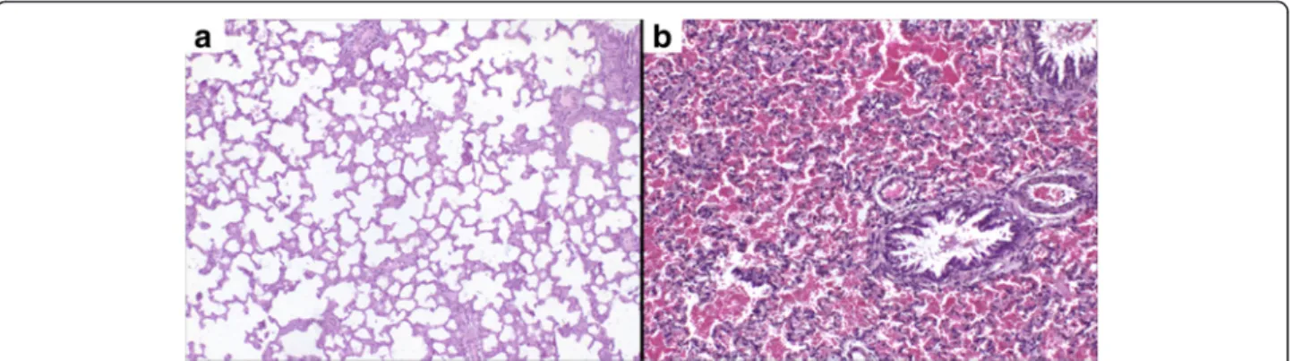
Lungs of animals from control and Hlsv groups were re-moved and analyzed microscopically. Only lungs of Hlsv treated rats showed significant alterations. Those of control animals were morphologically normal (Fig. 4a), whilst lungs of envenomed rats showed diffuse hemorrhage (Fig. 4b).
Immunofluorescence and calcium transient
To investigate whether Hlsv could bind to mouse ven-tricular cardiomyocytes, suggesting a direct cardiotoxic effect (Fig. 5), freshly isolated ventricular myocytes were treated withHlsvand then incubated with anti-HlsvIgG for evaluation. Fluorescent labeling was detected only in
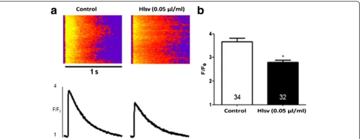
Hlsv treated cells (Fig. 5a). Results were analyzed using ImageJ software to quantify the fluorescence intensity (Fig. 5b). A concentration-dependent binding was observed. At last, it was investigated whether Hlsv could cause alterations in the calcium transient in isolated cardio-myocytes. Calcium transient amplitude, given by the ra-tio between maximal fluorescence (F) and baseline fluorescence (F0), was significantly reduced in cardiac
cells incubated with Hlsv compared to control cells
(Fig. 6). Thirty-four cardiomyocytes were analyzed in control group and 32 in theHlsvgroup (Fig. 6b).
Discussion
It is known that scorpion envenomations associated with cardiorespiratory alterations may culminate in heart fail-ure, pulmonary edema and even death [10, 17]. The present study is the first to evaluate the consequences of H. lunatusvenom with special attention to the cardiore-spiratory system. Other than some transcriptomic ana-lysis, little is known about H. lunatus and other non-Buthidae scorpion venoms and the potential harm caused by accidents with these less medically relevant scorpion species [18–21].
Since scorpion envenomation leads to adrenalin re-lease, the increase in the heart rate detected by ECG might be a consequence of its positive chronotropic ef-fect [22]. Venom interference on heart electric activity regulation results in ionic imbalance between intra and extracellular spaces. Consequently, the duration of cell depolarization phase is longer and leads to a hyperex-cited status [23]. This explains the premature complexes of atrial or ventricular origin detected on envenomed rats.
Electrolytic imbalance has already been reported as a systemic effect of scorpion envenomation [10]. Such al-teration may be diagnosed by ECG as the presence of in-creased T waves, which has been detected in victims of scorpionism [24]. T wave amplitude must be lower than a quarter of the R amplitude and higher values are a sensitivity parameter for electrolytic imbalance [23]. Therefore, increased T waves found in Hlsvgroup may be a consequence of electrolytic imbalance.
Moreover, rS wave detected onHlsvgroup suggests over-load of the right ventricle, due to pulmonary hemorrhage, which was confirmed by diffuse hemorrhage detected in lung histology. Occurrence of pulmonary alterations due scorpion envenomation is a common finding in the literature and pulmonary edema, hemorrhage and in-flammation have already been described, but predomin-antly in victims of stings by Buthidae family scorpions [10, 22, 25, 26]. Describing such alterations as a result of envenomation by a species considered not very toxic is re-markable and draws attention to the fact that accidents with this scorpion should receive medical attention, since
Table 1Heart rate and T wave amplitude alterations
Time (T) Heart rate (bpm) T wave amplitude (mV)
min Control Hlsv Control Hlsv
T0 398.1 ± 25.1 420.04 ± 36.9 0.07 ± 0.03 0.07 ± 0.03 T5 406.6 ± 22.1 463.37 ± 40.9ab 0.08 ± 0.04 0.09 ± 0.04 T10 420.1 ± 20.0 419.02 ± 29.3 0.08 ± 0.04 0.14 ± 0.03cd T15 426.6 ± 22.1 416.23 ± 24.0 0.08 ± 0.03 0.15 ± 0.02cd T20 443.3 ± 13.7 402.30 ± 17.4 0.07 ± 0.03 0.17 ± 0.03cd T25 450.0 ± 15.2 397.56 ± 20.8 0.06 ± 0.03 0.19 ± 0.02cde T30 446.6 ± 18.8 410.43 ± 37.5 0.06 ± 0.02 0.19 ± 0.03cde
Hlsvgroup presented significant increase of heart rate at T5 (highlighted in bold).avs. control group T5; T5bvs.Hlsvin all other time points.Hlsvgroup
presented increased T waves at all times after T10 (highlighted in bold).cvs.
lung complications are considered the main cause of death in victims of scorpion stings [25–27].
Some of these ECG findings were also detected in a similar animal model using Tityus fasciolatus venom, a member of the Buthidae family, one of the most toxic scorpions species [10]. Indeed, several ECG alterations
are also reported in patients severely envenomed by buthid scorpionsTityus[27].H. lunatusdoes not belong to this family and yet present similar alterations, which, once more, emphasizes the importance of studying the so-called less toxic venoms and providing careful med-ical support toHlsvenvenomed victims [28].
Fig. 4Histopathology of the lungs. (a) Lungs of control rats showing no alterations and (b) lungs with diffuse hemorrhage of animals injected with 750μg ofH. lunatusvenom. Magnification: 40x
Since cardiac alterations were detected by ECG and corroborated by prior enzymatic tests, we decided to evaluate the direct venom effect in cardiomyocytes [5]. Although the so-called “adrenergic storm”, caused by venom neurotoxin-induced discharge of catecholamines, is widely accepted as the main reason for the cardiore-spiratory impairment in scorpion envenomation, it is suggested that the release of cytokines and a direct effect of venom components on the heart can also account for this clinical condition [8, 29–31]. We have previously attested the presence of neurotoxins in Hlsv and the augment in inflammatory cytokines following Hlsv ad-ministration in mice [5, 6]. In the present work, it was shown that Hlsvis able to directly bind to mouse car-diomyocytes. Therefore, all the possible molecular mechanisms for the onset of cardiorespiratory syn-drome following scorpion envenoming seems to be present inHlsv.
The decrease in calcium transient amplitude observed in cells exposed toHlsvmight explain some of the ECG alterations detected on treated group. It was already re-ported that imperatoxin I (IpTxI), a 15 kDa phospholip-ase from Pandinus imperator scorpion, induces a fast and reversible blockade of ryanodine receptors (RYR) of skeletal and heart muscles. When injected into ventricu-lar cells, IpTxI leads to decreased amplitude of contrac-tion and intracellular calcium transient, which indicates a blockade of calcium release from the sarcoplasmic reticulum [32]. The lipolytic fraction M1 from Buthus occitanus tunetanusscorpion venom was also capable of decreasing calcium transient in isolated cardiomyocytes [33]. Although the specific toxin responsible for this
action was not identified, its lipolytic activity may sug-gest the presence of phospholipases in this fraction.
We have previously showed thatHlsvcontains remark-able phospholipase activity [5]. The presence of a high content of proteins between 12 and 17 kDa, compatible with PLA2molecular weight, was also attested by SDS-PAGE in the present work. Therefore, it is possible to suggest that an enzyme similar to IpTxI can be present in Hlsv and be involved in the effects observed in the present study. It has been indicated that scorpion venom
PLA2 can also be involved in the induction of lung
edema [34]. A component pertaining to this class of en-zymes inHlsvcan be the responsible for many of the al-terations described in this study. The isolation of Hlsv PLA2would help to elucidate its role in envenomation.
Conclusion
H. lunatusscorpion venom (Hlsv) induced cardiorespira-tory alterations in experimentally envenomed rodents. The study of the pathogenesis of systemic effects pro-voked by this venom, and the involvement of individual venom components in the complex alterations detected, will be useful for identifying suitable therapeutic agents for treating the clinical symptoms caused byHlsv.
Abbreviations
DMEM, Dulbecco’s modified Eagle’s medium; ECG, electrocardiography;Hlsv,
Hadruroides lunatusscorpion venom; HR, heart rate Acknowledgements
We would like to thank Dra. Cleida Aparecida de Oliveira and Dr. Geovani Cassali for the histological analysis.
Fig. 6Cardiomyocytes exposed toHlsvdisplay reduced Ca2+transient amplitude.a(Top) Representative confocal images of electrically stimulated
intracellular Ca2+transient recordings in ventricular myocytes. (Bottom) Ca2+transient line-scan profile.bSignificant reduction in peak Ca2+transient
amplitude was observed in freshly isolated adult ventricular myocytes incubated with 0.05μg/mL ofHlsvvenom for 5 to 20 min. Numbers
Funding
This work was supported by the Brazilian Coordination for the Improvement of Higher Education Personnel (CAPES–project“Toxinologia”no. 23038000825/ 2011-63), by Brazilian National Council for Scientific and Technological Development (CNPq– “Chamada Bilateral”no. 17/2013, process: 490269/ 2013-3) and by funds of the INCTTOX Program of CNPq.
Authors’contributions
FCO and CGD contributed equally to this work. They designed and developed the experiments, analyzed the results and prepared the manuscript for publication. MSO conducted the electrocardiogram experiments, analyzed the results and helped revising the manuscript for publication, under the supervision of MMM. MSO and MMM helped with organ collection, slides preparation and statistical analysis. KLPC assisted with the preparation of samples and animals, being responsible for their maintenance during all the work. AL and ERMG worked on the isolation of the cardiomyocytes and performed confocal microscopy and calcium transient experiments, under the supervision of SG, who was responsible also for manuscript drafting. LLS helped conducting the immunofluorescence assays. CB collected, maintained the scorpions and worked in the extraction of venom by the electrical stimulation method. CCO is the corresponding author and designer of the research. All authors read and approved the final manuscript.
Competing interests
The authors declare that they have no competing interests.
Ethics approval and consent to participate
All experimental procedures performed were in accordance to the guidelines of the Institutional Committee for Animal Care and Use of UFMG, Brazil, and the Guide for the Care and Use of Laboratory Animals (NIH, 8thedition, 2011).
The present study was approved by the Ethics Committee on the Use of Laboratory Animals of UFMG (092/11) (CETEA-UFMG).
Author details
1Department of Biochemistry and Immunology, Institute of Biological
Sciences, Federal University of Minas Gerais, Belo Horizonte CP: 486 CEP: 31270-901, MG, Brazil.2College of Veterinary Medicine, Federal University of
Minas Gerais, Belo Horizonte, MG, Brazil.3Department of Physiology and
Biophysics, Institute of Biological Sciences, Federal University of Minas Gerais, Belo Horizonte, MG, Brazil.4Instituto Nacional de Salud, Universidad Nacional
Mayor de San Marcos y Universidad Científica del Sur, Lima, Peru.
Received: 22 February 2016 Accepted: 6 July 2016
References
1. Brito G, Borges A. A Checklist of the scorpions of Ecuador (Arachnida : Scorpiones), with notes on the distribution and medical significance of some species. J Venom Anim Toxins incl Trop Dis. 2015;21:23. doi:10.1186/ s40409-015-0023-x.
2. Ochoa JA, Prendini L. The genusHadruroidesPocock, 1893 (Scorpiones: Iuridae), in Peru: new records and descriptions of six new species. Am Mus Novit. 2010;3687(3687):1–56.
3. Zavaleta A, Navarro J, Castro De La Mata R. Pharmacological effects of a Peruvian scorpion (Hadruroides lunatus) venom. Toxicon. 1981;19(6):906–9. 4. Andina. Reportan 41 casos de personas picadas por alacranes en Lima.
2010. http://www.andina.com.pe/agencia/noticia-reportan-41-casos-personas-picadas-alacranes-lima-276761.aspx. Accessed 14 July 2016. 5. Costal-Oliveira F, Duarte CG, Machado de Ávila RA, Melo MM, Bordon KCF,
Arantes EC, et al. General biochemical and immunological characteristics of the venom from Peruvian scorpionHadruroides lunatus. Toxicon. 2012;60(5):934–42. 6. Costal-Oliveira F, Guerra-Duarte C, Castro KLP, Tintaya B, Bonilla C, Silva W,
et al. Serological, biochemical and enzymatic alterations in rodents after experimental envenomation withHadruroides lunatusscorpion venom. Toxicon. 2015;103:129–34.
7. Possani LD, Alagon AC, Fletcher Jr PL, Erickson BW. Purification and properties of mammalian toxins from the venom of the Brazilian Scorpion
Tityus serrulatusLutz & Mello. Arch Biochem Biophys. 1977;180(2):394–403. 8. Isbister GK, Bawaskar HS. Scorpion Envenomation. N Engl J Med. 2014;
371(5):457–63. doi:10.1056/NEJMra1401108.
9. Couraud F, Jover E. Mechanism of action of scorpion toxins. In: Tu AT editor. Handbook of Natural Toxins, Insects Poisons, Allergens and Other Invertebrate Venoms. New York: Marcel Dekker Inc. 1984; p. 659–78. 10. Pinto MCL, Borboleta LR, Melo MB, Labarrére CR, Melo MM.Tityus fasciolatus
envenomation induced cardio-respiratory alterations in rats. Toxicon. 2010; 55(6):1132–7.
11. Oukkache N, Chgoury F, Lalaoui M, Cano AA, Ghalim N. Comparison between two methods of scorpion venom milking in Morocco. J Venom Anim Toxins incl Trop Dis. 2013;19(1):5. doi:10.1186/1678-9199-19-5. 12. Lowry OH, Rosebrough NJ, Farr AL, Randall RJ. Protein measurement with
the Folin phenol reagent. J Biol Chem. 1951;193(1):265–75.
13. Laemmli UK. Cleavage of structural proteins during the assembly of the head of bacteriophage T4. Nature. 1970;227(5259):680–5.
14. Flecknell P. Laboratory Animal Anaesthesia. San Diego: Academic; 1996. 15. Prophet EB, Mills B, Arrington JB, et al. Afip Laboratory Methods in
Histotechnology. Washington: American Registry of Pathology; 1992. 16. Guatimosim S, Sobie EA, dos Santos CJ, Martin LA, Lederer WJ. Molecular
identification of a TTX-sensitive Ca(2+)current. Am J Physiol Cell Physiol.
2001;280(5):C1327–39.
17. Cupo P, Hering SE. Cardiac troponin I release after severe scorpion envenoming byTityus serrulatus. Toxicon. 2002;40(6):823–30. 18. He Y, Zhao R, Di Z, Li Z, Xu X, Hong W, et al. Molecular diversity of
Chaerilidae venom peptides reveals the dynamic evolution of scorpion venom components from Buthidae to non-Buthidae. J Proteomics. 2013;89: 1–14. doi:10.1016/j.jprot.2013.06.007.
19. Ma Y, Zhao R, He Y, Songryong L, Liu J, Wu Y, et al. Transcriptome analysis of the venom gland of the scorpion Scorpiops jendeki : implication for the evolution of the scorpion venom arsenal. BMC Genomics. 2009;15:1–15. doi: 10.1186/1471-2164-10-290.
20. Quintero-Hernández V, Ramírez-Carreto S, Romero-Gutiérrez MT, Valdez-Velázquez LL, Becerril B, Possani LD. Transcriptome analysis of scorpion species belonging to the Vaejovis genus. PLoS One. 2015;10(2):e0117188. 21. Schwartz EF, Diego-Garcia E, Rodríguez de la Vega RC, Possani LD.
Transcriptome analysis of the venom gland of the Mexican scorpion
Hadrurus gertschi(Arachnida: Scorpiones). BMC Genomics. 2007;8:119. doi:10. 1186/1471-2164-8-119.
22. Paneque-Peres AC, Nonaka PN, de Carvalho PT, Toyama MH, Silva CAM, Vieira RP, et al. Effects ofTityus serrulatusscorpion venom on lung mechanics and inflammation in mice. Toxicon. 2009;53(7–8):779–85. 23. Tilley LP. Essentials of canine and feline electrocardiography interpretation
and treatment. 3rd ed. Philadelphia: Lea & Febiger. 1992.
24. Kumar CM, Naveen Prasad SV. Echocardiologic evaluation and follow - up of cardiovascular complications in children with scorpion sting in coastal South India. Indian J Crit Care Med. 2015;19(1):42–6. doi:10.4103/0972-5229. 148645.
25. Bahloul M, Chaari A, Dammak H, Samet M, Chtara K, Chelly H, et al. Pulmonary edema following scorpion envenomation: mechanisms, clinical manifestations, diagnosis and treatment. Int J Cardiol. 2011;162(2):86–91. 26. Saidi H, Adi-Bessalem S, Hammoudi-Triki D, Laraba-Djebari F. Effects of
atropine and propranolol on lung inflammation in experimental
envenomation: comparison of two buthidae venoms. J Venom Anim Toxins incl Trop Dis. 2013;19(1):8. doi:10.1186/1678-9199-19-8.
27. Cupo P. Clinical update on scorpion envenoming. Rev Soc Bras Med Trop. 2015;48(6):642–9.
28. Agrawal A, Kumar A, Consul S, Yadav A. Scorpion Bite, a sting to the heart! Indian J Crit Care Med. 2015;19(4):233–6. doi:10.4103/0972-5229.154570. 29. Andrade MV, Lisboa FA, Portugal AL, Arantes RM, Cunha-Melo JR. Scorpion
venom increases mRNA expression of lung cytokines. Comp Biochem Physiol A Mol Integr Physiol. 2007;146(4):581–7. doi:10.1016/j.cbpa.2006.01.031. 30. Teixeira Jr AL, Fontoura BF, Freire-Maia L, Machado CRS, Camargos ERS,
Teixeira MM. Evidence for a direct action ofTityus serrulatusscorpion venom on the cardiac muscle. Toxicon. 2001;39(5):703–9.
31. Cupo P, Figueiredo AB, Filho AP, Pintya AO, Tavares Jr GA, Caligaris F, et al. Acute left ventricular dysfunction of severe scorpion envenomation is related to myocardial perfusion disturbance. Int J Cardiol. 2007;116(1):98– 106. doi:10.1016/j.ijcard.2006.02.015.
32. Valdivia HH, Kirby MS, Lederer WJ, Coronado R. Scorpion toxins targeted against the sarcoplasmic reticulum Ca(2+)-release channel of skeletal and cardiac muscle. Proc Natl Acad Sci USA. 1992;89(24):12185–9.
M1 following muscarinic receptors interaction involving adenylate cyclase pathway. Toxicon. 2006;48(4):373–87. doi:10.1016/j.toxicon.2006.06.016. 34. Kanoo S, Deshpande SB. Involvement of phospholipase A2 pathway for the
Indian red scorpion venom-induced augmentation of cardiopulmonary reflexes elicited by phenyldiguanide. Neurosci Lett. 2008;440(3):242–5. doi:10.1016/j.neulet.2008.05.088.
• We accept pre-submission inquiries
• Our selector tool helps you to find the most relevant journal
• We provide round the clock customer support
• Convenient online submission
• Thorough peer review
• Inclusion in PubMed and all major indexing services
• Maximum visibility for your research
Submit your manuscript at www.biomedcentral.com/submit