AUTOLOGOUS BONE TISSUE ENGINEERING
STRATEGIES ENVISIONING THE REGENERATION OF
CRITICAL SIZE DEFECTS USING CELL-SEEDED
SCAFFOLDS AND A NEWLY DEVELOPED PERFUSION
BIOREACTOR.
LEANDRO GARDEL
TESE DE DOUTORAMENTO EM CIÊNCIAS VETERINÁRIAS
LEANDRO GARDEL
AUTOLOGOUS BONE TISSUE ENGINEERING STRATEGIES
ENVISIONING THE REGENERATION OF CRITICAL SIZE
DEFECTS USING CELL-SEEDED SCAFFOLDS AND A NEWLY
DEVELOPED PERFUSION BIOREACTOR.
Tese de Candidatura ao grau de Doutor em Ciências Veterinárias, submetida ao Instituto de Ciências Biomédicas Abel Salazar da Universidade do Porto.
Orientador–Doutor Rui Luís Reis Categoria–Professor Catedrático
Afiliação–Departamento de Engenharia de Polímeros da Universidade do Minho
Co-orientador–Doutor Luís Alvim Serra Categoria–Professor Catedrático Convidado Afiliação–Faculdade de Medicina do Porto Co-orientador–Doutora Manuela Estima Gomes Categoria–Professora Assistente Convidada MIT-Portugal
Afiliação–Departamento de Engenharia de Polímeros da Universidade do Minho
É AUTORIZADA A REPRODUÇÃO PARCIAL DESTA TESE APENAS PARA EFEITOS DE INVESTIGAÇÃO, MEDIANTE DECLARAÇÃO ESCRITA DO INTERESSADO, QUE A TAL SE COMPROMETE
DATA:
ASSINATURA:___________________________________________________ ___
TO ALL THAT SOMEHOW
CONTRIBUTED TO THIS THESIS
In the last few years had the privilege of knowing and working with many exceptional and talented people that contributed to the accomplishment of the work described in this thesis. To them, I would like to deeply express my gratitude and dedicate this thesis.
My first acknowledgments are made to my supervisor Prof. Dr. Rui Reis which gave me the privilege of being a part of the 3B’s Group some years ago. During this period he always transmitted to me motivation and as natural leader showed me that it is always possible to do better. His accomplishments, professionalism and leadership are motivation and make me feel privileged by working under his supervision.
To Profª. Drª. Manuela Gomes, my co-supervisor, for her knowledgeable and careful supervision of my work. She showed always to be available and, above all, she constantly gave me important advices and inputs that resulted in the improvement of my work. I’m also thankful to Profª Manuela for the time that she patiently lost with me explaining me how to overcome my limitations and doubts.
To Prof. Dr. Luís Alvim Serra, my co-supervisor, for guiding my scientific work and help. I really enjoyed all our discussions around orthopedics science. I also acknowledge the way Prof. Luís received me at the department of the orthopedics- Santo António General hospital, during this last ten years. I’m sincerely grateful.
I would also like to express my gratitude to Prof. Dr. António Pereira and Prof. Dr. António Fonseca, my directors in the ICBAS-UP.
I also thank Prof.ª Drª. Isabel Malheiro, Profª Drª Beatriz Porto and team (Rosa, Lara and Filipa) for giving me the opportunity to develop part of my thesis in cytogenetic laboratory. ICBAS-UP.
I would also like to express my gratitude to Prof. Dr. António Oliveira director of orthopedics services of General Hospital of Santo António. ICBAS-UP.
I also thank Prof. Dr Eduardo Rocha and your team (Fernanda and Célia) for giving me the opportunity to carry out part of my thesis in Histology laboratory. ICBAS-UP.
I’m especially grateful to my department colleagues Prof. Dr. Augusto Matos, Profª. Drª. Ana Lúcia, Prof. Dr. Plabo Payo, Profª Drª Claúdia Baptista, and Prof. Dr Miguel Farias.
I want show my deepest gratitude to Maria Afonso, at the beginning of project my student, today my colleague (DVM) for his precious help in revising the English of all the work of this thesis, thank you.
To Profª Carla Gomes for you help in statistical analysis, thank you.
I would like to dedicate a special thank two people (friends) without them this thesis would have been very difficult mission to accomplish, the technical experts Sr. Carlos Frias (U.Porto) and Sr. Marques (U.Minho), a big thank for your help, ideas and availability.
I want to thank all the ICBAS staff who in one way or another during these years helped me, Drª Jorge; Drª Joana, Drª Liliana, D. Maria do Ceú (could not forget), Costa
e Silva, Engª Ana, D. Vitória, Carla, Verônica,Madalena(AEICBAS),
Lena(reprographic), João e Joana Carvalheiro, hope I'm not forgetting anyone.
I would like to dedicate a special thank to 3B’s people João Oliveira, Tommaso Rada, Pedro Costa, Márcia Rodrigues, Ana Dias, Dennis Link, Alexandra Marques, Ana Frias, Miguel Oliveira, Tírcia Santos, Mariana Cerqueira, Albino Martins, Isabel Leonor, João Requicha, Rógerio Pirraço, Wojtek and Simone Silva.
I would like to dedicate a special thanks to the worldwide spread 3B’s colleagues.
I cannot forget and express particular gratitude to the all members of the 3B’s management team.
I should also acknowledge the Portuguese Foundation for Science and technology (FCT) for my PhD grant SFR/BD/66714/2009.
children Vítor and Rafael. For your love, patience, dedication and for letting I am part of their lives. You are the inspiration that makes me have a happy life. I dedicate this thesis to you.
Leandro S. Gardel was born in Niterói, Rio de Janeiro, Brasil, His background includes a five-years graduation in Veterinary Medicine in 1993, University Federal Rural of Rio de Janeiro, Brasil and he started his career in Portugal, as a Lecture/Assistant Researcher in 1996, in the College of Veterinary Medicine of the University of Trás os Montes e alto Douro (UTAD), working in area of Surgery and Anaesthesiology Veterinary, In 1998, Leandro Gardel assumed the same position at the College of the Veterinary Medicine of University of Porto- ICBAS, where he also had the opportunity to supervise several students/trainnes. In 2003 defended the thesis entitled:” Repair of fractures femurais oblique in dogs” a review work, integrated in the evaluation of pedagogical and scientific capacity to that allowed him to become Assistant Professor. In the same year he started his research work in 3B’s Research Group-Biomaterials, Biodegradables, Biomimetics, of the University of Minho (Supervised by Prof. Rui Reis and Profª. Manuela Gomes) and in 2005 he enrolled as a PhD student of University of Porto, developing his research work at the 3B´s Group- Headquarters of the European Institute of Excellence on Tissue Engineering and Regenerative Medicine in collaboration of University of Porto- ICBAS and General Hospital of Santo António – University of Porto- ICBAS. His main research topics are related to bone tissue engineering strategies, namely, adult stem cells isolation and differentiation from different sources and design of dynamic bioreactors to obtain highly in vivo functional tissue engineered substitutes. Currently, Leandro Gardel is responsible for the Orthopedics Surgery service of the Hospital Veterinary Medicine of ICBAS (UP VET). Leandro S. Gardel is author and co-author papers international referred journals, one European patent application (Patent pending), communications (oral presentation) and poster presentation at Worlds and European Congres
LIST OF PUBLICATIONS
The work performed during this PHD resulted in the following publications
PATENT PENDING
Gardel, LS. Gomes ME, Reis RL. PTC: Bi-Diretctional Continuous Perfusion Bioreactor for Tri-Dimensional Culture of Mammal Tissue Substtitutes
(A4TEC) Association for the Advancement of Tissue Engineering Cell Based Technologies & Therapies. Nº: 104278; Date: 04 of December 2008.
PAPERS INTERNATIONAL JOURNAL WITH REFEREES
Gardel LS, Dias A, Link D, Serra LA Gomes ME, Reis RL. Development of a novel bidirectional continuous perfusion bioreactor (BCPB) for culturing cells in 3D scaffolds. Histology and Histopathology Cellular and Molecular Biology, 26, 2011. (supplement 1) : 70-71.
ORAL COMMUNICATIONS IN INTERNACIONAL CONFERENCES
Gardel LS; Afonso M, Frias, C, Correia-Gomes C, Serra LA, Gomes ME, Reis RL Osteogenic properties of osteoblast-like cells loaded on starch poly(caprolactone) dishes perfused on a new perfusion bioreactor system in a goat critical sized tibial defect. 16th ESVOT Congress, Bologna, Italy, September, 2012
Gardel LS, Dias A, Link D, Serra LA Gomes ME, Reis RL. Development of a novel bidirectional continuous perfusion bioreactor (BCPB) for culturing cells in 3D scaffolds. TERMIS EU 20011, Annual Meeting, Granada, Spain, June, 2011.
Gardel LS, Frias C Afonso M, RadaT, Serra LA Gomes ME. Reis, RL. A regenerative Medicine Aproach for the Treatment of Large Gap on Bone Fracture in the Cat Using Mesenchimal Stem Cells: A Case Report. 3rd World Veterinary Orthopaedic Congress, ESVOT-VOS, 15th ESVOT Congress, Bologna, Italy, September, 2010
stem Cell Therapy (ASCT) for the treatment of bone fractures in cat: A case report. World Conference on Regenerative Medicine and Veterinary Stem Cell Consortium, Germany, Leipzig, October, 2009.
POSTER PRESENTATION IN INTERNACIONAL CONFERENCES
Gardel LS, Dennis P. Link, Ana F. Dias, Manuela E. Gomes, Rui L. Reis. Development of an continuous perfusion bi-directional bioreactor for large-sized constructs. 2nd IBB Scientific Meeting, Braga, Portugal, October 2010
PAPERS INTERNATIONAL JOURNAL WITH REFEREES
Link D, Gardel LS, Correlo VM, Gomes ME, Reis RL. Osteogenic properties of osteoblast-like cells loaded on starch poly(Ű-caprolactone) fiber meshes in a rat critical-sized cranial defect. Histology and Histopathology Cellular and Molecular Biology, 26,2011. (supplement 1): 386.
Oliveira, JT, Gardel, LS, Rada, T, Martins L, Gomes ME, Reis RL. Injectable gellan gum hydrogels with autologous cells for the treatment of rabbit articular cartilage defects. J Orthop Res. 28, 2010. (9):1193-9.
Costa, PF, Gardel, LS, Rada, T, Gomes ME and Reis RL, Stimulating adult stem cells from different origins for bone TERM approaches, Experimental Pathology and Health Sciences, 2, 2008. (1): 45.
ORAL COMMUNICATIONS IN INTERNACIONAL CONFERENCES
M. Afonso, L. Gardel Stem cell therapy for the treatment of canine osteoarthritis: application in four patients. 16th ESVOT Congress, Bologna, Italy, September, 2012
Link D, Gardel LS, Correlo VM, Gomes ME, Reis RL. Osteogenic properties of osteoblast-like cells loaded on starch poly (Ű-caprolactone) fiber meshes in a rat critical-sized cranial defect. TERMIS EU 20011, Annual Meeting, Granada, Spain, June, 2011.
Oliveira JT, Gardel LS, Martins L, Santos TC, Rada T, Silva MA, Malafaya PB, Marques AP., Gomes ME, Sousa RA, Castro AG, Neves NM, Reis RL The Use of Gellan Gum Hydrogels for Cartilage Tissue Engineering Applications: In vitro characterization and In vivo evaluation. ICRS - 8th World Congress of the International Cartilage Repair Society. Biscayne Bay Miami, FL, USA. May, 2009.
Oliveira JT, Gardel LS, Martins L, Rada T, Gomes ME, Reis RL. Injectable Gellan Gum Hydrogels With Encapsulated Adipose Derived Cells For Cartilage Tissue Engineering Applications: Performance In A Rabbit Knee Defect, 3rd International Meeting of the Portuguese Society for Stem Cells and Cell Therapies (SPCE-TC, University of Algarve, Faro, Portugal. ), April, 2008.
Costa P., Gardel LS, Rada T, Gomes ME, Reis RL. Stimulating adult stem cells from different origins for bone TERM approaches, IX International Symposium on Experimental Techniques, UTAD, Vila Real, Portugal. October, 2008.
Rada T, Gardel LS, Serra LA, Gomes ME,. Reis RL. A novel cell-specific isolation method for Adipose derived Stem cell. In: 1st Annual International Meeting of the Portuguese Society for Stem Cells and Cell Therapies, Madeira, Portugal, May, 2006.
POSTER PRESENTATION IN INTERNACIONAL CONFERENCES
Oliveira JT, Gardel LS, Martins L, Santos TC, Rada T, Silva MA, Malafaya PB, Marques AP, Gomes ME, Sousa RA, Castro AG, Neves NM, Reis RL. The Use of Gellan Gum Hydrogels for Cartilage Tissue Engineering Applications: In vitro characterization and In vivo evaluation. 8th World Congress of the International Cartilage Repair Society (ICRS), Miami, Florida, United States (electronic poster
presentation), May 2009.
Oliveira JT, Gardel LS, Serra LA, Gomes ME,. Reis RL .Adipose Tissue Derived Cells and Articular Chondrocytes Combined with Injectable Gellan Gum Hydrogels in the Treatment of Full Thickness Articular Cartilage Defects in Rabbits. 6th Annual Meeting of the International Federation of Adipose Therapeutics and Science (IFATS 08), Toulouse, France. October 2008.
Bone is a dynamic living tissue and thus, upon trauma, it continuously undergoes absorption, reconstruction and remodelling. However, in the case of severe bone lesions, this repairing mechanism might fail, leading to a biomechanical failure of the tissue. In such clinical situations, tissue engineering offers the ultimate possibility of providing alternative successful therapies for the regeneration of injured bone tissue, through the creation of a functional tissue substitute in vitro. This is generally achieved through a specific interplay between cells and supportive materials – scaffolds, which can be further modulated by the culturing system used. A major challenge in the translation of such tissue engineered bone substitutes to viable clinical treatments consists in the need to grow large, fully viable grafts that are several centimetres in size that can be used for the regeneration of large critical size defects. The use of flow perfusion bioreactors has shown evident advantageous over static culturing, as these systems usually allow a more efficient transfer of nutrients and oxygen, enabling an uniform distribution of cell seeded on the 3D scaffold, thus stimulating the development of homogeneous tissue for subsequent implantation. Some of these systems have also demonstrated the ability to provide mechanical stimulus to cell – seeded scaffolds, leading to higher functionality of the resulting constructs. However, up to now, these studies have focused on culturing small constructs, which do not address the clinical need for developing grafts that can be used in the regeneration of bone critical sized defects
The main objective of this thesis was to study the importance of scaffolds and cells in the regeneration of critical size defects and to develop a new flow perfusion bioreactor enabling to culture large sized defects with improved functionality for the treatment of large bone defects.Therefore, the main specific objectives proposed for this thesis were:
- Studying the importance of the scaffold material and autologous stem cells obtained from the bone marrow in the regeneration of critical sized orthotopic defects.
- Development of a new flow perfusion bioreactor system enabling the culture of
3D-scaffold with large dimensions, up to 120 mm in length and 60 mm in diameter, allowing maintenance of cell viability/functionality in the interior of the scaffold, and also enabling mechanical stimulation through fluid flow induced shear stresses.
- Analysing the feasibility of the developed bioreactor, studying the behaviour of the three dimensional starch-based scaffolds seeded with bone marrow MSCs and cultured under static and flow perfusion culturing conditions.
- Assessing the influence of three dimensional cell-scaffold constructs in vitro cultured in the newly developed bioreactor on the regeneration of bone critical sized defects performed in a large animal model
- Studying the use of bone marrow stromal cells (from rat, goat and cat) as a reliable cell source for bone tissue engineering application, as they can be readily available and obtained by simple harvesting procedures from the same patient, avoiding the risk of disease transmission and/or immune rejection. These cells can be used in tissue engineering strategies but also in other regenerative medicine approaches, such as the direct implantation into a tissue defect to stimulate healing
Under the scope of this thesis it was created, designed, built, developed and optimized a Bi-Directional Continuous Perfusion Bioreactor (patent pending) that enables culturing three dimensional cell-scaffold constructs of large dimensions, for the reconstruction of critical size defects for bone tissue engineering. The feasibility of the system was demonstrated by the functionality of the tissue engineering constructs obtained under in vitro culturing conditions provided by the bioreactor, that allows to mitigate diffusion limitations typical of static culturing and simultaneously provide physiological-like stimulus to the seeded cells.
The 3D scaffolds studied in this thesis, namely fiber mesh based on a biodegradble polymeric blend of starch with polycaprolactone (SPCL) were able to promote adhesion, proliferation and differentiation of marrow stromal cells (of different animal species) towards the osteoblastic phenotype, particularly when cultured under flow perfusion conditions. Furthermore, the in vivo studies demonstrated that these 3D scaffolds, play an important role both in the formation of bone tissue and neovascularisation, conditions important for the development and sustainability of cell-scaffolds of large dimensions, but also provided an excellent biomechanical environment for the regeneration of critical sized defects. In summary, the culture of an appropriate scaffold material with bone marrow cells, cultured in the new bioreactor herein developed may provide an important step forward in the development of bone tissue engineered grafts of large dimensions that are highly in demand in the orthopedical clinical field.
O osso é um tecido dinâmico que, quando traumatizado, sofre um processo contínuo de absorção, reconstrução e remodelação. Contudo, no caso de lesões ósseas severas, este mecanismo de regeneração pode falhar, levando à falência biomecânica do tecido. Nestas situações, a engenharia de tecidos oferece uma solução terapeutica alternativa, capaz de levar à regeneração do tecido ósseo danificado através da criação in vitro de um substituto de tecido funcional. Isto é conseguido através da interação entre células e um material de suporte tridimensional (“scaffold”), que podem ser posteriormente modulados através do sistema de cultura usado. Um dos maiores desafios para a translação clínica destes materiais hibridos (constituídos pelos scaffolds e células) produzidos por metodologias de engenharia de tecidos, é a necessidade da produção de materiais híbridos de grande dimensão (vários cm) totalmente viáveis para a regeneração de defeitos de tamanho crítico. A utilização de biorreatores de perfusão de fluxo permite uma maior eficácia na transferência de nutrientes e oxigénio e possibilita uma distribuição uniforme das células no biomaterial de suporte tridimensional levando assim, à estimulação do desenvolvimento de tecido homogéneo para subsequente implantação. A utilização deste tipo de bioreactores tem-se revelado vantajosa quando em comparação com sistemas de cultura estática sendo que, alguns destes sistemas demonstram serem também capazes de proporcionar estímulos mecânicos às células semeadas no biomaterial de suporte tridimensional, proporcionando uma maior funcionalidade dos materiais híbridos obtidos. Até ao momento, estes estudos têm-se focado na cultura de scaffolds de reduzida dimensão, que não permitem a sua utilização clínica na regeneração de defeitos ósseos de dimensão crítica.
O principal objetivo desta tese consiste em estudar a importância dos scaffolds e das células na regeneração de defeitos de dimensão crítica e desenvolver um novo biorreator de perfusão de fluxo que possibilite a cultura de scaffolds de grandes dimensões conducentes ao tratamento de defeitos ósseos de dimensões críticas. Assim, os principais objetivos específicos desta tese foram:
- Estudar a importância do material do biomaterial de suporte tridimensional e das células estaminais autológas obtidas por aspiração da medula óssea na regeneração de defeitos ósseos ortotópicos de tamanhos críticos.
- Desenvolver um novo bioreactor de perfusão de fluxo que possibilite a cultura de biomaterial de suporte tridimensional de grandes dimensões (até 120 mm de comprimento e 60 mm de diâmetro), que permita a manutenção da viabilidade/funcionalidade celular no interior do biomaterial de suporte tridimensional e possibilitar estímulos mecânicos, através das forças de tensão de corte induzidas pela perfusão do meio de cultura.
- Analisar a funcionalidade do biorreator desenvolvido através do estudo do comportamento de scaffolds baseados numa mistura polimérica de amido de milho e policaprolactona (starch/polycaprolactone - SPCL), e semeados com células estaminais mesenquimatosas, originárias da medula óssea, após cultura no bioreactor de perfusão de fluxo ou cultura em condições estáticas.
- Avaliar a influência dos materiais hibridos constituidos pelo scaffolds e células após cultura in vitro no novo biorreator, na regeneração em defeitos ósseos de tamanho crítico, através da implantação em modelos animais de grande porte (cabra).
- Estudar a utilização da medula óssea (de rato, cabra e gato) enquanto fonte segura de células estaminais para aplicações em engenharia de tecido ósseo, uma vez que estão disponíveis em quantidades interessantes e podem ser obtidas através de procedimentos de recolha simples a partir do próprio paciente, evitando assim o risco de transmissão de patologias e/ou rejeição imune. Estas células podem ser usadas em estratégias de engenharia de tecidos mas também noutras técnicas de medicina regenerativa, como é o caso da implantação direta em defeitos tecidulares para estimulação da cicatrização óssea.
No âmbito desta tese, foi criado, desenhado, construído, desenvolvido e otimizado um Biorreator de Perfusão Contínua Bi-direcional (patente pendente) que permite a cultura de scaffolds de grandes dimensões para a reconstrução de defeitos de tamanho crítico na área da engenharia de tecidos ósseos. A viabilidade do sistema foi demonstrada pela funcionalidade das estruturas de engenharia de tecidos obtidas em condições de cultura in vitro proporcionadas pelo biorreator que permite mitigar as limitações de difusão típicas de sistemas de cultura estática fornecendo, simultaneamente, um estímulo às células em cultura, semelhante ao existente em condições fisiológicas.
malha de fibra baseada numa mistura polimérica biodegradável de amido de milho com policaprolactona (SPCL), possibilitaram a adesão, proliferação e diferenciação de células medulares estromais (de diferentes espécies animais) em células de fenótipo osteoblástico, particularmente quando submetidas a condições de cultura em perfusão de fluxo. Paralelamente, o estudo in vivo demonstrou que estes biomateriais de suporte tridimensional têm um importante papel não só na formação de tecido ósseo e
na neovascularização (condições importantes para o desenvolvimento e
sustentabilidade de biomateriais de suporte tridimensional celulares de grandes dimensões) como também mostraram proporcionar um excelente ambiente biomecânico, fator importante no que diz respeito ao uso de biomateriais em defeitos de dimensão crítica. Em síntese, a cultura de biomateriais de suporte tridimensional apropriados, com células da medula óssea cultivadas neste novo biorreator pode providenciar um importante passo em frente no desenvolvimento de enxertos ósseos de grandes dimensões, desenvolvidos através de técnicas de engenharia de tecidos que constituem uma importante necessidade para o campo da ortopedia clínica.
TABLE OF CONTENTS
Ack nowledegements IV
Short Curriculum vitae VIII
List of publication IX Abstract XII Resumo XIV Table of contents XVII List of figures XXIV List of tables XXVIII List of abbreviation XXIX
SECTION 1 1
CHAPTER I. GENERAL INTRODUCTION: Bone biology 1.1. Introduction 3
1.2. Normal vascularization of bone 4
1.3. Structure and composition of bone tissue 4
1.3.1. Macroscopic organization of bone tissue 4
1.3.2. Microscopic organization of bone tissue 5
1.4. Cells of bone tissue 5
1.5. Biochemical composition of extracellular bone matrix 8
1.6. Functions of bone tissue 9
1.7. Remodelling of bone tissue 9
1.8. Biology of bone fracture healing 9
1.8.1. Inflammatory phase 10
1.8.2. Reparative phase 10
1.8.3. Remodelling phase 11
1.8.4. Primary bone healing 11
1.8.5. Secondary bone healing 11
1.8.6. Intramembranous bone formation 12
1.9. Bone regeneration 13
1.10. Bone mesenchimal stem cells (MSC) 13
1.11. Osteogenesis 15
1.14. Osteopromotion 18
References 20
CHAPTER II 27
REVIEW: Use of perfusion bioreactors and large animal models for long bone tissue engineering. Abstract 29
2.1. Introduction 30
2.2. Bioreactors for bone tissue engineering 31
2.3. Perfusion bioreactors 32
2.3.1. Perfusion bioreactors with individual culture chamber for culturing small 33
dimension constructs 2.3.2. Single culture chamber perfusion bioreactors that enable culturing large sized constructs 42
2.4. Large animal models 47
2.4.1. Selection of appropriate animal models 47
2.4.2. Animal models in bone repair research 48
2.4.3. Ovine models 49
2.4.4. Caprine models 50
2.5. Critical/cylindrical size defects 51
2.6. Creation of the cylindrical critical size defects in large animals 52
2.7. Fixation of the cylindrical critical size defects in large animals 53
2.8. Conclusion 59
2.9. Acknowledgments References 61
SECTION 2 75
CHAPTER III MATERIALS AND METHODS 3.1. Developed of a new Flow Perfusion Culture System 76
3.1.1. Design philosophy 76
3.1.2. Materials used in constituents parts 76
3.2. Scaffolds 81 3.2.1. Starch-poly (ε-caprolactone)(SPCL) fiber-meshs scaffolds 81 3.3. Harvesting, isolation and culture of bone marrow mesenchymal stromal cells 83 3.3.1. Harvesting, isolation and culture of rat bone marrow stromal cells 83 (RBMSC’s)
3.3.2. Harvesting, isolation and culture of goat bone marrow stromal cells 84 (GBMSC’s)
3.3.2.1 Cryopreservation of cells 84 3.3.2.2 Replating cells after cryopreservation 85 3.3.3 Harvesting, isolation and culture of cat bone marrow stromal cells 85 (CBMSC’s)
3.4. Osteogenic differentiation 86 3.5 “In-Vitro” biological testing 86 3.5.1 Culture of Goat bone marrow stromal cells in SPCL constructs 86 3.5.1.1 Seeding GBMSC’s onto SPCL constructs 86 3.5.1.2 Static culture 87 3.5.1.3 Bioreactor culture 87 3.6 “In-Vitro” characterization of the cultured cell-scaffolds constructs 88 3.6.1 Cell morphology – Scanning Electron Microscopy (SEM) 88 3.6.2 Cell viability assay 88 3.6.3 Cell proliferation assay 89 3.6.4 Determination of alkaline phosphatase activity in cells – scaffold 90 constructs
3.6.5 Histological analysis 90 3.6.6 Light microscopy 90 3.6.7 Statistical analysis 91 3.7 “In-Vivo” studies 91 3.7.1 Rat animal model 91 3.7.1.1 Seeding rat bone marrow stromal cells in SPCL constructs 91 3.7.1.2 Characterization of DNA in cell-seeded meshes 91 3.7.1.3 Animal surgical procedures 92 3.7.2 Characterization of the samples retrieved from in vivo study performed 93 in a rat model
3.7.2.1 Micro-CT analysis 93 3.7.2.2. Histology 93 3.7.2.3. Statistical analyses 93
3.7.3.1 Cell seeding onto SPCL scaffolds 94 3.7.3.2 Static Culture Groups 94 3.7.3.3 Bioreactor culture groups 94 3.7.3.4 Animal surgical procedures 95 3.7.4 Characterization of the samples retrieved from in vivo study performed 96 in a goat model 3.7.4.1 Digital radiography 96 3.7.4.2 Micro-CT 96 3.7.4.3 SEM 97 3.7.4.4 Histological analysis 97 3.7.4.5 Light microscopy 97 3.7.4.6 Statistical analysis 97 3.7.5 Cat animal model 98 3.7.5.1 Cells harvesting, culturing and implantation 98 3.7.5.2 Osteogenic differentiation - Determination of the alkaline 98 phosphatase activity
3.7.5.3 Osteogenic differentiation- Alizarin red staining 99 3.7.6 Characterization performed upon cells implantation 99 3.7.6.1 Radiography 99 3.7.6.2 Determination of alkaline phosphatase activity in cat blood serum 99 References 100
SECTION 3 103
CHAPTER IV
Osteogenic properties of starch poly (ε-caprolactone) (SPCL) fiber meshes loaded with rat bone marrow stromal stem cells (RBMSCs) in a rat critical-sized cranial defect
Abstract 105 4.1. Introduction 106 4.2. Material and Methods 107 4.2.1 Scaffolds preparation 107 4.2.2 Cell isolation and seeding into scaffolds 108 4.2.3 Characterization of cell-seeded meshes 108 4.2.3.1 DNA quantification 108 4.2.4Surgery 109 4.2.5 Characterization of the explants110
4.2.5.1MicroCT 110 4.2.5.2 Histology 111 4.2.6 Statistical analyses: 111 4.3 Results 111 4.3.1 In vivo experiment 112 4.3.1.1 General observations 112 4.3.1.2 MicroCT 112 4.3.1.3 Histology 114 4.4 Discussion 115 4.5 Conclusions 117 References 118 SECTION 4 122 CHAPTER V
A novel bidirectional continuous perfusion bioreactor for mammalian tissue growth in 3D scaffolds.
Abstract 124 5.1. Introduction 125 5.2 Materials and methods 126 5.2.1. Flow perfusion bioreactor design 126 5.2.2. Construction and functioning of the BCPB 127 5.2.3 Preliminary evaluation of the bioreactor functionality 129 5.2.3.1Scaffolds design and preparation 129 5.2.3.2 Cell seeding of scaffolds 130 5.2.3.3 Bioreactor culture 131 5.2.4. –Characterization of cell-scaffolds constructs 131 5.2.4.1 Cell proliferation and viability analysis 131 5.2.4.2 Determination of alkaline phosphatases (ALP) activity of cells on 132 scaffolds
5.2.4.3 Cell morphology analysis 132 5.2.4.4 Histological analysis 133 5.2.5 Statistical Analysis 133 5.3. Results 133 5.3.1 Alkaline phosphatises activity 133 5.3.2 Cell proliferation 134 5.3.3 Cellular viability 135
5.3.5 Histological analysis 137 5.4. Discussion 139 5.5. Conclusions 142 References 143 SECTION 5 148 CHAPTER VI
Assessing the repair of critical size bone defects performed in a goat tibia model using tissue engineered constructs cultured in a bi-directional flow perfusion bioreactor
Abstract 150 6.1. Introduction 151 6.2. Materials and methods 152 6.2.1 GBMSC’s harvesting, isolation and culture 152 6.2.2 Scaffold design and preparation 153 6.2.3 Flow-perfusion bioreactor design 153 6.2.4 GBMSC’s seeding onto SPCL scaffolds and bioreactor culture 154 6.2.4.1 Characterization of cell viability in seeded scaffolds 155 6.2.5 Animal surgical procedures 155 6.2.6 Characterization of explants from in vivo studies 157 6.2.6.1 Radiographic Analysis 157
6.2.6.2 Scanning Electron Microscopy (SEM) and Energy-Dispersive 158 X-ray Spectroscopy (EDS) analysis
6.2.6.3 Micro-CT analysis 158 6.2.6.4 Histological analysis 159 6.2.7 Statistical analysis 159
6.3. Results 159 6.3.1 Cell adhesion and viability of scaffolds 159
6.3.2 Radiographic analysis 160 6.3.3 SEM analysis 162 6.3.4. Micro-CT analysis 165 6.3.5 Histological analysis 166 6.4. Discussion 169 References 172
SECTION 6 176 CHAPTER VII
Autologous stem cell therapy for treatment of a tibial fracture gap in a cat. A case report. Abstract 178 7.1. Introduction 179 7.2. Case report 180 7.3. Results 182 7.4. Conclusion 186 Reference 187 SECTION 7 190 CHAPTER VIII 8.1- General Discussion 192
LIST OF FIGURES
SECTION 1CHAPTER I. GENERAL INTRODUCTION: Bone biology
Figure1.1. Schematic representation of the principal source of adult stem cells. 14
Adapted from:(57)
Figure1.2. Schematic representation of the osteogenic lineage. Adapted from:(57) 16 Figure1.3. Schematic representation of an ideal bone graft. Adapted from:(57) 19
SECTION 2
CHAPTER III: MATERIALS AND METHODS
Figure3.1. Design of the cellular culture chamber with the scaffold and culture 78
medium.
Figure3.2.Overview of the design of the BCPB. 79 Figure3.3.Two parts of extremities of the cellular chamber culture. 79 Figure3.4. Coaxial perforated tube in polycarbonate or stainless steel and 80
scaffold.
Figure3.5. Scaffold size of the same size of the perforations in the tube. 80 Figure3.6. Overview of the BCPB circular flow system. 81 Figure3.7. Schematic representation of the SPCL fiber-meshs scaffolds. 87
SECTION 3
CHAPTER IV: Osteogenic properties of starch poly(ε-caprolactone) (SPCL) fiber meshes loaded with rat bone marrow stromal stem cells (RBMSCs) in a rat critical-sized cranial defect
Figure4.1.Surgical approach and implantation of the scaffold. 110
Figure4.2. Implant characterization and SEM picture before implantation after 111
1 day of culturing.
Figure4.3. MicroCT reconstructions of the various implant formulations. 113
Figure4.4.MicroCT quantification of the new bone formation of the various 113
implant formulations.
Figure4.5. Histological sections stained with hematoxylin and eosin 114
(first two rows) and Masson’s trichrome (bottom row) of SPCL fiber meshes loaded with MSCs after four (A) and eight (B) weeks of implantion. Four (C) and eight (D) weeks of implantation of SPCL fiber meshes and empty controls
(E) after eight weeks were stained as well. C = cranial bone; F = fibrous tissue; N = newly formed bone; S = SPCL fibers
SECTION 4
CHAPTER V: A novel bidirectional continuous perfusion bioreactor for mammalian tissue growth in 3D scaffolds.
Figure5.1. A) Coaxial perforated tube in polycarbonate and scaffold. B) Scaffold 127
positioned in the tube.
Figure5.2. A) Representation of the cell culture chamber with the scaffolds 128
Inserted in the perforated tube and filled with medium culture. B) Picture of the complete BiDirectional Continuous Perfusion Bioreactor (BCPB) system under operation in a CO2 incubator.
Figure5.3. Schematic representation of the flow system “circuit” that is 129
Associated with the continuous bi-directional flow bioreactor
Figure5.4. Schematic representation of the SPCL fiber-mesh scaffolds 130
samples used in this study.
Figure5.5. ALP activity in static and flow culture (A), and maximum and 134
minimum ALP activity values expressed (B).
Figure5.6. DNA concentration in static and flow culture (A) and 135
maximum and minimum DNA concentration values expressed (B).
Figure5.7. MTS quantification in static and flow culture. 136 Figure5.8. A) Cells in the interior of the scaffolds in day 0, x1000. 136 Figure5.9. A) Cells in the interior of the scaffolds in day 14 flow, x 200. B) 137
Day 14 flow, x1000.
Figure5.10. A) Cells in the interior of the scaffolds in day 21 flow, x200.B) 137
Day 21 flow, x1000.
Figure5.11. A) Cells in the interior of the scaffolds in day 14 static, x200.B) 137
Day 21 static, x1000.
Figure5.12. Sections of cell-scaffolds constructs cultured in the bioreactor 138
and stained with H&E: (A) 14 days; (B) 21 days of culture. S: Scaffold.
Figure5.13. Sections of cell-scaffolds constructs cultured in static conditions 138
and stained with H&E: (A) 14 days; (B) 21 days of culture. S: Scaffold.
Figure5.14. Graphical representation of the scaffolds volume cultured in 140
CHAPTER VI: Assessing the repair of critical size bone defects performed in a goat tibia model using tissue engineered constructs cultured in a bi-directional flow perfusion bioreactor
Figure6.1. Schematic representation of the SPCL fiber-meshs stacked into a 153
scaffold of critical sized defect dimensions
Figure6.2. (A) Bone fragment removed together with the periosteum; (B) 156
Osteosynthesis material applied to the tibia; (C) Implantation of the biomaterial in tibia bone and fixation with osteosynthesis material;
(D) Empty defect (control group).
Figure6.3. X-ray obtained immediately after surgery, showing the correct 157 positioning of the bone fixation device. (A) Control group (empty defect);
(B) Static group; (C) Bioreactor group. In B and C images it is possible to visualize radiographically the cells-scaffolds constructs implanted within the defect.
Figure6.4. (A) Boxplot of MTS quantification from all groups, day 0;(B) Boxplot 160 of DNA quantification from all groups, day 0;(C) SEM photomicrographs obtained from cell seeded SPCL samples at day 0.
Figure6.5. Radiographs obtained after 6 weeks of implantation: 160 (A) Control- empty defect (B) Static group (C) Bioreactor group.
Figure6.6. Radiographs obtained after 12 weeks of implantation: 161 (A) Control-empty defects weeks; (B) Static; (C) Bioreactor.
Figure6.7. Radiographs obtained after implantation: (A) Static 6/ weeks group;(b) 162 Static/12 weeks group; (C) Bioreactor/6 weeks group;
(D) Bioreactor/12 weeks group.
Figure6.8. SEM photomicrographs of the cell-scaffolds constructs retrieved upon 163 implantation: (A) Static 6 weeks [A1 (x200),A2 (x500),A3 (x1000)];
(B) Static 12 weeks [B1 (x200),B2 (x500),B3 (x1000)]; (C) Bioreactor 6 weeks
[C1(x200),C2(x500),C3(x1000)] and (D) Bioreactor 12 weeks (D1 (x200),D2 (x500),D3 (x1000)].
Figure6.9. EDS spectra for Bioreactor 6 weeks group (A), Bioreactors 12 weeks 164 group (B), Static 6 weeks group (C) and Static 12 weeks group (D).
Figure6.10. µ-CT 3D virtual models images of the region corresponding to the 166 bone interfaces with SPCL-Cells constructs. A) Interface between proximal
fragment and scaffolds bioreactor 6 weeks group; B) Interface between distal fragment and scaffolds bioreactor 6 weeks group; C) Interface between proximal fragment and scaffolds bioreactor 12 weeks group and D) Interface among distal fragment and scaffolds bioreactor 12 weeks group.
Figure6.11. Light microscopy images of cell-scaffolds sections corresponding 167
to the bioreactor groups after 6 weeks implantation (HE (A) and MT (B) and
static groups after 6 weeks implantation ( H&E(C) and MT(D). Bone (B);
Matrix Deposition (MD); Fibrous Tissue (FT); Revascularization (R); Neovacularization (NV); SPCL fiber-meshs (S).
Figure6.12. Light microscopy images of cell-scaffolds constructs sections 168 corresponding to bioreactor groups after 12 weeks implantation (HE (E)
and MT (F), and static groups after 12 weeks of implantation. Bone (B); Bone Marrow (BM); Matrix Deposition (MD); Revascularization(R); Neovacularization(NV); SPCL fiber-meshs (S).
SECTION 6
CHAPTER VII: Autologous stem cell therapy for treatment of a tibial fracture gap in a cat. A case report
Figure7.1. Craneo-caudal radiography projections of the affected limb, where 180
it is observed a lateral displacement in the axial focus of fracture.
Figur7.2. Syringe containing the cells suspension, used for the application of 181
the cells in fracture gap.
Figure7.3. ALP activity in cells cultured in vitro, in osteogenic medium, at the 183
day of implantation (after expansion for 2 weeks in osteogenic medium) and at 7 and 21 days of subsequent culturing.
Figure7.4. Alizarin red staining of the cat bone marrow cells after 3 weeks of 183
in vitro culture in osteogenic medium
Figure7.5. ALP activity in blood serum of the cat, measured at the day of the 184
application of the cells (day 0) and 7 and 21 days after application.
Figure7.6. Craneo-caudal radiographic projections obtained at: (A) day 0 185
(day of cells application); (B) after 1 weeks of cells application in the fracture site; (C) after 3 weeks of cells application in the fracture site.
Figure7.7. Craneo-caudal and latero-medial radiographic projections showing 186 a medial deviation and recurvatum deformity of the tibial bone after eighteen
LIST OF TABLES
SECTION 1CHAPTER II: REVIEW: Use of perfusion bioreactors and large animal models for long bone tissue engineering.
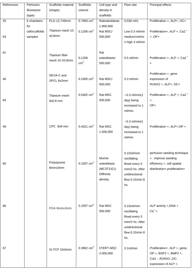
Table2.1.Summary table on research studies describing the development/use 34
of perfusion bioreactor of individual culture chambers in bone tissue engineering.
LIST OF ABBREVIATIONS
A
AdBMP7 adenoviral vector containing the bone morphogenetic protein 7 gene
AA ascorbic acid
ALP alkaline phosphatise
AO orthopaedic
association foundation
α MEM alpha modified eagle medium .
B
b-TCP b-tricalcium phosphate BCP biphasic calcium phosphate BCPB bi-directional continuous perfusion bioreactor BM bone marrowBMSCs bone marrow stromal cells BMP-2 bone morphogenetic protein 2 BSP bone sialoprotein BPT Thermoplastic Elastomer Tubing
C
CaCO3 calcium carbonate
CaP calcium phosphate
°C celsius degree
CG collagen-GAG
cm2 square centimeter cm3 cubic centimeter Col I collagen type I
CO2 dioxide carbon CT computed tomography cm2 square centimeter cm3 cubic centimeter C3H10T1/2 cell line
D
DCP dynamic compression plate DEX dexamethasoneDMEM dulbecco’s modified eagle’s medium
DMSO glycerol or dimethyl sulfoxide
DNA deoxyribonucleic acid
E
ECM extracellular matrix
EDS energy dispersive spectrometer
EO ethylene oxide
ESF external skeletal fixator
F
FBS Foetal bovine serum
FDA food and drug administration
FGF-2 fibroblast growth factor 2
FTIR fourier transform infra-red
G
GAGs glycosaminoglycans GBR guided bone regeneration GFP Green fluorescent proteinGMCs goat marrow stromal cells
GBMCs goat bone marrow mesenchymal cells
GMP good manufacturing practices
H
HASi triphasic ceramic-coated HA
hASCs human adipose-derived stem cells HE hematoxylin & Eosin
Hz hertz
hMSC humam mesenchymal stem cells
hFOB 1.19 Human fetal osteoblasts
hTERT human telomerase catalytic subunit
I
IGF-1 insulin growth factor-1
K
Kg kilograms
kPa kilopascal unit
L
LC-DCP limited contact dynamic compression plate LCP locking compression plateM
MATE mechanoactive transduction and evaluation bioreactor Micro-CT micro-computed tomographyMC3T3-E1 mouse immature
osteoblast-like cell line
mM milimolar
mm milimeters
mm3 cubic milimeters ml mililiters
imaging MSCs mesenchymal stem cells MT Masson Trichrome MTS/MTT 3-(4,5-dimethylthiazol- 2-yl)-5-(3-carboxymetyhoxyphen yl)-2(4-sulphophenyl)-2H tetrazolium µg micrograms µl microliters µm micrometers
N
N Newton NADPH nicotinamide adenosine dinuccleotide phosphateNADP nicotinamide adenine dinucleotide
NHPs nonhumans primates
O
OC osteocalcin
OP osteopontin
OP-1 osteogenic protein-1
O2 Oxigene
P
PA peptide–amphiphile PBS phosphate-buffered saline PCL polycaprolactonePET poly (ethylene- terepthalate)
PGA poly (glycolic acid)
PLGA poly (DL-lactic-co-glycolic acid)
pH potencial of hydrogen
PRP platelet rich plasma
E2 Prostaglandin E2
R
RBMCs rat bone marrow mesenchymal cells RGD Arg–Gly–Asp peptides rhTGFbeta-3 recombinant humam transforming growth factor-beta 3 RT-PCR reverse transcription polymerase chain reaction
RNA ribonucleic acid
S
SEM scanning electron microscopy SPCL starch-poly(ε -caprolactone)
T
TMJ temporomandibular joint TCP tricalcium phosphate cylindersT-CUP tissue culture under perfusion
TERM tissue engineering and regenerative medicine TGF-β1 Transforming growth factor beta1 TPS tubular perfusion system 2D two dimensional 3D three dimensional
U
UTN universal tibial nail
UV ultraviolet
V
VEGF vascular endothelial growth factor
X
SECTION 1
Chapter I
1.1 Introduction
The embryologic mechanisms of bone formation entail an orchestration of cellular humoral and mechanical factors resulting in the formation of skeletal bony tissues. Bone is a connective tissue with cells and fibres which are embedded in a hard substance that protects and supports it. In addition to these mechanical functions, bones serve an important chemical function, providing a reservoir for mineral hemeostasis (1, 2). Bone consists of several functionally distinct regions: At the articulating surfaces is the articular cartilage; Surrounding the entire bone is a membranous structure, the periosteum; Lining the area enclosing the cartilage (capsule) of the joints and also lining tendons sheaths are the synovial membranes, providing nutrition and lubrication to the articular cartilage while serving as a protective barrier; The woven lamellar, cancellous bone lies below the phyisis in a methaphysis, and the compact, cortical bone surrounds a marrow cavity in the disphyseal region(2). Bone is a dynamic tissue that, throughout life, is renewed and remodelled. It has a unique structure that provides a great tensile strength with the least amount of weight when compared to any other tissue, having the ability to adapt its internal structure to changes that might happen in the load environment (3). Structurally, bone is a dense material, or matrix, in which, as rule, spidery cells called osteocytes are embedded. The matrix is composed of collagen fibbers, crystals of a calcium-phosphate complex, and a ground substance, or cement, containing mucopolysaccharides, among many other components. The nucleated body of each osteocyte occupies a small cavity, or lacuna, in the matrix; and the long, branching, cytoplasmatic processes lie in fine tunnels, or canaliculi, which radiate from the lacuna.
Canaliculi from neighbouring lacunae anastomose freely; thus, bone is characteristically permeated throughout by an extremely rich system of communicating cavities and canals (4).Bone is a unique tissue that gives mechanical support to the body. It is a major reservoir of calcium and bone marrow, which produces the blood cells. Cells present in bone are the supporting cell, termed osteoblasts and osteocytes, and remodelling cells called osteoclasts. Osteoblasts are the skeletal cells responsible for synthesis, deposition, and mineralization of bone. Osteocytes are derived from osteoblasts. After secreting osteoid and mineral salts, osteoblasts become isolated in a cavity of bone matrix and become an osteocyte. Osteoclasts are constantly eroding deposited bone, while osteoblasts are constantly replacing osteoid and minerals.
In this way, bone is a dynamic tissue that is being formed and broken down in a continuous cycle in response to physical and hormonal factors (5, 6).
1.2 Normal Vascularization of Bone
In 1691, Clopton Havers described in his book how a large nutrient artery pieces the shaft of long bones and ramified in bone marrow. He presumed that an oily medullary substance was expelled into a system of straight pores within the bone, these pores becoming known as Haversian canals. It is now well accepted that the nutrient artery penetrating the nutrient foramen, divides into ascending and descending branches of the medullary artery and these are the main source of centrifugal blood flow in the cortical bone of long bones. The medullary blood vessels arborize and anastomose with both the metaphyseal vessels as well as the periosteal vessels in regions of the strong soft tissue attachment. This allows increased compensatory blood flow from the anastomossing supply should one system, such as the medullary supply be knocked out(7).
In Compact bones the vascularisation is performed between the afferent and efferent systems and function as the vascular lattice where critical exchange between the blood and surrounding living tissue occurs.
Cortical canal of Havers and Volkmann and the minute canaliculi make this system, which convey blood supply to the osteocytes(7).
1.3 Structure and composition of bone tissue
All bones – long, flat, intramembranous, woven and compact- are specialized forms of connective tissues, and their form and function, like those of other connective tissues, depend on the arrangement and interactions of the elements of the extracellular bone matrix that distinguishes it from other connective tissue matrices and enables it to perform its unique functions(2).
1.3.1 Macroscopic organization of bone tissue
Macroscopically, bone is considered to be a non-homogenous, porous (porosity varying from 5 to 95%) and anisotropic tissue (8).
Two main forms of bone may be seen in vertebrate organisms: woven bone (immature bone with randomly arranged collagen fibres in the osteoid, produced when osteoblasts rapidly produce osteoid) and lamellar bone (composed of regular parallel bands of collagen). The woven bone, which is rapidly formed, will eventually be remodelled and will form lamellar bone, a stronger type of bone (9, 10).
In the mature skeleton two types of lamellar bone may be found: cortical bone and medullary bone.
Compact or cortical bone (5-10% porosity) forms the walls of the diaphysis, it consists of parallel columns that individually are made of concentric lamellae disposed around Haversian channels (central channel with blood and lymphatic vessels and nerves, also called osteons) which, on their turn, connect with other Haversian channels and with periosteum and endosteum through Volkmann’s channels. The limits between osteons and surrounding bone are called cement lines.
Medullary bone or cancellous bone (50-95% porosity) occupies part of the central cavity of the bone and consists of trabeculae (a network of fine plates) which are separated by spaces that intercommunicate containing the medullary blood supply and filled mostly by fat tissue. An adult long bone, as for example the humerus or the femur, in a longitudinal cut consists of two enlarged ends called epiphyses which are connected by a hollow cylindrical shaft named diapysis. Between these, lies a very important cancellous type of bone named methapysis that contains important hematopoietic elements. Separating the epiphysis from metaphysis, we can distinguish a vital element for immature bone known as growth plate, wich is responsable for the longitudinal lengthening of long bones(9, 10).
1.3.2 Microscopic organization of bone tissue
Bone is composed of cells and an extracellular matrix predominantly collagenous called osteoid and it mineralizes after the deposition of calcium hydroxyapatite, which makes bone strong and rigid (10).
1.4 Cells of bone tissue
There are three principal cell types in all bones: osteoblasts, osteocytes, and osteoclasts.
Osteoblasts synthesize and mediate the mineralization of osteoid and are found lined along the bone surface (where new bone is deposited), ranging from columnar to squamous shape, the nucleus is located in the basal region and the cytoplasm is basophilic whit abundant endoplasmic reticulum. They are found on the surface of bone-forming regions, know as haversian systems, which surround blood vessels within the matrix of woven bone (2). These cells are of mesenchymal origin and are of great importance in various functions:
Matrix synthesis – being responsible for producing all matrix components and matrix mineralization – it is believed that initiation of mineralization is under control of the osteoblast within matrix vesicles (whose function is to destroy inhibitors of mineralization and to concentrate precipitates and crystals of calcium and phosphorus) facing the bone-forming surface (9, 10).
Initiation of resorption – receptors that activate osteoclastic bone resorption are found on osteoblasts: hormones (PTH, for example) and citocines that estimulate this process bind to osteoblasts. In the particular case of parathyroid hormone these receptors located on the cell membrane of osteoblasts induce an increase in intracellular cAMP that will phosporylate intracellular proteins consequently activating a physiological response (PTH will cause osteoblasts to round up and separate from one another and will also activate the release of collagenases that will then destroy the thin and unmineralized layer of collagen allowing osteoclasts to gain access to bone surface through gaps formed by this osteoblastic separation and destruction of the collagen layer). Communication with osteocytes (osteoblast-osteocyte network) – osteoblasts are able to send cytoplasmic projections into the matrix during the synthesis of the latter. Some of these projections extend through canaliculi and then from lining cells to the osteocytes and from osteocyte to osteocyte, forming gap junctions that allow small molecules and also ions to travel between these cells. Osteocytes are inactive osteoblasts trapped inside formed bone and are able to assist in the nutrition of the bone. These are the principal cell type in mature bone and they reside in lacunae, microscopic holes in the bone. These cells are able to send cytoplasmatic projections that are able to penetrate through the bone in canaliculi and gap junctions are present where the cell contacts processes of an adjacent bone. The long cellular projections are able to shorten and lengthen and this activity possibly allows the movement of pulsatile fluid flow through lacunae and canaliculi in order to transfer metabolites (10, 11). Osteocytes and bone-lining cells are presently thought to be the primary mechanosensory cells responsible for interpreting mechanical forces in bone tissue and translating them to osteoblasts and osteoclasts for bone remodelling (12-14).
Multiple investigators report evidence supporting the key mechanosensory role of osteocytes in bone formation as detected by changes in matrix protein expression, and production of nitric oxide (NO) and prostaglandin E2 (PGE2), a potent stimulator for bone formation (14, 15).
The osteocytes trapped deeper in the bone are older than the more superficial ones. Osteocytes play a role in the homeostasis of calcium and that changes in their metabolic activity are able to solubilise small amounts of mineral in the lacunar walls. Osteocytes are the most abundant differentiated cells. Numerous of these cells are released from the lacunae when osteoclast resorption is occurring (10, 11).
Osteoclasts are large phagocytic cells (20 to 100 µm in diameter) that have the capacity to erode the bone, and which are important in the process of bone turnover and refashioning. These cells are originated from the monocyte-macrophage system. Preosteoclasts are mononucleated cells that when activated will fuse with cells alike and when after accessing the bone surface, they differenciate further finally becoming multinucleated (15 to 30 nuclei per cell) osteoclasts. The activated ostoclasts have ruffled borders (is a resorbing organelle, and it is formed by fusion of intracellular acidic vesicles with the region of plasma membrane facing the bone (16) and secrete acid and lysosomal enzymes are directly responsible for removing the mineral and matrix bone (17, 18). After enzymatic and acid digestion (acid phosphatese, collagenase, cathepsins and neutral proteases) of the bone matrix the erosion lacune (Howship’s lacuna) is formed(2).Differences in mode of degradation have been observed as well. Calvarial (flat bone) osteoclasts use cysteine proteinases as well as matrix metalloproteinases (MMPs) to degrade the bone matrix, whereas resorption by long bone osteoclasts is accomplished mainly by cysteine proteinases (19, 20). This lacuna will remain even after the osteoclast is gone, which indicates prior areas of bone resorption.
Osteoclasts are also found as part of cellular complexes involved in bone remodelation and these cells are responsible for the release and activation of TGF-beta, and of mineral and protein components of bone, participating in the maintenance of blood calcium homeostasis by responding to parathyroid (which stimulates osteoclastic resorption and the release of calcium ions from the bones) an calcitonin (acts as a mitogenic agent on pre-osteoblastic cell lineage) (10).
Interleukine 1 (in vitro induces bone resorption and osteoclast-like cell formation in human and murine marrow cultures; in in vivo studies induced hypercalcemia and growth and differentiation of the earliest identifiable osteoclast precursor – CFU-GM), IL - 6 (controversial role, potentiates the effect of PTH on calcium homeostasis and bone resorption in vivo) and IL-11 (effects on osteoclast differentiation), M-CSF (important role on osteoclast development), TGF-α (proliferative factor, stimulates the growth of early osteoclast precursors), TNF-α and β (stimulate the formation of osteoclast like multinucleated cells in human marrow cultures, increase bone resorption, enhance the effect of IL-1) are soluble factors that increase osteoclast activity. TGF-β (modulates
osteoclastic bone resorption, migration and differentiation, inhibiting bone resorption)
IFN-γ (inhibitor of bone resorption in vitro, suppresses formation of and maturation of osteoclasts), IL-4 (high levels may inhibit bone formation and bone resorption), IL-18 (inhibits osteoclastic formation in vitro), nitric oxide (produces rapid contraction and detachment of osteoclasts in cell surfaces, inhibits bone resorption), sex steroids (estrogen inhibits osteoclast formation), and osteoprotegerin (disrupts an F-actin ring inhibiting osteoclastogenesis and bone resorption) inhibit osteoclastic activity (21). Bone lining cells consist of inactive osteoblasts not buried in new bone. After bone formation stops, these cells will remain on bone surface and will only be re-activated after chemical, mechanical stimuli or both (9, 22).
1.5 Biochemical composition of extracellular bone matrix
The mature compact bone is made of approximately 65% inorganic salts and 35% organic matrix (1, 23). Of this organic component, about 90% is collagen (specially type I fibers), the protein which gives strength to the bone; in mature bone collagen fibers are organized in concentric layers in osteons (cortical bone) or parallel the rounding surfaces of trabecullae (cancellous bone). The remaining 10% is made of ground substance proteoglycans (essentially chondroitin sulphate and hyaluronic acid forming aggregates) which besides controlling the content in water of the bone, are also supposed to be involved in the regulation of the formation of collagen fibers for ulterior mineralization of the matrix and non-collagen molecules which are supposed to be involved in the regulating the bone mineralization (osteocalcin – which is involved in binding calcium; osteonectin – which may have a bridging function between the mineral component and collagen; sialoproteins and other molecules) (8, 10). Differences in the organic matrix were observed in a study where cortical bone (midshaft of tibia) was compared with trabecular bone (lumbar spine), showing trabecular bone to have more osteonectin but less osteocalcin than cortical bone (24).
The inorganic component consists of hydroxiapatite crystals which are deposited within the collagen fiber network. The principal salts in bone are: Ca, CO3, PO4, and OH. There are also substancial amounts of Na, Mg and Fe present in bone, which is also considered a “storehouse” for calcium and phosphorus (23). Although only one percent of the amount of calcium-phosphorus existing in the matrix can be rapidly deployed in ion (22)The inorganic component of bone controls the compression strength and stiffness, whereas the collagen is believed to contribute to the ultimate strength and toughness of bone (25-27).
1.6 Functions of bone tissue
The most evident functions of bone tissue are obviously to support to the body organs and tissues, so that it maintains its basic structure, and to enable movement/locomotion. Moreover bones provide attachment points for the muscles, transmitting forces from one part of the body to another under controlled strain and to protect vital organs and structures (8). Within the bone it is found the bone marrow, an essential structure for hematopoiesis and immune function. In addition to this, bone also plays a function in mineral, specially calcium, homeostasis (this feature is however rather small when compared to other organs such as the kidney) (10).
1.7 Remodelling of bone tissue
The remodelling process consists in the activation of resorption -proteases that dissolve bone matrix and acid that will release bone mineral into the extracellular space beneath the ruffled border are secreted by osteoclasts (21) followed by formation of new bone. Theoretically, the total amount of new bone replaced is equal to the total amount of removed bone (so that the volume difference is equal to zero). This process allows that microfactures and other small damages suffered with time are removed and replaced with new tissue(9).
Bone remodelling only happens in the internal surface of trabecular surfaces (cancellous bone) and of Haversian systems (cortical bone). The process is not performed individually by each different cell type, but actually by “basic multicellular units” (BMUs) which are groups of organized cells that operate replacing old bone by new bone in a discrete fashion, in a sequence that usually follows a similar pattern: activation – resorption – formation(28).
1.8 Biology of bone fracture healing
Fracture healing represents a complex series of responses incluiding inflammation, wound repair, developmental responses and bone remodelling. Biologically, fracture healing has been classically divided into several stages, which include haematoma formation, inflammation, angiogenesis, cartilage formation, endochondral ossification and remodelling. The early stages are all non-specific to bone repair in that they are wound repair phenomena, common to soft tissue injury also. The specific bone repair phenomena include endochondral and intramembranous ossification and bone remodelling.
Basically, there are three stages in the process of post-fracture bone healing, which are similar to what occurs in other tissues.
1.8.1 Inflammatory Phase
After a trauma to the bone, which results in a fracture, the periosteum and the muscles around the bone are torn and many blood vessels are damaged. The hematoma (which will later organize itself and will serve as a fibrin scaffold for repair cells to perform their function) accumulates inside the medullary canal, between fracture ends and beneath elevated periosteum. This vascular damage will deprive osteocytes of nutrition which will cause their death. Thus, the edges of the fractures are dead. The presence of necrotic material in large quantities leads to an intense acute inflammatory response characterized by vasodilatation, plasma exudation and acute edema. Inflammatory cells, polymorphonuclear leukocytes and macrophages will then migrate to the region(22).
1.8.2 Reparative Phase
The cells directly involved in this process are pluripotent cells of mesenchymal origin. During this period the gap fracture is filled with bony like material and the revascularization of bone fragments occurs by a temporary formation of an extraosseous blood supply by connecting the surrounding soft tissues to the bone fragments and by establishment of a centripetal flow circulation.
Under natural circumstances, the periosteal vessels will contribute to the majority of capillary buds early in bone healing, with the medullary artery being of greater importance later in the process. As soon as the intramedullary blood supply is re-established the afferent vascular system returns to its normal pattern. All tissues require microvasculature to heal beyond scar, with the exception of cartilage, which makes microvasculature a good indicator of bone healing (29).
It is know that the microenvironment around the fracture gap is acid, and this may stimulate the cell behaviour during this phase. Over time, as the reparative phase occurs, the pH will return to a slightly alkaline level. The hematoma, which provides some mechanic support on the site, tends to be invaded by granulation tissue composed mostly by fibrous tissue and cartilage known as fibrous callus. This process enhances the stability in the fracture ends leading to the next phase called mineralized callus. This occurs when there is a high quantity of collagen which leads to deposition of calcium hydroxyapatite crystals (30).