Ana Luísa Lopes Santos
Optimization of in vitro fertilization using
cryopreserved sperm of Crassostrea angulata and
establishment of a cryopreservation protocol for
Chamelea gallina
UNIVERSIDADE DO ALGARVE
FACULDADE DE CIÊNCIAS E TECNOLOGIA 2018
ii
Ana Luísa Lopes Santos
Optimization of in vitro fertilization using
cryopreserved sperm of Crassostrea angulata and
establishment of a cryopreservation protocol for
Chamelea gallina
Thesis for Master Degree in Aquaculture and Fisheries
Specialization Aquaculture
Thesis supervision by: Professora Doutora Elsa Cabrita, CCMAR, Universidade do Algarve Dra Catarina Anjos, CCMAR, Universidade do Algarve
UNIVERSIDADE DO ALGARVE
FACULDADE DE CIÊNCIAS E TECNOLOGIA 2018
iii
“Declaro ser a autora deste trabalho, que é original e inédito. Autores e trabalhos consultados estão devidamente citados no texto e constam da listagem de referências incluída.”
________________________________________________
Ana Luísa Lopes Santos
Copyright ©
“A Universidade do Algarve tem o direito, perpétuo e sem limites geográficos, de arquivar e publicitar este trabalho através de exemplares impressos reproduzidos em papel ou de forma digital, ou por qualquer outro meio conhecido ou que venha a ser inventado, de o divulgar através de repositórios científicos e de admitir a sua cópia e distribuição com objetivos educacionais ou de investigação, não comerciais, desde que seja dado crédito ao autor e editor.”
iv
Eles não sabem, nem sonham, que o sonho comanda a vida, que sempre que um homem sonha o mundo pula e avança como bola colorida entre as mãos de uma criança.”
v
This work was supported by
VENUS Project (0139_VENUS_5_E)
“Estudio Integral de los bancos naturales de moluscos bivalvos en
el Golfo de Cádiz para su gestión sostenible y la conservación de
vi
Table of Contents
Acknowledgements/Agradecimentos ... viii
Abbreviations and acronyms ... ix
Index of figures ... xi
Index of tables ... xiii
Abstract ... xiv
Resumo ... xv
Introduction ... 1
1. Bivalves aquaculture... 1
2. Cryopreservation ... 2
2.1. Extenders and Cryoprotectants ... 4
2.2. Freezing and Thawing conditions ... 5
2.3. Methods for damage analysis ... 6
2.3.1. Spermatozoa motility ... 6
2.3.2. Plasma membrane integrity (Viability) ... 7
2.3.3. DNA integrity ... 8
3. Fertilization ... 9
4. Species in study ... 11
4.1. Portuguese oyster (Crassostrea angulata) – An endangered species with genetic resources need to be preserved ... 11
4.2. Striped venus (Chamelea gallina) – A new aquaculture candidate for preservation of natural populations ... 12
Objectives ... 14
Material and methods ... 15
1. Broodstock conditioning ... 15
2. Sperm collection ... 15
3. Optimization of a cryopreservation protocol for Crassostrea angulata... 16
3.1. Motility analysis and cell concentration ... 16
3.2. Viability analysis ... 17
3.3. DNA Integrity analysis (Comet assay) ... 17
4. Fertilization trials using cryopreserved sperm of Crassostrea angulata ... 18
4.1. Trial 1: Sperm-to-egg ratio ... 18
4.2. Trial 2: Fertilization volume ... 19
5. Establishment of a cryopreservation protocol for Chamelea gallina ... 20
5.1. Motility analysis and cell concentration ... 20
5.2. Extenders and cryoprotectants ... 20
vii
5.4. Cryopreservation assays ... 20
5.5. Viability analysis ... 21
6. Statistical analysis ... 21
Results ... 23
1. Optimization of a cryopreservation protocol for Crassostrea angulata... 23
1.1. Motility analysis ... 23
1.2. Viability analysis ... 24
1.3. DNA integrity analysis (Comet assay) ... 25
2. Fertilization trials for Crassostrea angulata ... 26
2.1. Trial 1: Sperm-to-egg ratio ... 26
2.2. Trial 2: Fertilization volume ... 27
3. Establishment of a cryopreservation protocol for Chamelea gallina ... 28
3.1. Toxicity trials ... 28
3.2. Cryopreservation assays ... 29
3.2.1. Motility analysis ... 29
3.2.2. Viability analysis ... 30
Discussion ... 31
1. Optimization of a cryopreservation protocol for Crassostrea angulata... 31
1.1. Motility, Viability and DNA Integrity analysis ... 31
2. Fertilization trials for Crassostrea angulata ... 32
2.1. Trial 1: Sperm-to-egg ratio ... 32
2.2. Trial 2: Fertilization volume ... 33
3. Establishment of a cryopreservation protocol for Chamelea gallina ... 35
3.1. Toxicity trials ... 35
3.2. Cryopreservation assays ... 35
Conclusions ... 38
viii
Acknowledgements/Agradecimentos
Antes de mais, este trabalho é inteiramente dedicado a uma pessoa muito especial, que apesar de não ter estado presente fisicamente foi sempre a minha estrela guia. Avô, esta é para si!
No entanto a realização desta tese não teria sido possível se não tivesse tido a ajuda preciosa de várias pessoas e entidades, portanto quero agradecer:
À minha orientadora, professora Elsa Cabrita (para mim será sempre minha professora), por ter confiado em mim, pela oportunidade que me deu, por toda a ajuda e conhecimento transmitido durante a realização deste trabalho. Agradeço também à minha co-orientadora, Catarina, por todo o conhecimento transmitido, toda a ajuda e incentivo durante os dias e noites passadas no laboratório.
A todos os membros do AquaGroup, pela simpatia e por me aturarem.
À Patrícia pela ajuda preciosa com o microscópio de fluorescência, cometas, estatística, por todos os concelhos dados e por ouvir as minhas lamúrias quando as coisas estavam a correr menos bem.
Ao Miguel Correia e Jorge Palma por me permitirem conhecer o mundo da aquacultura, por todos os ensinamentos e conselhos dados ao longo destes três últimos anos.
À equipa do Centro Ciência Viva do Algarve, em especial à professora Cristina Veiga-Pires e ao Tiago Gomes, por permitirem a guiarem a minha descoberta pelo mundo fascinante dos aquários, o que deu o empurrão final para a realização deste mestrado.
Aos meus amigos, por estarem sempre presentes para me ouvir e por todo o apoio dado ao longo destes últimos 8 anos.
À minha família, principalmente aos meus pais e irmão, pois sem eles não seria o que sou hoje. Aos meus tios, pela ajuda que me deram durante a minha estadia em Lisboa, sem eles a realização deste mestrado não teria sido possível.
À Estação Experimental de Moluscicultura de Tavira (IPMA), pela manutenção dos reprodutores, extremamente necessária à realização deste trabalho e pela disponibilização das suas instalações durante a realização de algumas experiências.
Por último ao ASSEMBLE+ JRA2-H2020-INFRAIA-2016-2017 (No 730984) e ao EBB-EAPA_501/2016 (Interreg Atlantic Area) e ao projeto VENUS (0139_VENUS_5_E (Interreg POCTEP)) que financiou a bolsa de técnica de investigação que permitiu com que eu conseguisse acabar este mestrado.
A todos os que contribuíram para que a concretização de mais um sonho, o meu MUITO OBRIGADA!
ix
Abbreviations and acronyms
ANOVA – Analysis Of Variance ATP – Adenosine TriphosphateCASA – Computer Assisted Sperm Analysis ºC – Degrees Celcius
cm – Centimeters
DMSO – Dymethyl Sulphoxide
DGRM – Direção Geral de Recursos Naturais, Segurança e Serviços Marítimos DNA – Deoxyribonucleic Acid
EG – Ethylene Glycol € - Euro(s)
FAO – Food and Agriculture Organization of the United Nations Gly - Glycol
HBSS – Hank´s Balanced Salt Solution Kg - Kilograms LIN – Linearity mM – Milimolar mA – Miliampères MetOH – Methanol mg – Miligrams min – Minutes ml – Millilitres nm – Nanometres
x
PI – Propidium Iodide ppt – Parts per thousand µl – Microliters
s – Seconds
SD – Standard Deviation
SCGE – Single Cell Gel Electrophoresis SCSA – Sperm Chromatin Structure Assay TMC – Tropical Marine Centre
TUNEL – Terminal deoxynucleotidyl transferase-nick-end-labelling V – Volts
VCL – Curvilinear Velocity VSL – Straight Line Velocity
xi
Index of figures
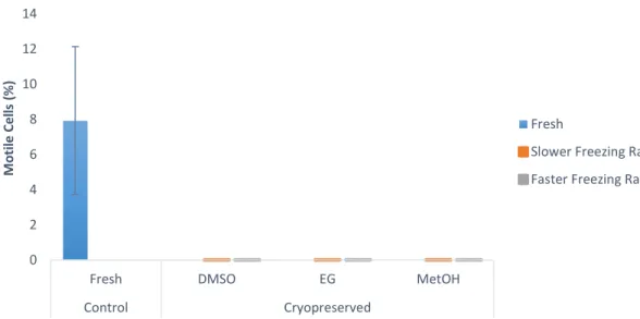
Figure 1 – (A) Capture production and (B) Aquaculture production for Chamelea gallina since 1950’s. (Source: FAO, 2018b). ... 12 Figure 2 – Percentage of motile cells in Crassostrea angulata (n=5) comparing cryopreserved sperm with different cryoprotectants (DMSO, DMSO+trehalose and DMSO+sucrose). Data are expressed as mean±SD. Statistical analysis was performed using a one-way ANOVA... 23 Figure 3 – Percentage of viable cells in Crassostrea angulata (n=5) comparing cryopreserved sperm with different cryoprotectants (DMSO, DMSO+trehalose and DMSO+sucrose). Data are expressed as mean±SD. Statistical analysis was performed using one-way ANOVA. ... 24 Figure 4 – Percentage of DNA fragmentation in Crassostrea angulata (n=5) comparing sperm cryopreserved with different cryoprotectants (DMSO, DMSO+trehalose and DMSO+sucrose). Data are expressed as mean±SD. Statistical analysis was performed using one-way ANOVA followed by Student-Newman-Keuls (SNK) as a post-hoc test. Different superscripts means significant differences (P<0.05). ... 25 Figure 5 – Fertilization rate using different sperm-to-egg ratios in Crassostrea angulata (n=9). Data are expressed as mean±SD. T-test was used to compare fresh and cryopreserved sperm and to compare the fertilization rate between each cryoprotectant for each sperm-to-egg ratio. A one-way ANOVA was used followed by Student-Newman-Keuls (SNK) as a post-hoc test to compare different sperm-to-egg ratios. The asterisk corresponds to significant differences between fresh and cryopreserved sperm and different letters means significant differences between sperm-to-egg ratios (P<0.05). ... 26 Figure 6 – Fertilization rate using different fertilization volumes (50ml, 100ml and 150ml) in Crassostrea angulata (n=9). Data are expressed as mean±SD. Statistical analysis was performed using Mann-Whitney test to compare fresh and cryopreserved sperm and Kruskal-Wallis test to compare the different volumes used in cryopreserved sperm. The asterisk means significant differences (P<0.05) between fresh and cryopreserved sperm. ... 27 Figure 7 – Percentage of motile cells in Chamelea gallina (n=5) after exposure to different cryoprotectants (DMSO, EG and MetOH) for 10 minutes. Statistical analysis was performed using Kruskal-Wallis test to compare all the treatments. Data are expressed as mean ± SD. Different superscripts means significant differences (P<0.05). ... 28 Figure 8 – Percentage of motile cells in Chamalea gallina (n=5) comparing fresh sperm (blue bar) and cryopreserved sperm with different cryoprotectants (DMSO, EG and MetOH) and two freezing rates (slower freezing rate – orange bars, faster freezing rate – grey bars). Data are expressed as mean±SD. ... 29
xii
Figure 9 – Percentage of viable cells of Chamelea gallina (n=5) in fresh sperm (blue bar) and cryopreserved sperm with different cryoprotectants (DMSO, EG and MetOH) using two freezing rates (slower freezing rate – orange bar, faster freezing rate – grey bars). Statistical analysis was performed using t-test to compare fresh and cryopreserved sperm and both freezing rates. A One-way ANOVA followed by Student-Newman-Keuls (SNK) as a post-hoc test was used to compare the viability of the three cryoprotectants for each freezing rate. Data are expressed as mean±SD. Different superscripts means significant differences (P<0.05). The asterisk corresponds to significant differences between fresh and cryopreserved sperm, different numbers means significant differences between freezing rates and different letters means significant differences between cryoprotectants (P<0.05) ... 30
xiii
Index of tables
Table 1 - Sperm-to-egg ratio that was used for each type of sperm (fresh and post-thawed sperm). ... 19
xiv
Abstract
The development of methods that allow the conservation of genetic resources as sperm cryopreservation are necessary to avoid the pressure on the natural populations and the risk of extinction of Crassostrea angulata and Chamelea gallina, improving the reproductive strategies for eventually restocking programs and even aquaculture production with commercial purposes. Therefore, this work aimed to optimize the cryopreservation protocol for C. angulata testing different combinations of cryoprotectants, to assess the fertilization conditions for oyster and to assess the sperm quality parameters before and after exposure of C. gallina sperm to cryoprotectants solutions, determining the best cryopreservation conditions in terms of freezing rates. For optimization of a cryopreservation protocol for C. angulata, motility, viability and DNA integrity analysis were performed for cryopreserved sperm with dimethyl sulfoxide (DMSO), DMSO+trehalose and DMSO+sucrose. Fertilization trials with different sperm-to-egg ratios and volumes were tested and the success of fertilization trials was determined. For the assessment of sperm quality parameters of C. gallina, toxicity tests and cryopreservation assays were performed. For toxicity tests, fresh sperm was exposed to cryoprotectants (DMSO, ethylene glycol and methanol) and motility analysis were recorded. For cryopreservation assays, the same cryoprotectant conditions and two freezing rates were analysed. Motility and viability analysis were performed in post-thaw sperm. Supplementation of DMSO with sugars did not optimize motility, viability or decrease DNA fragmentation in Crassostrea angulata sperm. In fertilization trials, the best sperm-to-egg ratio using post-thawed sperm was 50,000 spermatozoa per egg. Trehalose did not improve the fertilization rate and no effect of fertilization volume was seen between different volumes. For Chamelea gallina, the toxicity trials demonstrated that DMSO was the most suitable cryoprotectant. However, post-thawed sperm showed no motility but DMSO showed the higher viability results, being the best conditions for sperm cryopreservation achieved with the fastest freezing rate. More studies are necessary to develop successfully a cryopreservation protocol for C. gallina.
Keywords: Crassostrea angulata; Chamelea gallina; Cryopreservation; Sperm; Fertilization.
xv
Resumo
O desenvolvimento de métodos que permitam a conservação dos recursos genéticos, tais como a criopreservação de sémen, é necessário de modo a evitar a pressão exercida sobre as populações naturais e diminuir o risco de extinção de ostra portuguesa (Crassostrea angulata) e de pé-de-burrinho (Chamelea gallina). Estes métodos permitem melhorar as estratégias reprodutivas destas espécies permitindo eventuais programas de repovoamento e ainda a produção em aquacultura com fins comerciais. Portanto, a realização deste trabalho teve como objetivos a otimização do protocolo de criopreservação para a C. angulata testando diferentes combinações de crioprotetores internos e externos e avaliar as condições de fertilização desta espécie em condições experimentais. Outro dos objetivos foi avaliar os parâmetros de qualidade do sémen para a C. gallina antes e depois de ser exposto às soluções de criopreservação (crioprotetores) e determinar as melhores condições de criopreservação em relação a rampas de congelação. Para a otimização do protocolo de criopreservação de C. angulata, foram realizadas análises de mobilidade, viabilidade e de integridade de DNA (cometas) em sémen criopreservado com dimetilsulfóxido (DMSO) e duas combinações de DMSO com açucares (DMSO+trealose e DMSO+sacarose). Para esta espécie foram também realizados ensaios de fertilização onde foram testados diferentes rácios de espermatozóides por ovo e diferentes volumes de fertilização. Com isto, foi calculada uma taxa de fertilização para determinar o sucesso destes ensaios. Na avaliação dos parâmetros de qualidade de sémen de C. gallina, foram realizados testes de toxicidade e ensaios de criopreservação. Nos testes de toxicidade, o sémen fresco foi exposto durante 10 minutos a diferentes crioprotetores (DMSO, etilenoglicol e metanol), tendo sido realizadas análises de mobilidade. Nos ensaios de criopreservação, o sémen foi exposto às mesmas condições anteriormente descritas para os testes de toxicidade (mesmos crioprotetores e mesmo tempo de equilíbrio) tendo sido posteriormente exposto a duas rampas de congelação. Depois de descongelado, realizaram-se análises de mobilidade e a viabilidade. Com a realização destes procedimentos concluiu-se que a suplementação de DMSO com açucares não melhora a mobilidade, viabilidade ou diminui a fragmentação de DNA no sémen de Crassostrea angulata. Nos ensaios de fertilização, quando foi usado sémen criopreservado o melhor rácio de espermatozóide por ovo foi 50000:1, a trealose não melhora a taxa de fertilização e não foi observado qualquer efeito na variação do volume de água na fertilização. Para a Chamelea gallina, os testes de toxicidade demonstraram que o DMSO foi o crioprotector mais adequado. No entanto, o sémen
xvi
descongelado não apresentou qualquer mobilidade. Em termos de viabilidade, o DMSO mostrou resultados mais elevados, e as melhores condições para a criopreservação do sémen desta espécie foram atingidas com a rampa de congelação mais rápida. No entanto, serão necessários mais estudos para o desenvolvimento de um protocolo de criopreservação com sucesso para a C. gallina.
Palavras-chave: Crassostrea angulata; Chamelea gallina; Criopreservação; Sémen; Fertilização.
1
Introduction
1. Bivalves aquaculture
Nowadays, aquaculture is an important source of food that contributes to answer to the significant increase in demand for seafood, due to the fast demographic growth (FAO, 2014; 2016). World per capita fish supply is continuously increasing due to the increase in aquaculture, which provides half of all fish supply for human consumption (FAO, 2016). The aquaculture production includes finfishes, molluscs, crustaceans, amphibians, reptiles, aquatic invertebrates and algae (FAO, 2018a). At least 109 species produced in aquaculture belongs to molluscs group which is the second group most produced after finfishes (FAO, 2018a).
Most bivalve molluscs produced in aquaculture are oysters, mussels, clams and scallops (Helm et al., 2004; FAO, 2018a). China is known as the biggest producer of bivalves in the world, producing five times more than the rest of the world, obtaining 12 million tonnes of bivalves production in 2014, meanwhile Europe produced 632000 tonnes (FAO, 2016).
According to the last report of Direção-Geral de Recursos Naturais, Segurança e Serviços Marítimos (DGRM, 2018), in Portugal, the bivalves production corresponded to 56.4% of the total aquaculture production, where clams had the most relevant production with 3716 tones followed by mussels and oysters with 1474 and 1014 tones, respectively. The 87.9% of bivalves production related to aquaculture activities are located in Ria Formosa, and just the 2.1% of these activities are performed in floating structures. The production of bivalves is mostly running in extensive regime (DGRM, 2018), due to the fact that bivalves are filter-feeders and need low maintenance during grow out stage.However, in the production of some bivalve species, wild spat collection is necessary due to the lack in reproductive control or to higher cost that this technology involves (Suquet et al., 2014).
Nowadays, in some species, bivalve hatcheries are starting to appear, especially for oysters, where juveniles/spats (seed) can be provided for commercial production (Wallace et al., 2008) in farms, usually located in estuarine waters and in longlines (offshore production). The utilization of hatcheries for bivalve intensive production could be the solution to obtain seeds without exploiting the wild beds, or to obtain them out of
2
reproductive season or to improve genetic strains with specific biological characteristics (Helm et al., 2004).
Furthermore, keeping broodstocks in hatcheries, under controlled conditions (tanks, temperature, water quality, photoperiod, adequate feeding) and simulating natural conditions is crucial to obtain gametes and larvae of good quality (Joaquim et al., 2008). Therefore, gametogenesis is related to the broodstock maintenance in appropriate conditions, being the temperature and food availability the most important factors that affects it (Helm et al., 2004).
In the past decades, several techniques to improve the reproductive performance of species have been developed in order to enhance bivalve aquaculture. These techniques include broodstock conditioning with adequate diets (Anjos et al., 2017), genetic manipulation as polyploidy (Helm et al., 2004), methods for artificial spawning induction, such as stripping, non-lethal sperm collection (biopsy) and thermal stimulation; (Yang et al., 2015; Joaquim et al., 2016;). Another developed technology that can improve production of these bivalves is the cryopreservation of genetic resources. Using this methodology, gametes, mainly sperm, but also oocytes and embryos or larvae in the case of some invertebrates, may be preserved (Labbé et al., 2013) such as the Pacific oyster, Crassostrea gigas (Labbé et al., 2018; Paredes et al., 2013; Suquet et al., 2014; Tervit et al., 2005), mussels with the species - Mytilus galloprovincialis (Paredes et al., 2013) and Perna canaliculus (Paredes et al., 2012). According to Labbé et al. (2018), this technique associated to larvae cryopreservation can be a very valuable tool for oyster farming, helping hatcheries production out of the reproduction season when gametes already are not available, but food availability and water temperature still allow larval production.
2. Cryopreservation
Cryopreservation is one important technology developed to assist reproductive biology of the aquatic species, being an important tool that allow the preservation of genetic material that could be used as restocking of natural populations in the future (Riesco et al., 2017a). This technique consists in the preservation of live cells or tissues in liquid nitrogen at -196 ºC for a long period of time, without compromising the biological functions of these cells, maintaining cell viability and functionality (Labbé et al., 2013; Hassan et al., 2015).
3
If eggs and sperm can be cryopreserved, the storage of these biological materials during natural breeding season can be very useful in hatcheries, being possible to produce spat without the need to maintain a broodstock out of the reproductive season, once this imply more costs (Adams et al., 2008; Smith et al., 2012; Paredes et al., 2013).
Nowadays sperm cryopreservation is used as a complementary technique for artificial fertilization above all in species with high commercial interest, endangered species and species with an interesting genotype (Riesco et al., 2017a). It is a safety method for seed supply, once is possible to select disease-free material, ensuring sperm quality. It is also a safe method to store and preserve the male genetic material (Cabrita et al., 2014). Other advantages of sperm cryopreservation include the use of this stored material when eggs are available; the possibility of storing all sperm production, being used when it is difficult to obtain and when low volumes are stripped in captivity; the simplification of broodstock maintenance out of the spawning season and transport of gametes between farms avoiding animal transportation (Cabrita et al., 2010). However, there are several concerns related with post-thaw quality and a special interest in preventing and reducing cell damage (Chao & Liao, 2001), since losses of genetic information when the genetic material is altered by freezing and storage can occur (Labbé et al., 2013).
To perform sperm cryopreservation several steps should be performed properly, such as sperm collection, fresh sperm quality assessment, extender and cryoprotectant preparation, cooling and storage at very low temperatures, thawing and post-thaw sperm quality assessment (Liu et al., 2015). However, in the cryopreservation protocols mainly extenders and cryoprotectants, freezing and thawing rates and the containers for sperm storage need to be adapted for each species in study since it is highly species-specific (Cloud & Patton, 2008; Beirão, 2011). Furthermore, there is a variation on sperm characteristics (spermatozoa structural pattern, shape and size) between species, stocks, or samples of the same individual, which depends on the maturation period during reproductive season, maturation stage of the cell, evolutive position of the species (mammalian, fish, crustaceans) or even damaged provoked by environmental factors (Cabrita et al., 2008; 2010). All these factors can influence the success of any cryopreservation protocol.
4
2.1. Extenders and Cryoprotectants
Extenders and cryoprotectants are necessary for cryopreservation, however, as stated before, these solutions are species-specific (Chao & Liao, 2001).
During preparation for cryopreservation sperm is suspended in a balanced salt buffer solutions denominated extenders (Hassan et al., 2015). These solutions can be sterilized with or without antibiotic to avoid the bacterial growth that can affect sperm viability (Hassan et al., 2015). In fish, extenders are used to mimic the osmolality of seminal plasma and immobilize sperm, because this have low motility duration. However, in oysters it is not necessary to have this concern, because they have a sperm movement of several hours to days (Hassan et al., 2015). The most common extenders used for several bivalve species are: seawater (natural, sterilized or filtered and artificial), Hank´s balanced salt solution (HBSS) and calcium-free HBSS (Hassan et al., 2015).
Cryoprotectant agents are used to prevent ice crystals formation during freezing and other cell damage during freezing and thawing (Tiersch et al., 2007; Cloud & Patton, 2008). However, these compounds can be toxic for the cells, since the level of toxicity is dependent on concentration, the equilibrium time, and the temperature during loading (they are normally manipulated and added to sperm cells slowly and at low temperatures to reduce the toxicity effect) (Chao & Liao, 2001; Cloud & Patton, 2008; Beirão, 2011).
Several agents can be used as cryoprotectants, such as glycerol (Gly), ethylene glycol (EG), propylene glycol (PEG) and methanol (MetOH) (Hassan et al., 2015). However, dimethyl sulfoxide (DMSO) is the permeating cryoprotectant for excellence used for sperm cryopreservation. This is generally used with other non-permeable agents that act as membrane stabilizer, such as sugars (trehalose and sucrose) or lipoproteins as bovine serum albumin and egg yolk that allow to protect the cells reducing the extracellular ice crystals damage (Cloud & Patton, 2008; Paredes, 2015). Previous studies demonstrated that the addition of 0.45M of trehalose to DMSO can improve the viability and the fertility of cryopreserved sperm of Crassostrea gigas (Adams et al., 2004; Hassan et al., 2015).
Before cryopreservation, the sperm is exposed to cryoprotectants during a certain time (equilibrium time) to allow the penetration of permeating cryoprotectants by the cells (Tiersch et al., 2007). However, it is very important to analyse the toxicity of cryoprotectants because these solutions, during pre-treatments and post-thawing can cause gamete mortalities. Therefore, the best option is to choose a cryoprotectant with non- or low toxicity and with high water solubility and permeation (Chao & Liao, 2001) which in turn is capable of reducing the freezing rate and avoiding ice crystal formation.
5
For cryopreservation of any material (sperm, oocytes, embryos or even tissues), after the preparation with extenders and cryoprotectants, but before freezing it is necessary to pack this material in containers. Straws with 0.25ml and 0.5ml are the most used, once occupy less space being easy to transport, allowing the storage of higher amount of different samples, and obtaining higher cooling and thawing rates (Paredes, 2015). On the other hand, cryovials (1-4.5ml) also can be used, storing higher volumes than straws and allowing the cryopreservation of material with bigger size (tissues). These containers, contrary to the straws have lower cooling and thawing rates (Adams et al., 2008; Paredes, 2015).
2.2.Freezing and Thawing conditions
Freezing is one of the most stressful steps during sperm cryopreservation, due to cold shock, intracellular ice formation and solute concentration, causing cell damage (Chao & Liao, 2001). This step of cryopreservation can be performed using two methods, using a programmable biofreezer or a non-programmable method. The programmable biofreezer has the advantage of controlling temperature during all the process; however, is an expensive equipment. On the other hand, the non-programmable method is usually performed using liquid nitrogen in a styrofoam box with racks where a large number of samples can be freeze with lower costs (Liu et al., 2015).
During freezing several mechanisms can occur becoming a critical step, such as loose of water by the cells (dehydration) with the temperature decreasing and with this, the increasing of extracellular osmolarity due to salt concentration (Cloud & Patton, 2008).
To prevent damage a slow freezing rate is ideal, where cells are capable of suffer enough dehydration to avoid intracellular ice formation. However as stated before cells will be exposed to an increase of osmolarity of the surrounding media, with consequent toxic effects. To solve this problem an increase of the freezing rate (with step-wise rates) or a faster freezing would be the solution (Hassan et al., 2015). Nevertheless, the optimal rate needs to be slow enough to allow the water outflow of the cells, minimising intracellular ice formation but fast enough to minimize the time of exposure of sperm to extracellular cryoprotectants (Cloud & Patton, 2008; Tiersch et al., 2007).
Thawing is usually performed in a water bath, but this step needs to be performed under controlled conditions to be homogeneous and the samples cannot to be in direct contact with the thawing water (Herráez et al., 2012). A heterogeneous thawing of samples should be avoided in order to reduce the risk of overheating the cells closest to
6
the container wall (Herráez et al., 2012). This process is also a stressful step for sperm caused by increasing temperature and recrystallization of water molecules (Hassan et al., 2015). Chao & Liao (2001) suggested that water temperature for thawing cryopreserved sperm could be similar to that used during spawning season. Tiersch et al. (2007) suggested that to decrease the damage associated with recrystallization a rapid thawing is preferred than a slow one. However, both freezing and thawing rates are dependent on the size (volume) and type of container used for sperm storage (straws or vials) due to the rate of heat transfer for the cells (Hassan et al., 2015).
Even with all the indications previously mentioned, post-thaw sperm can have lower quality than fresh sperm both in fish as in shellfish, being this fact explained by the occurrence of some cell damage during the cryopreservation process (Chao & Liao 2001).
2.3. Methods for damage analysis
As stated before, the spermatozoa may be damaged during cryopreservation at several levels, functional (fertilization capacity) and structural (plasma membrane, chromatin, mitochondria and ATP content) (Cabrita et al., 2010; Riesco et al., 2017b). Motility, viability and DNA integrity are the most usual analysis performed to evaluate spermatozoa damage.
2.3.1. Spermatozoa motility
Motility is the most used biomarker for sperm quality and for testing the effect of different cryoprotectants (Cabrita et al., 2008). Mitochondria status, ATP production, plasma membrane integrity and spermatozoa flagellar structure are important aspects of which depends motility (Cabrita et al., 2010). Sperm motility also depends on species, therefore, contrary of the most group species studies (e.g. fish), that have a very short sperm motility duration after its activation (few minutes) (Cabrita et al., 2008), oyster sperm can have a high motility duration. This can be observed in C. angulata sperm that can be stored, at least for 3 days, at 4ºC without losing motility characteristics (Riesco et al., 2017a).
There are several methods for motility evaluation including the subjective methods, the semi-quantitative methods and the quantitative computer assisted methods (Rurangwa et al., 2004). The subjective methods analyse sperm motility using scales for motility classification and the percentage of motile cells and type of movement is estimated by the operator, however these methods have low reproducibility and variable results, according
7
to the interpretation made by different operators, overestimation the motile population due to the influence of motile spermatozoa in immotile one (Rurangwa et al., 2004; Cabrita et al., 2008; 2010;). The semi-quantitative methods analyse sperm motility using a videotape recordings viewed by two or more independent observers, however these methods are time consuming and operator dependent (Rurangwa et al., 2004). Regarding quantitative methods, nowadays, these systems also called CASA (computer assisted sperm analysis) are a way to evaluate motility in sperm from several species, analysing several motility parameters which subjective methods cannot register. However, it is need specialized and expensive equipment and training (Rurangwa et al., 2004; Cloud & Patton, 2008; Cabrita et al., 2010;).
During sperm analysis using CASA system, among other parameters, total motile cells, progressive motile cells, straight-line velocity, curvilinear velocity, linearity and sperm concentration can be evaluated. The evaluation of motility is an important process, once according to Gwo (2009) it can be also used as an indicator of sperm status.
2.3.2. Plasma membrane integrity (Viability)
Plasma membrane of spermatozoa is a very sensitive structure responsible for the exchanges between the surrounding media and the cell interior, reception of stimulus and triggering of responses (motility activation). The integrity and permeability are important factors for cell survival, therefore cells with damaged membrane cannot develop the previous functions (Cabrita et al., 2008; 2010). It is also the most vulnerable structure to suffer injuries during cryopreservation (Cabrita et al., 2010).
Viability is the common name used to the procedure where plasma membrane integrity is tested. There are several methods to perform this analysis, such as the use of fluorescent microscope or flow cytometry, followed by the use of selective dyes or fluorescent probes, which specifically label viable or non-viable cells. It is possible to analyse sperm viability both in fresh and cryopreserved, being DNA staining evaluated according to the plasma membrane condition (Cabrita et al., 2008). The fluorescent probes commonly used are: propidium iodide (PI), hoescht 33258 and rhodamine 123 (R123) which are non-permeable dyes, that only penetrate dead cells, staining the DNA of cells with damaged membrane. SYBR Green is a permeable dye that penetrate and stain the DNA of viable cells, with (Cabrita et al., 2008). SYBR-14 and propidium iodide in combination can be used to assess viability in flow cytometry allowing to distinguish more easily non-viable and viable cells due to dual staining (Cabrita et al., 2005).
8
2.3.3. DNA integrity
DNA is an important structure to preserve in any cell and specially in spermatozoa (Robles et al., 2017), since it allows the transmission of male genetic information to the next generation, contributing to embryo development which is the ultimate goal of spermatozoa. Therefore, the complete preservation of spermatozoa genomic information is important during cryopreservation process (Cabrita et al., 2008; 2010), because cryopreserved sperm carrying damaged DNA have the capacity to fertilize the oocytes, however inducing abortions during embryonic development (Pérez-Cerezales et al., 2010).
There are several methods to evaluate DNA integrity. This can be detected using comet assay also known as single cell gel electrophoresis (SCGE), terminal deoxynucleotidyl transferase-nick-end-labelling (TUNEL) and sperm chromatin structure assay (SCSA) (Cabrita et al., 2010). All of those methods detect simple and double strand breaks in the DNA double helix (Cabrita et al., 2008). However, to assess DNA fragmentation, Gwo et al. (2003) described comet assay as an “economic, rapid, simple, visual and sensitive technique”, being tail length the appropriate indicator of DNA damage. This technique is based on the migration of DNA fragments according to the size when cells are immobilized over an agar layer, lysed and submitted to electrophoresis, allowing the formation of a comet-like with two distinguish structures (head – non-fragmented DNA and tail – fragmented DNA) (Cabrita et al., 2014). For comet analysis, DNA need to be stained and the comet tail length can be measured manually but image analysis software can be used (Komet or other free software: http://cometassay.com). This method allows an objective evaluation of each single cell, and several parameters (as total intensity (DNA content), tail length, tail percentage and tail moment) of DNA fragmentation can be evaluated (Olive & Banáth, 2006; Cabrita et al., 2008; 2014; Martínez-Páramo et al., 2009).
Furthermore, DNA integrity can also be assessed using TUNEL assay, where a fluorescent labelled nucleotide is added to 3’ end of the strand. For TUNEL analysis fluorescent microscopy or flow cytometry can be used and if there is a higher amount of free 3’ ends, more fragmentation exists, increasing the fluorescence emitted by the nucleus (Cabrita et al., 2014).
As stated before, sperm chromatin structure assay (SCSA) is also another method to evaluate DNA integrity, mostly used in mammals to assess the vulnerability of sperm chromatin status using the metachromatic properties of acridine orange. This is detected
9
using flow cytometry where cells without DNA fragmentation emit green fluorescence while cells with damaged DNA fluoresces red (Cabrita et al., 2008; 2010; 2014).
3. Fertilization
Fertilization process implies sperm attraction to the oocyte and penetration, for this reason, the fertilization of oocytes depends on spermatozoa functionality, being this a critical process on the sexual reproduction (Boulais et al., 2017). In order to achieve the oocyte, it is necessary that spermatozoa maintain their moving capacity (Cabrita et al., 2010; Boulais et al., 2017), as stated before. DNA is also an essential molecule, once its damage can cause lower fertilization rates and impaired embryo development (Cabrita et al., 2010; Robles et al., 2017). Consequently, to have a successful embryo development after fertilization, the guarantee of DNA integrity is mandatory (Cabrita et al., 2010). There are other factors that may influence fertilization ability, such as the sperm concentration in the extender dilution (Glenn et al., 2011), and spermatozoa viability (Boulais et al., 2017), since samples with high concentration of non-viable cells may interfere with functional spermatozoa to reach the micropyle and the proportion of sperm-to-egg ratio. ATP content also influence fertilization ability of spermatozoa, being directly related with spermatozoa motility (Boulais et al., 2017).
It is necessary to test the spermatozoa ability to fertilize the oocytes, due to cell damage provoked by cryopreservation process. Therefore, the fertilization process is the last and the most reliable assay for determination of sperm quality after cryopreservation (Honeyfield & Krise, 2000; Riesco et al., 2017a) ensuring the successful of a sperm cryopreservation protocol. Fertilization is also the measuring of the sperm ability to produce viable embryos with good survival and growth (Honeyfield & Krise, 2000; Nahiduzzaman et al., 2012; Liu et al., 2015). Thus, the first demonstration of cryopreservation success, is a well-succeeded fertilization (Tiersch, 2011).
The in vitro fertilization using cryopreserved sperm is performed in the post-thawed phase once the sperm that survived cryopreservation can be immediately used (Chao & Liao, 2001). However, it is important to take into account the sperm motility determination (allowing the estimation of fertilization success), the time between thawing and fertilization (due to sperm motility duration), and the dilution ratio used during cryopreservation as described by Chao & Liao (2001).
As mentioned previously, cryopreserved sperm can have lower quality than fresh sperm (Chao & Liao, 2001) having thus lower fertilization capacity (Gwo et al., 2002).
10
Furthermore, fertilization rates also depend on sperm quality, on species characteristics and on the volume of oocytes used. It is common to use 100-fold the number of cryopreserved spermatozoa compared with fresh sperm (Cabrita et al., 2008). However, it is important to optimize the sperm-to-egg ratio in order to avoid several problems such as (Stephano & Gould, 1988; Gwo et al., 2003; Cabrita et al., 2008; Nahiduzzaman et al., 2012):
1) spermatozoa competition and polyspermy;
2) difficulty in distinguish different treatments, since excess of sperm may compensate the low quality;
3) the difficulty that spermatozoa can have to reach the oocyte due to low concentrations, since sperm is previously diluted in extender solutions during cryopreservation;
4) the use of excess amount of sperm unnecessarily, losing in some species important vital material;
5) the production of higher percentage of unviable eggs provoked by lysis of the egg membrane due to high sperm concentration.
Depending on species, other factors can also be studied to improve in vitro fertilization, such as the oocytes conservation after collection, the oocyte concentration, the fertilization volume, the gamete contact time, the effect of temperature and salinity in sperm motility as well as the quality of oocytes, since can be variable and can affect fertilization success (Rurangwa et al., 2004; Song et al., 2009; Diogo, 2010).
To determine the success of fertilization, the proportion of fertilized and unfertilized eggs is quantified, and a fertilization rate is calculated (Cloud & Patton, 2008). When an oocyte is fertilized by spermatozoa the egg starts to suffer some transformations which allow distinguish fertilized and unfertilized eggs. The determination of fertilization is different between groups of organisms. While in fish the initial cleavage divisions are endpoints, in bivalve’s fertilization can be determined by the observation of exclusion of polar body and the first embryonic divisions (Cloud & Patton, 2008; Riesco et al., 2017a).
11
4. Species in study
4.1. Portuguese oyster (Crassostrea angulata) – An endangered species with genetic resources need to be preserved
The Portuguese oyster is one of the species that have been caught from the wild and cultivated during centuries in Portugal as well as in France (Boudry et al., 1998; Batista et al., 2005;). But, in the late 1960’s and early 1970’s, the populations were severely affected by mass mortalities leading to the collapse of natural populations and almost extinction (Comps, 1988). However, natural populations are still present and can be found in the Guadalquivir river (Michinina & Rebordinos, 1997), and Sado and Mira rivers (Fabioux et al., 2002). Therefore, Batista et al. (2005) described C. angulata as “a valuable genetic resource for the development of selective breeding as well as in the context of production and biodiversity preservation”.
Cryopreservation can be an alternative to preserve the genetic material and restore natural populations of this species in risk, however Crassostrea angulata have only one protocol for sperm cryopreservation developed by Riesco et al. (2017a). On the other hand, Crassostrea gigas (Pacific oyster) is the most studied species in sperm cryopreservation with at least 12 developed protocols (Hassan et al., 2015; Riesco et al. in press) being the first developed by Lannan (1971).
12
4.2. Striped venus (Chamelea gallina) – A new aquaculture candidate for preservation of natural populations
The striped venus is a bivalve species which supports important fisheries in peninsula Iberica as in the Algarve Coast (Portugal) as well as in Gulf of Cádiz (Spain) (Gaspar et al., 2004; Delgado et al., 2013). This species is captured by fisheries, however with instable production over the years (figure 1A; FAO, 2018b). According to Delgado et al. (2013) fishing pressure or environmental variables could be responsible for captures decreasing. One of the solutions to deal with high fishing pressure is the production of this species in aquaculture, preserving natural populations or restocking areas where striped venus is highly captured (Joaquim et al., 2016). However, there is few information about the production of this species. FAO statistics reports production levels only in 1998, 1999 and 2000 (figure 1B; FAO, 2018b), and the only study found regarding this species was developed by Joaquim et al., (2016).
In terms of conservation of genetic resources (cryopreservation) of this species or the same genus, any study is known until now, but already exists studies in clams (macha clam Mesodesma donacium) performed by Joo & Dupré (2002) and Dupré & Joo (2006) and Dupré & Guerrero (2011) belonging to a different genus.
Figure 1 – (A) Capture production and (B) Aquaculture production for Chamelea gallina since 1950’s. (Source: FAO, 2018b).
A
13
To avoid the pressure on the natural populations and the risk of extinction of C. angulata and C. gallina it is necessary to develop methods that allow the conservation of genetic resources of these two species. Sperm cryopreservation could be applied for preserving genetic material and conserving native populations (Riesco et al., 2017a), aiming to reconstruct the original strain, population or diversity (Martínez-Páramo et al., 2017). This tool can be used also to improve reproductive strategies (Riesco et al., 2017a) in C. angulata and C. gallina production for eventually restocking programs and even aquaculture production with commercial purposes. Besides, sperm cryopreservation could also be very useful to control the biological and commercial issues provoked by diseases and market variations (Paredes et al., 2013).
14
Objectives
The two main objectives of this work were to optimize the in vitro fertilization using cryopreserved sperm of Crassostrea angulata and to establish a cryopreservation protocol for Chamelea gallina. Therefore, in order to achieve these objectives was necessary:
1. To optimize the cryopreservation protocol for C. angulata established by Riesco et al. (2017a), testing different combinations of internal and external cryoprotectants;
2. To assess the fertilization conditions for C. angulata under experimental conditions, since fertilization is the last process that proves the successful of a sperm cryopreservation protocol;
3. To assess the sperm quality parameters before and after exposition of C. gallina sperm to cryoprotectants solutions and to determine the best cryopreservation conditions in terms of freezing rates.
15
Material and methods
1. Broodstock conditioning
Crassostrea angulata (Portuguese oyster) broodstock (n=200) was collected with commercial weight (>70g) in two commercial facilities located in Mira River (Sociedade Agrícola Herdade das Moitas) and in Sado River (Monte da Pedra), where this species naturally occurs and is produced.
Chamelea gallina (striped venus) broodstock (n=100) was collected through fisheries from natural beds located in southern coast of Portugal (Algarve Coast) and in Doñana park (Spain). The individuals were harvested with commercial size (>2.5cm).
The organisms were transported to the IPMA Experimental Hatchery at Tavira (37°7´17´´N and 7°37´12´´W), where they were maintained in a flow-through circuit under controlled conditions until further use. According to Anjos (2014) a diet formulated with diatoms maximizes reproductive effort and output performance of C. angulata. Therefore, C. angulata was fed with Skeletonema costatum and Chaetoceros calcitrans. On the other hand, C. gallina diet was constituted by Isochrysis galbana and Chaetoceros calcitrans as suggested by Joaquim et al., (2016).
2. Sperm collection
Using an oyster knife, individuals were opened and identified as female and male in a microscope (100x magnification) using a sample of gametes collected directly from gonad.
For both species, sperm was collected, by a dry method, directly from gonad using a scalpel doing small incisions in the gonad and using a micropipette to collect sperm into an Eppendorf tube. However, in oyster, sperm was collected from individual males, while in striped venus sperm was collected from pools of four males.
Immediately, sperm was sieved with a 35µm sieve to eliminate pieces of gonad and other debris. Thereafter an aliquot was diluted 1:9 (10x) in artificial seawater (TMC, UK) at a concentration of 32ppt, and for each fresh sperm sample, concentration and total motility were recorded.
Previous studies evidence problems with sperm agglutination in oyster, related with high concentrations (Dong et al., 2007; Riesco et al., 2017a). For this reason, each sample was diluted in order to have a final concentration between 1 to 2 x10⁹ spermatozoa/ml as
16
fixed by Riesco et al. (2017a). For C. angulata, only samples with total motility higher than 40% were used for further cryopreservation.
3. Optimization of a cryopreservation protocol for Crassostrea angulata Sperm collected from five males (n=5) of C. angulata, was diluted in cryoprotectant solution (1:1) and cryopreserved using three different cryoprotectants: dimethyl sulfoxide (DMSO), DMSO+trehalose and DMSO+sucrose, at a concentration of 10% for DMSO and 0.45M for sugars, after 10min of equilibrium time. Sugar were added to the extender with the idea of protecting the cells against ice crystal formation and to avoid plasma membrane destabilization.
Taking into account the study made by Riesco et al. (2017a) and the description by Paredes (2015), 0.5ml French straws were used as container to cryopreserve the sperm samples.
A fast-freezing rate of 6ºC/min from 0 to -70ºC using a portable programmed biofreezer (Asymptote Grant EF600, UK) was performed as described in Riesco et al. (2017a). Samples were then plunged into liquid nitrogen and stored in a liquid nitrogen container.
Sample thawing was performed at 37ºC for 10s as described in Riesco et al. (2017a) and sperm motility analysis, cell viability analysis and comet assay were performed. In quality analysis of cryopreserved sperm, DMSO treatment was used as control.
3.1. Motility analysis and cell concentration
Total motility and cell concentration of C. angulata sperm was determined using computer assisted sperm analysis (CASA) and ISAS software (ISAS, Proiser R+D, S.L., Spain). Post-thawed sperm was diluted 1:9 in artificial seawater and each sample was assessed in triplicate placing 10µl of diluted post-thawed sperm in a Makler chamber using a phase-contrast microscope (Nikon Eclipse E200, Japan) with a 10x negative contrast objective and a digital camera (Basler A312f C-mount, Germany) set for 25 fps.
The CASA software settings were adapted to oyster sperm (1<particles area (in microns2)<90, progressivity 80%, connectivity 14, minimum number images to calculate ALH:10) and after that, the percentage of motile cells and the concentration in millions of spermatozoa per millilitre (M/ml) were assessed. To assess cell concentration, the dilution factor was taken in account. A total of 15 samples were analysed.
17
3.2. Viability analysis
For viability analysis, the settings for flow cytometer were adjusted previously for C. angulata sperm according cell size and complexity using a positive control (100% dead cells). The positive control was prepared adding 500µl of 1% NaCl solution, 5µl of sperm previously exposed to several cycles of freezing and thawing and 2.5µl of propidium iodide (PI-Sigma, Spain) inside a cytometer tube (BD Biosciences, CA, USA).
Thereafter, 500µl of 1% NaCl solution, 5µl or 10µl of post-thawed sperm (it depends on cell concentration) and 2.5µl of propidium iodide (PI-Sigma, Spain) at a concentration of 2.4 mM were added inside a cytometer tube to detect cells permeable to the dye (dead cells). The samples were incubated for 5min in the dark before the analysis in a flow cytometer (FACSCalibur, BD Biosciences, CA, USA) adjusted for blue excitation (488nm) line for the detection of PI (670/30). A total of 10000 cells were counted for each sample and the percentage of viable cells was determined with BD CellQuest Pro (version 8.7, BD Biosciences, CA, USA) software acquisition. A total of 15 samples were analysed.
3.3. DNA Integrity analysis (Comet assay)
For DNA integrity analysis a comet assay was performed determining DNA fragmentation. For this, a positive control was prepared with 10µl post-thawed sperm and 150µl hydrogen peroxide at 60mM for 10 min and the reaction was stopped with 75µl PBS.
For each male 5µl post-thawed sperm was diluted in 150µl PBS, thereafter 15µl of this solution were introduced in 300µl agarose low melting point. Two slides were prepared with 75µl agarose solution being immediately covered with coverslips and placed in the fridge for 15 min. Coverslips were removed and slides were introduced into coplin jars with lysis solution (100mM Na2EDTA, 2.5M NaCl, 10mM Tris, 1% lauril sarcosine and 1% triton X-100) for 1h. When lysis was completed, slides were introduced into an electrophoresis cube (subCell GT, BioRad, Portugal) with electrophoresis solution (1mM Na2EDTA and 0.3M NaOH). Electrophoresis was performed at 25V and 300mA for 10min at 4ºC. After electrophoresis, samples were neutralized in a neutralization solution (0.4M Tris) with two baths of 5min each and then fixed with pure ethanol for 3min.
Samples visualization was performed with 25µl propidium iodide pre-diluted in 500µl PBS and observed at 600x in a fluorescent microscope (Olympus IX 81, Olympus, Japan) with blue excitation 450–480 nm. Around 100 comets per sample were photographed and
18
recorded with a digital camera (F-view, Olympus, Japan), processed with the CellF image software (Olympus, Japan) and analysed using Komet 6.0 software (Andor Technology Ltd., United Kingdom). A total of 15 samples per duplicate were analysed and the percentage of tail DNA was the parameter used to determine the amount of DNA fragmentation (%).
According to motility and viability analysis and comet assay results, the best cryoprotectant was used to cryopreserve the sperm samples that were used in fertilization trials.
4. Fertilization trials using cryopreserved sperm of Crassostrea angulata
For cryopreserved sperm C. angulata two trials were performed to identify the optimal sperm-to-egg ratio and the volume of fertilization. For each fertilization trial, oocytes were collected from 27 females to perform 9 oocyte pools of three different females. Oocyte were collected by gonadal stripping, sieved with a 100µm and 20µm mesh and stored in 500ml of seawater to mature for 1h. While oocytes maturation occurs, three 50µl drops were collected and introduced in a Sedgewick rafter counting chamber to determine oocyte concentration for each pool of females. For fertilization trials, an initial fertilization volume of 50ml and an oocyte concentration of 500 oocytes/ml was used, according to Song et al. (2009). Sperm concentration was determined using CASA system.
For each fertilization trial (n=9) approximately 20 straws per treatment were thawed. The diameter of mature oocytes (small oocytes were not mature) and structural integrity were the criteria used to evaluate oocytes quality (Cannuel & Beninger, 2005). Nine pools containing oocytes from three different females were used in each fertilization trial.
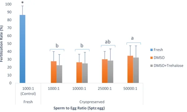
4.1. Trial 1: Sperm-to-egg ratio
Different sperm-to-egg ratios 1000:1, 10000:1, 25000:1, 50000:1 (table 1) were tested in cryopreserved samples with DMSO and DMSO+trehalose. A control group using fresh sperm was used with a sperm-to-egg ratio of 1000:1 (Riesco et al., 2017a). After 1h of oocyte maturation, oocytes were distributed in 50ml of artificial seawater at a final concentration of 500 oocytes/ml.
19
According to the concentration of post-thawed sperm, determined using CASA system as previously described, the volume of sperm necessary for each sperm-to-egg ratio was calculated and added to the eggs. The fertilization trials were done in triplicate and for each individual male, one pool of eggs from three females was used.
Table 1 – Sperm-to-egg ratio that was used for each type of sperm (fresh and post-thawed sperm).
4.2. Trial 2: Fertilization volume
The second experiment was set to test the variation of the water volume inside the beakers before the addition of sperm to oocytes. In this experiment the best cryoprotectant (DMSO) and sperm-to-egg ratio (5000:1) obtained in the anterior experiment was used and a fixed number of oocytes (25000 oocytes) and spermatozoa was introduced in each beaker, changing only the volume of water during fertilization.
Three different water volumes (50ml, 100ml and 150 ml) were tested.
As previously, the fertilization trials were done in triplicate, and for each individual male (n=9) one pool of eggs from three females was used. The fresh sperm was used as control with the same sperm-to-egg ratio previously used for control group (1000:1).
To measure the success of trials, the fertilization rate was calculated after at least 1h post-fertilization according to Anjos et al. (2017). Eggs were considered fertilized when the exclusion of polar body or the first embryonic stages were observed. This was done by the method used by Riesco et al. (2017a) (Formula 1).
(1) Fertilization rate (%) = Number of fertilized eggs
Total number of incubated eggs × 100%
Sperm-to-egg ratio (Sperm:Egg)
Fresh sperm (Control) 1000:1
20
5. Establishment of a cryopreservation protocol for Chamelea gallina
5.1. Motility analysis and cell concentration
For motility analysis and cell concentration evaluation in C. gallina, the same procedure done for C. angulata was used. Although a different species, the CASA software settings were the same used for C. angulata sperm characteristics (1<particles area (in microns2) <90, progressivity 80%, connectivity 14, minimum number images to calculate ALH:10). Each sample of C. gallina sperm was diluted in order to have a final concentration between 1 to 2 x10⁹ spermatozoa/ml to avoid putative agglutination.
5.2. Extenders and cryoprotectants
For the development of a cryopreservation protocol for C. gallina, seawater was used as an extender. As cryoprotectants, DMSO, ethylene glycol (EG) and methanol (MetOH), were tested at a concentration of 10%. Sperm was diluted 1:1 in cryoprotectants. For each collected sample a total of three treatments were tested. A total of five pools (n=5) of four animals each were used due to the few amount of collected sperm in each male. These combinations were tested for toxicity studies and cryopreservation assays.
5.3. Toxicity trials
In toxicity trials, 150µl of sperm collected by gonadal stripping, as previously described, was exposed to the experimental conditions for 10min (equilibrium time) in Eppendorf tubes and motility analysis was performed using CASA system. This parameter was also evaluated for fresh sperm, which was used as control.
5.4. Cryopreservation assays
For cryopreservation assays the containers used were the same used for sperm cryopreservation of C. angulata (0.5ml French straws) and two freezing rates were tested: a slower freezing rate of 0.3ºC/min from 0ºC to -30ºC and a faster freezing rate of 6ºC/min from 0ºC to -70ºC were tested using portable programmed biofreezer (Asymptote Grant EF600, UK). After removing samples from the biofreezer, they were directly introduced in liquid nitrogen at -196ºC.
To evaluate the quality of cryopreserved sperm, motility and viability analysis were performed for fresh and cryopreserved sperm using the same method previously used for
21
C. angulata. These parameters were also evaluated for fresh sperm, which was used as control.
A total of five pools of four animals (n=5) were analysed for each cryopreservation protocol. In order to compare results, the same samples were used for the different tested conditions.
5.5. Viability analysis
For viability analysis, the settings of flow cytometer were previously adapted to C. gallina sperm characteristics, as previously made for C. angulata. However, inside a cytometer tube, 500µl of 1% NaCl solution, 2µl of sperm (fresh or cryopreserved sperm) and 1.5µl of propidium iodide (PI-Sigma, Spain) at a concentration of 2.4 mM were mixed and incubated for 5min in the dark. After this time, cell viability was assessed. For C. gallina 10000 events were recorded.
6. Statistical analysis
The statistical analysis were performed with SPSS Statistics version 25 (IBM, USA) software, being the results expressed as mean± standard deviation (SD).
According to the results obtained, a normal distribution using Shapiro-wilk test and homogeneity variance using Levene´s test were tested. If data sets on this model, a one-way ANOVA was used to compare the different treatments in cryopreservation assays of C. angulata in terms of motility and viability analysis and comet assays. A post-hoc test Student Newman Keuls (SNK) was performed to find the main differences. In the fertilization trials, t-test was used to compare fresh and cryopreserved sperm. To test the influence of two cryoprotectants in trial 1 a t-test was used for each sperm-to-egg-ratio. To compare the different sperm-to-egg ratio in cryopreserved sperm a one-way ANOVA followed by SNK was used. The influence of the water volume in fertilization was analysed using Mann-Whitney parametric test followed by a Kruskal-Wallis non-parametric test.
To analyse the data obtained for C. gallina, a Kruskal-Wallis test was followed comparing data collected in toxicity tests. For cryopreservation assays a t-test was performed to compare fresh and cryopreserved sperm and to compare the two freezing rates. A one-way ANOVA, to compare the cryoprotectants of each freezing rate was followed.
22
Only if data did not follow a normal distribution or homogeneity variance, a non-parametric test was applied (Mann-Whitney or Kruskal-Wallis test).
For all statistical tests, the significance level at P ≤ 0.05 was set.
Before statistical analysis, percentage data suffered angular transformation according to formula 3 (Sokal & Rohlf, 1981).
(3) 𝜃 = 𝑎𝑟𝑐𝑠𝑖𝑛√𝑝
23
Results
1. Optimization of a cryopreservation protocol for Crassostrea angulata As described before, motility and viability analysis and comet assay were performed to test a putative effect of sugars (trehalose and sucrose) in the improvement of post-thaw sperm quality. The best cryoprotectant was used to cryopreserve the samples used in fertilization trials.
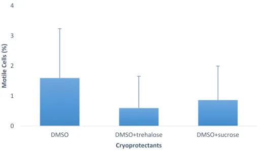
1.1. Motility analysis
In post-thaw sperm motility of Crassostrea angulata there were no significant differences. However, DMSO had a higher percentage of motile cells (1.6±1.6%) comparing with DMSO+trehalose (0.6±1.1%) and DMSO+sucrose (0.9±1.1%) (figure 2).
Figure 2 - Percentage of motile cells in Crassostrea angulata (n=5) comparing cryopreserved sperm with different cryoprotectants (DMSO, DMSO+trehalose and DMSO+sucrose). Data are expressed as mean±SD. Statistical analysis performed using a one-way ANOVA.
0 1 2 3 4
DMSO DMSO+trehalose DMSO+sucrose
M o ti le C e lls (% ) Cryoprotectants
24
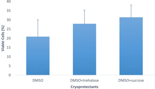
1.2. Viability analysis
Post-thaw viability of Crassostrea angulata spermatozoa did not show any significant differences between the tested cryoprotectants. Nevertheless, cryopreserved sperm with DMSO+sucrose had higher percentage of viable cells (31.5±6.6%) than sperm cryopreserved with the other two cryoprotectants (figure 3). Therefore, according to these results, sugar supplementation did not improve sperm quality in terms of viability.
Figure 3 – Percentage of viable cells in Crassostrea angulata (n=5) comparing cryopreserved sperm with different cryoprotectants (DMSO, DMSO+trehalose and DMSO+sucrose). Data are expressed as mean±SD. Statistical analysis was performed using one-way ANOVA.
0 5 10 15 20 25 30 35 40
DMSO DMSO+trehalose DMSO+sucrose
V iab le C e lls ( % ) Cryoprotectants
25
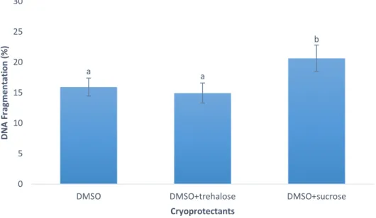
1.3. DNA integrity analysis (Comet assay)
DNA fragmentation was significantly higher in cryopreserved sperm with DMSO+sucrose when compared to DMSO and DMSO+ trehalose treatments. However, there were no significant differences between DMSO and DMSO+trehalose, obtaining a percentage of fragmented DNA of 15.9±1.5% and 14.9±1.7%, respectively. DMSO+sucrose was the treatment with worst results for DNA integrity (20.6±2.2%) as can be observed in figure 4.
Figure 4 – Percentage of DNA fragmentation in Crassostrea angulata (n=5) comparing sperm cryopreserved with different cryoprotectants (DMSO, DMSO+trehalose and DMSO+sucrose). Data are expressed as mean±SD. Statistical analysis was performed using one-way ANOVA followed by Student-Newman-Keuls (SNK) as a post-hoc test. Different superscripts means significant differences (P<0.05).
Since there were no significant differences between DMSO and DMSO+trehalose in this set of sperm analysis (motility, viability and DNA integrity), these cryoprotectants were used to cryopreserve the sperm samples used in further in vitro fertilization trials for Crassostrea angulata. DMSO supplemented with sucrose was excluded since showed the highest levels of DNA fragmentation.
a a b 0 5 10 15 20 25 30
DMSO DMSO+trehalose DMSO+sucrose
D N A F rag me n tati o n ( % ) Cryoprotectants