The Effect of Low-Level Laser Irradiation on
Sperm Motility, and Integrity of the Plasma
Membrane and Acrosome in Cryopreserved
Bovine Sperm
Guilherme Henrique C. Fernandes1, Paulo de Tarso Camillo de Carvalho1,2 *, Andrey Jorge Serra1, André Maciel Crespilho3, Jean Pierre Schatzman Peron4, Cristiano Rossato4, Ernesto Cesar Pinto Leal-Junior1,2, Regiane Albertini1,2
1Postgraduate Program in Rehabilitation Sciences, Universidade Nove de Julho (UNINOVE), São Paulo, SP, Brazil,2Postgraduate Program in Biophotonics, Universidade Nove de Julho (UNINOVE), São Paulo, SP, Brazil,3Postgraduate Program in Veterinary Medicine—Universidade de Santo Amaro (UNISA) São Paulo, São Paulo, SP, Brazil,4Instituto de Ciências Biomédicas da Universidade de São Paulo—USP— São Paulo, São Paulo, SP, Brazil
Abstract
Background and Objective
Freezing changes sperm integrity remarkably. Cryopreservation involves cooling, freezing, and thawing and all these contribute to structural damage in sperm, resulting in reduced fer-tility potential. Low-level laser irradiation (LLLI) could increase energy supply to the cell and cause reactive oxygen species reduction (ROS), contributing to the restoration of oxygen consumption and adenosine triphosphate synthesis (ATP) in the mitochondria. Our goal was to analyze the effects of low-level laser irradiation on sperm motility and integrity of the plasma membrane and acrosome in cryopreserved bovine sperm.
Study Design/Materials and Methods
We analyzed 09 samples of bull semen (Bos taurus indicus), divided into three groups: a con-trol group without laser irradiation, a 4J group subjected to a laser irradiation dose of 4 joules, and a 6J group subjected to dose of 6 joules. Samples were divided for the analysis of cell via-bility and acrosomal membrane integrity using flow cytometry; another portion was used for motion analysis. Irradiation was performed in petri dishes of 30 mm containing 3 ml of semen by an aluminum gallium indium phosphide laser diode with a wavelength of 660 nm, 30 mW power, and energy of 4 and 6 joules for 80 and 120 seconds respectively. Subsequently, the irradiated and control semen samples were subjected to cryopreservation and analyzed by flow cytometry (7AAD and FITC-PSA) using the ISAS - Integrated Semen Analysis System.
Results
Flow cytometry showed an increase in the percentage of live sperm cells and acrosome in-tegrity in relation to control cells when subjected to irradiation of low-power laser in two
OPEN ACCESS
Citation:Fernandes GHC, de Carvalho PdTC, Serra AJ, Crespilho AM, Peron JPS, Rossato C, et al. (2015) The Effect of Low-Level Laser Irradiation on Sperm Motility, and Integrity of the Plasma Membrane and Acrosome in Cryopreserved Bovine Sperm. PLoS ONE 10(3): e0121487. doi:10.1371/journal. pone.0121487
Academic Editor:Roberto Amendola, ENEA, ITALY
Received:December 8, 2014
Accepted:February 2, 2015
Published:March 17, 2015
Copyright:© 2015 Fernandes et al. This is an open access article distributed under the terms of the
Creative Commons Attribution License, which permits unrestricted use, distribution, and reproduction in any medium, provided the original author and source are credited.
Data Availability Statement:All Data are available from the Dryad database (accession number(s) doi:
10.5061/dryad.c9k00).
Funding:These authors have no support or funding to report.
different doses of 4 and 6 joules (p<0.05). In the analysis of straightness, percentage of
cell movement, and motility, a dose of 4 joules was more effective (p<0.05).
Conclusion
We conclude that LLLI may exert beneficial effects in the preservation of live sperm. A dose of 4 joules prior to cryopreservation was more effective than a dose of 6 joules in preserving sperm motility.
Introduction
Techniques in animal reproduction, such as artificial insemination (AI), are constantly used to
increase the quality and quantity of genetic and phenotypically superior calves [1], [2]. The
Ar-tificial Insemination in Fixed Time (TAI) allows cows to be inseminated without determining whether they are in heat, because the technique itself induces ovulation. The TAI is widely used to improve genetic quality and herd production volume. The efficiency of the technique is de-pendent on the semen quality fertilizer and pre-frozen state for post-thaw, which lead to
ade-quate semen motility, vigor, and high viability [3], [4], [5] [6].
Sperm integrity shows remarkable changes with freezing. The cooling, freezing, and thawing involved in cryopreservation all contribute to structural damage and reduced function in
sperm, resulting in reduced fertility potential [6], [7], [8].
The cryopreservation process induces morphological changes in semen and damages the plasma membrane, acrosome, and mitochondria. These changes are sufficient to adversely af-fect the fertilizing capacity of the semen and they significantly accentuate the production of
adenosine triphosphate (ATP) and lead to cell death [9]. The sperm plasma membrane
regu-lates the intracellular calcium concentration, particularly the calcium pump by ATP-dependent
sodium/calcium and the voltage-dependent calcium channel. [10]. Intracellular calcium
move-ments play a vital role in cell proliferation and in mammalian spermatozoa have a pivotal role
in control of sperm motility and acrosome reaction. [11]
Cryopreservation of bull semen adversely affects the sperm membrane integrity, thereby
re-ducing the ability to fertilize the processed sample [12]. In this perspective, effective techniques
are needed to protect sperm from adverse effects of cryopreservation.
The low- power laser irradiation of spermatozoa can increase sperm motility as well as ve-locity can be improved by He-Ne laser irradiation. The first published studies dating back to
the year 1984. [13] According to Huang et al. [14], the first Law of Photochemistry says that
light photons are absorbed by photoreceptors or chromophores. The low-level laser mecha-nism at the cellular level has been attributed to the absorption of monochromatic visible radia-tion and near infrared (NIR) radiaradia-tion by the cell respiratory chain components. The low-level laser has become an alternative to modulate various biological processes. Depending on the wavelength, dosage, and condition of the irradiated tissue, the laser can induce an
anti-inflammatory effect, reducing pain, and accelerating cell proliferation [15].
The biological mechanisms of interaction of the low-power laser aren’t totally known,
how-ever, it is known that different kinds of cells don’t behave at same way when irradiated by the
same wavelength. For this reason, it is difficult to extrapolate the effects from one cell type to another, but it can be said that at the molecular level, the activation of certain receptors and
Therefore, the present study aimed to investigate the effects of low-level laser irradiation on sperm motility, and integrity of the plasma membrane and acrosome in cryopreserved bovine sperm.
Methods and Materials
All semen handling procedures were performed in accordance with standards established by the Brazilian College of Animal Reproduction (CBRA). The experimental procedures were ap-proved by the Research Ethics Committee of the Universidade Nove de Julho- UNINOVE n.0047/2014 and are in accordance with current legislation.
Animals
We used 09 semen samples from Nelore bulls (Bos taurus indicus), with ages ranging from 24
to 50 months, from the Central Artificial Insemination TAIRANA, (Rod Raposo Tavares,
563 KM.—Presidente Prudente, SP—Brazil). The animals were fed all year round with mineral
supplementation and balanced feed and water ad libitum.
Collection and processing of semen samples
The semen collection was performed according to the health and safety criteria established by the Brazilian College of Animal Reproduction (CBRA). After collection, the semen was taken immediately to the laboratory for sample preparation and physical and morphological analysis. Semen samples were stored in formalin-saline for evaluation of concentration and sperm
mor-phology. The samples of semen used were those at the 50thpercentile and above in motility,
sperm concentration greater than 200 × 106/mL, and ejaculate volume greater than 5.0 ml.
Evaluation of sperm concentration
Sperm concentration was determined using Neubarhemocytometer. Samples of semen were
di-luted with BotuBOV (Botupharma, Botucatu SP—Brazil), until obtaining a final concentration
25 × 106sperm / mL. The number of spermatozoa was counted in 10 squares with the help of
manual counter grid was located with 200X magnification under a phase contrast microscope.
Experimental design
Viable semen samples obtained from 9 animals were divided into 3 groups: a control group without LLLI (n = 18 Straws); a 4J group treated with LLLI with 4 joules (n = 18 Straws); and a 6J group, exposed to LLLI with 6 joules (n = 18 Straws). The resulting samples were divided into 27 samples to examine cell viability and acrosomal membrane integrity and 27 samples for motion analysis.
Laser irradiation
The laser used was the Aluminum gallium indium phosphide (AlGaInP) DMC brand, model
Photon Laser III (DMC, São Carlos, SP—Brazil) with a wavelength of 660 nm, and variable
power from 30 to 100 mW. For this study the equipment was programmed for a power of 30 mW, beam area 0.028 cm and 4 joules of energy for the 4J group (133 seconds of tion), and 6 joules for the 6J group (200 seconds of irradiation). Since we performed irradia-tions directly to petri dishes, we decided to use lower power output of laser device we had in laboratory. The irradiation procedure was carried out in petri dishes of with a total area of
23.6 cm2, containing 3 mL of semen with the laser probe was positioned perpendicularly to the
spreading to the outside area, resulting in a power density of 0.0012 W / cm2. We choose to perform the irradiation before freezing of samples in order to protect samples of freezing pro-cedures. In order to ensure a uniform procedure was made a protective Ethylene Vinyl Acetate (EVA) black, involving the entire outer area of the plate. The measurement of delivered energy was performed using the Newport multifunction optical meter (Model 1835C, Newport Corpo-ration, Irvine, CA, USA). The samples of control group were not exposed to irradiation, but were exposed to same experimental conditions of all other groups, including the time before freezing procedure start to be performed.
Cryopreservation
The samples were packaged in French 0.5-mL straws (medium), previously identified with the animal number, the group to which it belongs and collection date. Filling and sealing of the straws was done by an automated system. The semen was cryopreserved (196°C negative)
using the Digitcool IMV (IMV—L'Aigle, France). The straws were removed from the machine
and immersed in liquid nitrogen, placed in identified according raquis with the experimental group and stored in cryogenic cylinders.
Analysis of sperm acrosomal integrity and living cells by flow cytometry
The samples were thawed in a water bath at 37°C for 30 seconds and semen placed in a micro-fuge preheated to 37°C. A 0.5-mL aliquot was removed from each treatment and added to
1.5 mL of 1× PBS solution. An aliquot of 300μL was withdrawn from the first solution and
added to 1.0 mL then centrifuged at 300 g for 10 minutes (Model Mini Spin Minicentrífuga Plus, Eppendorf) in microtubes. The supernatant was discarded and the pellet resuspended in
240μL based on the second solution and 80μL resuspended in PBS. Thus, the samples showed
a concentration of 25 × 106sperm/mL. Then 2μL of 7-amino actinomycin (7AAD) was added,
associated with 2μL of fluorescein isothiocyanate (FITC) + Pisium Sativum Agglutinin (PSA)
in the 80μL sample, and incubated for 8 minutes at room temperature and protected from
light. [17], [18]
The analysis of acrosome integrity and live sperm cells were analyzed by flow cytometry that was performed using an Accuri C6 flow cytometer (Accuri Cytometers, Inc. Ann Arbor, MI USA), equipped with a blue and a red laser, two scatter detectors, and four fluorescence
de-tectors (FL1 533/30 nm; FL2 585/40 nm; FL3>670 nm and FL4 675/25 nm) whose range
dis-played data across 6.2 logs. For this we use fluorescein isothiocyanate (FITC) + Pisium Sativum
Agglutinin (PSA) fluorescence was detected at 515–545 nm Fluorescence detector 1 (Fl 1) and
7-amino actinomycin (7AAD) fluorescence was detected at 640 and 680 nm Fluorescence de-tector 3 (Fl 3). The data analyzed using the Accuri software (CFlow Plus, Ver. 1.0.202.1). The forward scatter and side scatter were plotted, as well as florescence detected by plotting detec-tion on FL-1 versus FL-3. Gating and fluorescence compensadetec-tion values were set after data col-lection. A total of 10000 events were analyzed for each sample.
Sperm motility analysis
Sperm motility was evaluated with the ISAS—Integrated Semen Analysis System. The samples
were thawed in a water bath at 37°C for 30 seconds, and 2μL of the sample were placed in the
previously heated reading chamber. Images were captured by a camera attached to a micro-scope connected to a computer and then analyzed in real time by the software. For this, each sperm cell was identified and its trajectory reconstructed. Parameters analyzed were fast cells;
total motility (MT-%); progressive motility (MPRO-%); path velocity (VAP—microns / s)
velocity (VSL—microns / s), which is the distance traveled between the beginning and end of
the path divided by the elapsed time; curvilinear velocity (VCL—microns / s); the lateral
dis-placement of the head (ALH—microns), the average width of the head and the oscillation of its
movement; beat frequency (BCF—Hz), the frequency at which the sperm crosses a path in
each direction; straightness (STR-%), measuring the straight path of the sperm cell, is the ratio of VSL / VAP; and linearity (LIN-%), corresponding to the direction of travel, is the ratio of
VSL / VCL. [19], [20]
Statistical Analysis
The Kolmogorov specification test was used to verify the normal statistical distributions and all data were expressed with means ± standard deviation. One-way ANOVA followed by the New-man-Keuls post-hoc test were used for the comparisons with GraphPad Prism software
(ver-sion 5.0, GraphPad Software, Inc., La Jolla, CA, USA). Apvalues of p0.05 was
considered significant.
Results
Sperm viability and acrosome membrane integrity determined by flow
cytometry
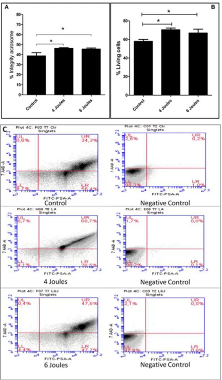
Irradiation with a low-power laser significantly increased (p<0.05) the percentage of live
sperm cells as evaluated by flow cytometry, both in the 4 joule group (70.3 ± 4.8) and in the 6
joule group (66.9 ± 11.7) compared to the control group (57.9 ± 5.4), as shown inFig. 1A).
Sim-ilarly, low-power laser irradiation at both 4 joules (46.2 ± 2.2) and 6 joules (45.5 ± 3.3) main-tained the integrity of the acrosome membrane of living cells, as assessed by flow cytometry,
significantly better (p<0.05) when compared to the results obtained in the control group
(38.7 ± 9.8) (Fig. 1B).
Evaluation of sperm motility
In comparing laser irradiation at doses of 4 and 6 joules, and a non-irradiated control group, the following variables were analyzed: curvilinear speed, rectilinear speed, average value, linear-ity index, oscillation index, head side movement, beat frequency, mobile progressive, and straightness index. On the straightness index variable, there was a statistical difference between the control group and the group irradiated with 4 joules and also between the two treatment
groups, 4 and 6 joules (p<0.05). We also found statistical differences for the variable mobile
progressive when comparing the control group (30.4 ± 10.5) with the 4 joules group (43.5 ±
7.7). The straightness index result also showed statistical significance (p<0.05) between the
control group (68.7 ± 3.7) and the 4 joules group (73.1 ± 3.0), and between the 4 joules group
(73.1 ± 3.0) and the 6 joules group (68.7 ± 3.8) withp<0.05 (Fig. 2). The results of the other
outcomes related to motion analysis are summarized inTable 1.
Discussion
Fig 1. Representative Low Level Laser Irradiation in joules 4 and 6 and analyzed by flow cytometry.
(A) Shows the percentage of sperm with acrosome integrity under the total of living cells. (Newman-Keuls Test*p<0.05). (B) Shows the percentage of live sperm cells relative to the total cells analyzed. (C) Shows
representative plots of the respective conditions when exposed to 7AAD and FITC-PSA. The quadrants indicate: UL—sperm dead (plasmatic membrane injured (MP) and acrosome membrane integrate (MA), RH—live sperm (plasmatic membrane injured (MP) and acrosome membrane integrate); LL—live (plasmatic membrane integrate (MP) and Membrane damaged acrosome (MA); LR—(plasmatic membrane injured (MP) and acrosome membrane integrate (MA), in the control, 4 joule and 6 joules groups.
Fig 2. Evaluation of sperm movement through the Integrated Semen Analysis System in (A) Straightness Index percentage of sperm analysis presenting the non-irradiated control group and groups subjected to irradiation with low-power laser with dose 4:06 joules.Data shown are the mean± standard deviation. One-way ANOVA and Newman-Keuls post hoc analysis were applied.*p<0.05 control
group vs. 4 Joules group and # p<0.05. group 6 Joules vs. 4 Joules group. (B) Mobile percentage of
progressive sperm analysis presenting the group not irradiated groups and subjected to low-level laser irradiation dose 4 and 6 joules group. Data shown are the mean±standard deviation. One-way ANOVA and Newman-Keuls post hoc analysis were applied.*p<0.05 control group vs. 4 Joules group. (C) Graphical
representation of sperm movement by Integrated Semen Analysis System, The red lines show the movements of the sperm in nm/s and the yellow lines represent the static sperm.
doi:10.1371/journal.pone.0121487.g002
Table 1. Results of the evaluation of sperm movement by the Integrated Semen Analysis System.
Control 4J 6J
Curvilinear Speed (μ/ s) 81±20 80±20 77±21
Rectilinear Speed (μ/ s) 35±6 36±7 32±8
Average Value (μ/ s) 50±10 50±10 47±12
Linearity Index % 42±3 46±5 41±6
Oscillation Index % 61±3 63±5 60±4
Head lateral moving (μ) 2.9±0.7 2.8±0.7 2.8±0.7
Beat Frequency (Hz) 10±1 10±1 9±2
Values are expressed as mean and standard deviation.
two different doses (4 and 6 joules). The analysis of straightness and the percentage of cell mo-tility show that a dose of 4 joules is more effective.
In vitro production (IVP) of bovine embryos using frozen/thawed semen is used around the world for commercial purposes. Sperm cells are exposed to a series of potential risks during
cryopreservation [21]. The freeze-thaw process damages the plasma membrane and the
acro-some of the sperm [22]. One of the reasons proposed to explain this variation is a change in the
integrity of the sperm chromatin [21]. Cryopreservation also leads to a reduction in size of the
head of the sperm as compared to fresh semen, perhaps because of damage or loss of the
acro-some or overcondensation of the nuclear chromatin of sperm [6].
Cryopreservation also significantly increases the production of reactive oxygen species (ROS) in sperm. ROS have two effects on sperm function: at low concentrations they induce sperm capacitation, hyperactivation of the acrosome, and sperm-oocyte fusion and, on the other hand, excessive amounts of ROS damage DNA, inhibit sperm-oocyte fusion, and reduce
sperm motility [23].
Several studies have shown that LLLI accelerates wound healing [24], enhances repair of
bone defects [25], modulates the production of inflammatory mediators in joint inflammation
[26], and decreases oxidative stress and muscle fatigue [27]. Others have shown the LLLI
im-proves the activation of anti-inflammatory vasoactive peptides [15] and increases cell energy
and viability [13]. The mechanism of photobiostimulation by LLLI is still unclear. It has been
suggested that reactive oxygen species (ROS), which can be produced by photosensitization of endogenous chromophores such as cell cytochromes, flavins/riboflavins, and NADPH, may
have an important role in this light/tissue interaction [23]. Additionally, Albuquerque-Pontes
et al. [27] demonstrated that cytochrome c oxidase (complex IV of mitochondrial respiratory
chain) is modulated by different wavelengths and doses of LLLT at different time-intervals. We previously noted that LLLI with the wavelength of 660 nm, power 30 mW and doses of 4 and 6 joules was able to improve the percentage of live sperm cells evaluated by flow cytome-try and maintain acrosomal membrane integrity. A dose of 4 joules also increased the percent-age of mobile progressive sperm and the straightness index.
The improvement in semen quality after LLLI Has Been illustrated previously described in
several species: dog [16], bovine [28], [29], rabbit [30], and turkey. [31] Using the same energy
and wavelength as in previous similar studies, we show additional evidence that LLLI may re-sult in a significant increase in the percentage of live sperm cells, integrity of acrosome mem-brane, and higher sperm motility.
According to Karuet al. [13], the possible primary mechanisms of light activation of
sper-matozoids suggest that photoacceptors are connected with oxygen metabolism and, in particu-lar, with respiratory chains. It is important to recall that respiratory chain molecules in eukaryotic as well as prokaryotic cells are considered photoacceptors and photosignal
trans-ducers in these cells.[13]. On the other hand Lubarte et al. [32] report that LLLI inhibits
calci-um uptake by mitochondria and stimulates calcicalci-um to connect the vesicles of the plasma membrane of the sperm, promoting better cell maintenance.
The analysis of the percentage of live sperm cells and the acrosome membrane integrity have been used in several other studies because they are important for the diagnosis of the
via-bility of semen after cryopreservation [7], [9], [10], [22], [33]. However, few studies with LLLI
[16], [30], [31] have used these factors to analyze the improved quality of semen. It is
notewor-thy that these studies also differ from our study in the form of measurements used, since we
have fluorescein isothiocyanato-labeledPisum sativumagglutinin (FITC-PSA) to detect the
ac-rosome integrity by flow cytometry. Sperm motility after LLLI has been investigated in several
studies [16], [30], [31], [33], [34]. However, some studies [30], [35] showed negative outcomes
irradiation time, as well as the experimental analysis conditions. Considering the data pre-sented in our study and the current state of knowledge regarding the efficacy of LLLI in im-proving the quality of semen, we conclude that LLLI may exert beneficial effects on both the preservation of live sperm and sperm motility after cryopreservation.
Perspectives and Limitations
The low-level laser is used medically to accelerate repair processes of various types of tissue as well as to treat pain and inflammation. Pre-clinical studies have demonstrated several other possible uses. However, in vitro improvement in the quality of semen for artificial insemination has not been translated into actual practice. Possible mechanisms of low-level laser effects on the oxidative stress generated by cryopreservation include the following: (i) Superoxide anions induce hyperactivation and capacitation and are being altered by LLLI; (ii) capacitating sper-matozoa produce elevated concentrations of superoxide anions themselves; and (iii) if the LLLI is capable of superoxide dismutase by removal of this ROS.
Acknowledgments
The authors would like to thank Tairana Central de Congelamento de Semên Ltda., repre-sented by Dr. Vet. Tatiana Isaa Uherara Berton, for the donation of semen samples and techni-cal support and suggestions. The authors would also like to thank the CAPES scholarship of Guilherme H. C. Fernandes, and UNINOVE, for all support.
Author Contributions
Conceived and designed the experiments: PTCC RA ECLJ AJS. Performed the experiments: GHCF JPSP CR AMC. Analyzed the data: AJS PTCC RA. Contributed reagents/materials/anal-ysis tools: JPSP AMC CR. Wrote the paper: PTCC GHCF ECLJ. Edited the final format of the paper: GHCF PTCC. Discussion of the paper: PTCC GHCF. Edited the final version of the manuscript: PTCC GHCF ECLJ.
References
1. Thibier M (2005) The zootechnical applications of biotechnology in animal reproduction: current meth-ods and perspectives. Reprod Nutr Dev. May Jun; 45(3):235–42. PMID:15982450
2. Torres-Júnior JR, Penteado L, Sales JN, Sá Filho MF, Ayres H, Baruselli PS (2014) A comparison of two different esters of estradiol for the induction of ovulation in an estradiol plus progestin-based timed artificial insemination protocol for suckled Bos indicus beef cows. Anim Reprod Sci. Oct 2. pii: S0378–4320(14)00295–4.
3. Dahlen C, Larson J, Lamb GC (2014) Impacts of reproductive technologies on beef production in the United States. Adv Exp Med Biol.; 752:97–114. doi:10.1007/978-1-4614-8887-3_5PMID:24170356 4. Büyükleblebici S, Tuncer PB, Bucak MN, Eken A, Sarıözkan S, Taşdemir U, et al. (2014)
Cryopreserva-tion of bull sperm: Effects of extender supplemented with different cryoprotectants and antioxidants on sperm motility, antioxidant capacity and fertility results. Anim Reprod Sci. Sep 22. pii: S0378–4320(14) 00282–6.
5. Zeng C, Peng W, Ding L, He L, Zhang Y, Fang D, et al. (2014) A preliminary study on epigenetic changes during boar spermatozoa cryopreservation. Cryobiology. Aug; 69(1):119–27. doi:10.1016/j.
cryobiol.2014.06.003PMID:24974820
6. Ramón M, Pérez-Guzmán MD, Jiménez-Rabadán P, Esteso MC, García-Álvarez O, Maroto-Morales A, et al. (2013) Sperm cell population dynamics in ram semen during the cryopreservation process. PLOS One.; 8(3):e59189. doi:10.1371/journal.pone.0059189PMID:23544054
7. Forero-Gonzalez RA, Celeghini EC, Raphael CF, Andrade AF, Bressan FF, Arruda RP (2012) Effects of bovine sperm cryopreservation using different freezing techniques and cryoprotective agents on plasma, acrosomal and mitochondrial membranes. Andrologia. May; 44 Suppl 1:154–9. doi:10.1111/j.
8. Barbas JP, Mascarenhas RD (2009) Cryopreservation of domestic animal sperm cells. Cell Tissue Bank. Feb; 10 (1):49–62. doi:10.1007/s10561-008-9081-4PMID:18548333
9. Celeghini EC, de Arruda RP, de Andrade AF, Nascimento J, Raphael CF, Rodrigues PH (2008) Effects that bovine sperm cryopreservation using two different extenders has on sperm membranes and chro-matin. Anim Reprod Sci. Mar 3; 104(2–4):119–31.
10. Gadella BM, Luna C (2014) Cell biology and functional dynamics of the mammalian sperm surface.
Theriogenology. Jan 1; 81(1):74–84. doi:10.1016/j.theriogenology.2013.09.005PMID:24274412 11. Lubart R, Levinshal T, Cohen N, Friedmann H, Breitbart H (1996) Changes in Calcium Transport in
Mammalian Sperm Mitochondria and Plasma Membrane due to 633 nm and 780 nm Irradiation. Laser in der Medizin / Laser in Medicine, 449–453.
12. Rodriguez-Martinez H, Larsson B, Pertoft H (1997) Evaluation of sperm damage and techniques for sperm clean-up. Reprod Fertil Dev, 9(3):297–308. PMID:9261878
13. Karu TI (2012) Lasers in infertility treatment: irradiation of oocytes and spermatozoa. Photomed Laser Surg. May; 30(5):239–41. doi:10.1089/pho.2012.9888PMID:22551048
14. Huang YY, Sharma SK, Carroll J, Hamblin MR (2011) Biphasic dose response in low level light therapy— an update. Dose Response. 9(4):602–18. doi:10.2203/dose-response.11-009.HamblinPMID:
22461763
15. Manchini MT, Serra AJ, Feliciano R dos S, Santana ET, Antônio EL, de Tarso Camillo de Carvalho P, et al. (2014) Amelioration of cardiac function and activation of anti-inflammatory vasoactive peptides ex-pression in the rat myocardium by low level laser therapy. PLOS One. Jul 3; 9(7):e101270. doi:10. 1371/journal.pone.0101270PMID:24991808
16. Corral-Baqués MI, Rivera MM, Rigau T, Rodríguez-Gil JE, Rigau J (2009) The effect of low-level laser irradiation on dog spermatozoa motility is dependent on laser output power. Lasers Med Sci. Sep; 24(5):703–13 doi:10.1007/s10103-008-0606-7PMID:18787758
17. Motta JP, Paraguassú-Braga FH, Bouzas LF, Porto LC (2014) Evaluation of intracellular and extracellu-lar trehalose as a cryoprotectant of stem cells obtained from umbilical cord blood. Cryobiology. Jun; 68(3):343–8. doi:10.1016/j.cryobiol.2014.04.007PMID:24769312
18. Graham JK (2001) Assessment of sperm quality: a flow cytometric approach. Anim Reprod Sci. Dec; 68(3–4):239–47. PMID:11744274
19. Verstegen J, Iguer-Ouada M, Onclin K (2002) Computer assisted semen analyzers in andrology re-search and veterinary practice. Theriogenology. Jan 1; 57(1):149–79. PMID:11775967
20. Kathiravan P, Kalatharan J, Edwin MJ, Veerapandian C (2008) Computer automated motion analysis
of crossbred bull spermatozoa and its relationship with in vitro fertility in zona-free hamster oocytes. Anim Reprod Sci. Feb 1; 104(1):9–17. PMID:17254723
21. Simões R, Feitosa WB, Siqueira AF, Nichi M, Paula-Lopes FF, Marques MG, et al. (2013) Influence of bovine sperm DNA fragmentation and oxidative stress on early embryo in vitro development outcome. Reproduction. Oct 1; 146(5):433–41. doi:10.1530/REP-13-0123PMID:23940385
22. Shahverdi A, Rastegarnia A, Rezaei Topraggaleh T (2014) Effect of extender and equilibration time on
post thaw motility and chromatin structure of buffalo bull (bubalus bubalis) spermatozoa. Cell J. Fall; 16(3):279–88. PMID:24611139
23. Awda BJ, Mackenzie-Bell M, Buhr MM (2009) Reactive oxygen species and boar sperm function. Biol Reprod. Sep; 81(3):553–61. doi:10.1095/biolreprod.109.076471PMID:19357363
24. Aparecida Da Silva A, Leal-Junior EC, Alves AC, Rambo CS, Dos Santos SA, Vieira RP, et al. (2013) Wound-healing effects of low-level laser therapy in diabetic rats involve the modulation of MMP-2 and MMP-9 and the redistribution of collagen types I and III. J Cosmet Laser Ther. Aug; 15(4):210–6. doi:
10.3109/14764172.2012.761345PMID:23463906
25. Denadai AS, de Carvalho Pde T, dos Reis FA, Belchior AC, Pereira DM, Dourado DM, et al. (2009) Morphometric and histological analysis of low-power laser influence on bone morphogenetic protein in bone defects repair. Lasers Med Sci. Sep; 24(5):689–95. doi:10.1007/s10103-008-0595-6PMID:
18787760
26. Alves AC, Vieira R, Leal-Junior E, dos Santos S, Ligeiro AP, Albertini R, et al. (2013) Effect of low-level laser therapy on the expression of inflammatory mediators and on neutrophils and macrophages in acute joint inflammation. Arthritis Res Ther. 15(5):R116. PMID:24028507
27. Albuquerque-Pontes GM, Vieira R de P, Tomazoni SS, Caires CO, Nemeth V, Vanin AA, et al. (2015) Ef-fect of pre-irradiation with different doses, wavelengths, and application intervals of low-level laser thera-py on cytochrome c oxidase activity in intact skeletal muscle of rats. Lasers Med Sci. 30(1):59–66. doi:
28. Breitbart H, Levinshal T, Cohen N, Friedmann H, Lubart R (1996) Changes in calcium transport in mam-malian sperm mitochondria and plasma membrane irradiated at 633 nm (HeNe laser). J Photochem Photobiol B. Jul; 34(2–3):117–21. PMID:8810541
29. Lubart R, Friedmann H, Levinshal T, Lavie R, Breitbart H (1992) Effect of light on calcium transport in bull sperm cells. J Photochem Photobiol B. Sep 15; 15(4):337–41. PMID:1432397
30. Iaffaldano N, Rosato MP, Paventi G, Pizzuto R, Gambacorta M, Manchisi A, et al. (2010) The irradiation
of rabbit sperm cells with He-Ne laser prevents their in vitro liquid storage dependent damage. Anim Reprod Sci. May; 119(1–2):123–9.
31. Iaffaldano N, Meluzzi A, Manchisi A, Passarella S (2005) Improvement of stored turkey semen quality as a result of He-Ne laser irradiation. Anim Reprod Sci. Feb; 85(3–4):317–25. PMID:15581516 32. Lubart R, Friedmann H, Sinyakov M, Cohen N, Breitbart H (1997) Changes in calcium transport in
mammalian sperm mitochondria and plasma membranes caused by 780 nm irradiation. Lasers Surg Med.; 21(5):493–9 PMID:9365961
33. Gillan L, Evans G, Maxwell WM (2005) Flow cytometric evaluation of sperm parameters in relation to fertility potential. Theriogenology. Jan 15; 63(2):445–57. PMID:15626410
34. Salman Yazdi R, Bakhshi S, Jannat Alipoor F, Akhoond MR, Borhani S, Farrahi F, et al. (2014) Effect of 830-nm diode laser irradiation on human sperm motility. Lasers Med Sci. Jan; 29(1): 97–104. doi:10.
1007/s10103-013-1276-7PMID:23407899