Printed version ISSN 0001-3765 / Online version ISSN 1678-2690 www.scielo.br/aabc
http://dx.doi.org/10.1590/0001-3765201520140251
Correspondence to: Márcia Queiroz Latorraca E-mail: mqlator@terra.com.br
Short-term low-protein diet during pregnancy alters islet area and protein content of phosphatidylinositol 3-kinase pathway in rats
CRISTIANA S.B. SALVATIERRA1, SÍLVIA R.L. REIS2, ANA F.M. PESSOA1, LETÍCIA M.I. DE SOUZA2, LUIZ F. STOPPIGLIA3, ROBERTO V. VELOSO2, MARISE A.B. REIS2, EVERARDO M. CARNEIRO4, ANTONIO C. BOSCHERO4, EDSON M. COLODEL5, VANESSA C. ARANTES2 and MÁRCIA Q. LATORRACA2
1Universidade Federal de Mato Grosso, Faculdade de Nutrição, Laboratório de Avaliação
Biológica de Alimentos, Av. Fernando Correa da Costa, 2367, 78060-900 Cuiabá, MT, Brasil
2
Universidade Federal de Mato Grosso, Faculdade de Nutrição, Departamento de Alimentos e Nutrição, Av. Fernando Correa da Costa, 2367, 78060-900 Cuiabá, MT, Brasil
3Universidade Federal de Mato Grosso, Instituto de Educação, Departamento de Psicologia,
Av. Fernando Correa da Costa, 2367, 78060-900 Cuiabá, MT, Brasil
4
Universidade Estadual de Campinas, Instituto de Biologia, Biologia Celular e Fisiologia e Biofísica, Departamento de Anatomia, Av. Bertrand Russel, s/n, 13083-865 Campinas, SP, Brasil
5Universidade Federal de Mato Grosso, Faculdade de Agronomia e Medicina Veterinária, Departamento de
Clínica Médica Veterinária, Av. Fernando Correa da Costa, 2367, 78060-900 Cuiabá, MT, Brasil
Manuscript received on June 11, 2014; accepted for publication on December 15, 2014
ABSTRACT
The phosphatidylinositol 3-kinase and mitogen-activated protein kinase pathways mediate β cell growth, proliferation, survival and death. We investigated whether protein restriction during pregnancy alters islet morphometry or the expression and phosphorylation of several proteins involved in the phosphatidylinositol 3-kinase and mitogen-activated protein kinase pathways. As controls, adult pregnant and non-pregnant rats were fed a normal-protein diet (17%). Pregnant and non-pregnant rats in the experimental groups were fed a low-protein diet (6%) for 15 days. Low protein diet during pregnancy increased serum prolactin level, reduced serum corticosterone concentration and the expression of both protein kinase B/AKT1 (AKT1) and p70 ribosomal protein S6 kinase (p70S6K
), as well as the islets area, but did not alter the insulin content of pancreatic islets. Pregnancy increased the expression of the Src homology/collagen (SHC) protein and the extracellular signal-regulated kinases 1/2 (ERK1/2) independent of diet. ERK1/2 phosphorylation (pERK1/2) was similar in islets from pregnant and non-pregnant rats fed a low-protein diet, and was higher in islets from pregnant rats than in islets from non-pregnant rats fed a normal-protein diet. Thus, a short-term, low-protein diet during pregnancy was sufficient to reduce the levels of proteins in the phosphatidylinositol 3-kinase pathway and affect islet morphometry.
INTRODUCTION
Although pancreatic islet mass is relatively constant under normal conditions, it has a remarkable ability to adapt to metabolic changes. An example is pregnancy, when islet mass can increase markedly
due to both β-cell hyperplasia and hypertrophy
(Sorenson and Brelje 1997). An increase in
β-cell proliferation is first observed around day
10 of pregnancy, peaks around day 14 and then returns to control levels by the end of pregnancy at approximately 21 days (Parsons et al. 1992). Another example of the adaptation of pancreatic islets occurs upon malnutrition, which is associated with important structural changes in the endocrine
pancreas. Protein deficiency leads to reduced β-cell
mass due to a decrease in the proliferation rate and an increase in apoptosis (Swenne et al. 1992, Petrik et al. 1999). Additionally, when the data are
corrected for age and weight, a reduction in β-cell
size and total islet volume, a lower proportion of
β-cells per islet, a degranulation of β-cells and β-cells with a preponderance of abnormal pale
granules have been observed (Heard and Stewart 1971, Pimstone 1976, Weinkove et al. 1977).
Nutrients such as amino acids and glucose and certain growth factors such as insulin-like growth factor 1 (IGF-1), glucocorticoids, and the somatolactogenic hormones placental lactogen (PL), prolactin (PRL) and growth hormone (GH)
increase pancreatic β-cell mitogenesis (Parsons
et al. 1992, Swenne et al. 1992, Scharfmann and Czernichow 1996, Hogg et al. 1993, Hugl et al. 1998,
Brelje et al. 1993, Rafacho et al. 2007). 17β-estradiol (E2) does not increase β-cell proliferation but does
enhance the biosynthesis of insulin and is involved
in β-cell survival (Nadal et al. 2009).Progesterone
(P) appears to have a role in decreasing β-cell mass
(Picard et al. 2002).
In growing rats receiving a low-protein diet, serum corticosterone concentrations are increased, the plasma concentration of IGF-1 and the number of GH and PRL receptors (GH-R and PRL-R) are
reduced, at least in the liver (Herbert and Carrillo 1982, Prewitt et al. 1982, Maes et al. 1984). In rodents, the PL, PRL, E2 and P levels increase during the second half of pregnancy (Nadal et al. 2009), and elevated concentrations of corticosterone are observed during the later stages of pregnancy (Dupouy et al. 1975). Increases in the mRNA expression of GH-R and PRL-R (Moldrup et al. 1993) and the expression of PRL-R in the pancreas (Brelje and Sorenson 1997) have also been observed during pregnancy. Dexamethasone, a synthetic glucocorticoid, reverses the PRL-induced upregulation of islet mass by inhibiting cell proliferation and increasing apoptosis (Weinhaus et al. 2000).
PRL exerts its biological effects mainly by activating the Janus kinase 2/signal transducer and activator of transcription 5 (JAK2/STAT5) pathway (Ihle et al. 1998, Galsgaard et al. 1999, Levy and Darnell 2002). PRL can also stimulate insulin receptor substrate (IRS) 1/2, phosphatidylinositol 3-kinase (PI3K) and mitogen-activated protein kinase (MAPK) in rat Nb2 node lymphoma cells, in
COS cells, in JAK2-deficient cell lines (Rao et al.
1995, Yamauchi et al. 1998) and in cultured neonatal rat islets (Amaral et al. 2003). Moreover, PRL enhances the expression of several genes related to growth and differentiation but reduces the expression of genes involved in apoptosis in pancreatic islets from adult female rats (Bordin et al. 2004). PL acts by binding to PRL-R and triggering the activation of downstream signaling pathways, including the JAK2/STAT5, PI3K/protein kinase B/AKT (AKT), extracellular signal-regulated kinase 1/2 (ERK1/2), adenylate cyclase/cyclic adenosine monophosphate (AC/cAMP) and intracellular calcium pathways (Amaral et al. 2003, 2004, Brelje et al. 2002).
IGF-1 stimulates β-cell proliferation mainly via
Dickson et al. 2001). In contrast, the effects of GH are initiated via activation of the JAK2/STAT5 pathway (Cousin et al. 1999, Gahr et al. 2002). Crosstalk can occur at different levels between the PRL-R, GH-R, IGF-1-R and insulin-receptor (IR) signal transduction pathways (Hugl et al. 1998, Amaral et al. 2003, Cousin et al. 1999).
Changes in the protein expression and/or activation/phosphorylation of a protein common to these pathways could result in alterations in proliferation and apoptosis. These two events could be associated with the expansion and reduction of pancreatic islet mass that are observed during pregnancy with a low-protein diet. Thus, we investigated the effects of short-term protein restriction during pregnancy on the morphometry and the expression and phosphorylation of several proteins involved in the PI3K and MAPK pathways in pancreatic islets.Studies that evaluate the role of protein restriction on molecular and cellular
determinants involved in β-cell adaptation during
pregnancy are particularly important to identify possible causal factors of gestational diabetes and could contribute towards its prevention.
MATERIALS AND METHODS
ANIMALS AND DIET
The animal experiments were approved by the Institutional Ethics Committee in Animal Experimentation (Universidade Federal de Mato Grosso). Virgin female Wistar rats(90 days of age) were obtained from the University’s breeding colony. Mating was achieved by housing males with females overnight, and pregnancy was
confirmed by the examination of vaginal smearsfor
the presence of sperm. Pregnant and non-pregnant femaleswere each randomly assigned to two diet groups: control and low-protein. The control
non-pregnant (CNP) and non-pregnant (CP) groupswere fed a 17% protein diet, and the low-protein non-pregnant(LPNP) and pregnant (LPP) groups were
fed a 6% protein diet fromdays 1 to 15 of pregnancy. The diets were isocaloric, as previously described (Filiputti et al. 2008). During the experimental period, rats had free accessto food and water and were housed at 22°C with a 12h light:dark cycle. At the end of this experimental period, the rats were weighed and killed by decapitation. Blood samples were collected and allowed to clot; sera were stored at -20°C forthe subsequent measurement of
17β-estradiol and progesterone by EIA (BioChem
ImmunoSystems, Italy), prolactin by EIA (ALPCO Diagnostics, USA) and corticosterone by an enzyme-linked immunosorbent assay (Assay Design, USA). Serum albumin levels (Doumas et al. 1971)were determined immediately after decapitation. Since it was not possible to evaluate all variables in the same animals, the number of individual experiments varied among groups.
MORPHOMETRY AND IMMUNOHISTOCHEMISTRY
For morphological analyses, the pancreases of rats were kept for approximately 8h in 10% buffered formalin and then processed by routine methods for histological analysis. After histological sectioning (5-µm-thick sections) and mounting onto positivized slides (ImmunoSlide-EasyPath®), the anti-insulin immunohistochemistry technique was performed. The sections were de-waxed in xylene and rehydrated in decreasing concentrations of alcohol (in distilled water). The blocking of endogenous peroxidase was performed by incubating the slides in a 3% solution of hydrogen peroxide in distilled water for 15 min at room temperature. For antigen retrieval, the samples were microwaved for 2 min in a citrate buffer of pH 6.0.
Non-specific binding was minimized with a 15 min
incubation in 5% skimmed milk (Molico®) diluted
were then washed in distilled water and treated with an anti-IgG guinea pig biotinylated secondary antibody (61-4620, ZYMED, Invitrogen USA) for 30 min in a moist chamber at room temperature. Next, the sections were washed in distilled water and treated with a streptavidin-peroxidase conjugate (DAKO Corp., Carpinteria, CA) for 30 min in a moist chamber at room temperature. Staining was subsequently visualized with the application of red chromogen (VECTOR®NovaRED) for 5 min. The sections were then washed in distilled water and counterstained with Harris hematoxylin for 30s. Next, the samples were washed in water for 1-2 min, dehydrated in increasing concentrations of alcohol, cleared in xylene and mounted with Entellan (Merck, Darmstadt, Germany Sigma Chemical Co., Saint Louis, USA). Approximately 20 islets
were observed in one field of view for each slide.
The slides were evaluated by light microscopy, and images were captured with a digital camera (Sony Cyber-shot DSC-H5) and measured using the Motic® Images Plus 2.0 ML software. In the morphometric technique the observer was blinded as to the groups and the pancreatic tissue was assessed for the area of the islets.
WESTERN BLOTTING
After isolation by collagenase digestion of the pancreas and subsequent separation by handpicking, groups of islets were pelleted by centrifugation(15,000 x g) and then resuspended in 50-100 µL ofabuffer containing protease and phosphatase inhibitors (Amaral et al. 2003, Kelley et al. 1994).The islets were sonicated, and the total protein content was determined (Bradford 1976). Samples containing 50 µg of protein from each experimental group were incubated for 5 min at 80°Cwith a 4X concentrated Laemmli sample buffer (1 mmol sodium phosphate/L,pH 7.8; 0.1% bromophenol blue; 50% glycerol; 10% SDS; 2% mercaptoethanol)(4:1, v:v) and then run on 12% (ERK1, ERK2 and pERK1/2) or 10% (AKT, pAKT,
and SHC) polyacrylamide gels at 120 V for90 min. The electrotransfer of proteins to nitrocellulose membranes(Bio-Rad) was performed for 2h at 120 V in a buffer lackingmethanol and SDS.
After checking the transfer efficiency byPonceau
S staining, the membranes were blocked with 5% skimmed milk in Tween-Tris-buffered saline (TTBS) (10 mmol Tris/L; 150 mmol NaCl/L; 0.5% Tween 20) overnight at 4°C. AKT, pAKT, SHC, ERK1, ERK2 and pERK1/2,were detected on the membranes after a 2h incubation at room temperature with the primary antibodies: anti-AKT1 (mouse polyclonal sc5298), anti-phospho[Ser473]
AKT (rabbit polyclonal, sc7985), anti-SHC (rabbit polyclonal, sc288), anti-ERK1 (rabbit polyclonal, sc94), or anti-ERK2 antibodies (rabbit polyclonal, sc 154) from Santa Cruz Biotechnology, Inc. (Santa Cruz, CA, USA) or an anti-ERK1/2 (p44/42 MAPK mouse monoclonal, E10) antibody from Biolabs Inc (Beverly, MA, USA), diluted 1:500, v/v, in TTBS containing 3% albumin (Sigma). The membranes
were then incubated with a secondary specific IgG
antibody, diluted 1:5000, v/v, in TTBS containing 3% albumin (Sigma) for 2h at room temperature. After incubation with horseradish peroxidase-conjugated secondary antibody, enhanced chemiluminescence (SuperSignal West Pico) was used to detection, by autoradiography. Band intensities weredocumented
by digital scanning followed by quantification using
theScion Image analysis software. The numerical values obtained are expressed in arbitrary units.
To detect IRβ, IRS1, IRS2, PI3Kp85, p70S6K
and phospho p70S6K, samples containing 60 µg of
total protein were incubated with 10µl of the
anti-IRβ (rabbit polyclonal, sc 711), anti-IRS1 (rabbit
polyclonal, sc 559), anti-IRS2 (goat polyclonal, sc 1556), anti-p70S6K (rabbit polyclonal, sc 8418), or
complexes were then precipitated with protein A-Sepharose6 MB for 2h. The pellets were washed three times in a buffercontaining 100 mM Tris, 2 mM sodium vanadate, 1 mM EDTA and0.5% Triton X-100, resuspended in 18 µl of Laemmli sample buffer and boiled for 5 min prior to loading onto polyacrylamidegels (8% for IRS1/2 and 10% for IRβ, PI3Kp85, p70S6K and phospho p70S6K). Following electrophoresis, the proteins were transferred to nitrocellulosemembranes for 2h at 120 V. Non-specific protein binding tonitrocellulose was reduced by
pre-incubating the filters in blockingbuffer (5% BSA, 10
mM Tris, 150 mM NaCl, and 0.02% Tween 20)for
2h at 22°C. IRβ, IRS1, IRS2, PI3Kp85, p70S6K and
phospho p70S6K were detected in the nitrocellulose
membranes after 2h incubation at room temperature with primary antibodies (diluted 1:500, v/v, in TTBS containing 3% albumin). The membranes were then
incubated with a secondary specific IgG antibody,
diluted 1:5000, v/v, in TTBS containing 3% albumin (Sigma) for 2h at room temperature. After incubation with horseradish peroxidase-conjugated secondary antibody, enhanced chemiluminescence (SuperSignal West Pico) was used for detection by autoradiography. Band intensities weredocumented by digital scanning
followed by quantification using theScion Image
analysis software. The numerical values obtained are expressed in arbitrary units.
STATISTICAL ANALYSES
The results are expressed as the means ± SDs for thenumber of rats (n) indicated. For islets, n refers to the numberof experiments performed. Levene’s test for the homogeneityof variances was used
initially to check the fit of the datato parametric
ANOVA assumptions. When necessary, the data were log-transformed to correct for variance in heterogeneity or non-normality (Sokal and Rohlf 1995). Data were analyzed by a two-way ANOVA test (nutritional status andphysiological status). When necessary, these analyses were followed
by LSD’s honestly significantdifference test to
determine the significance of individualdifferences.
The level of significance was set at P<0.05.The
data were analyzed using the Statistic Software package(Statsoft).
RESULTS
Regardless of nutritional status, pregnant rats had higher weight gain and, consequently, higher
final bodyweights (F1,83= 236.3, P<0.0001 and
F1,83= 67.7, P<0.0001, respectively). Additionally,
enhanced serum P (F1,22= 85.8, P<0.0001 ) and
E2 (F1,23= 102.4, P<0.0001) levels were observed
in pregnant rats compared with non-pregnant rats. Serum prolactin concentrations were higher in LPP than in LPNP, CP or CNP (P<0.001) rats. In contrast, serum corticosterone levels were lower in LPP rats than in CP, LPNP or CNP rats. Serum albumin concentrations did not differ among groups (Table I).
Detection of IR-β, IRS-1, IRS-2 and PI3K-p85
obtained through immunoprecipitation showed
no significant differences in the levels of all four
proteins observed among the groups (Fig. 1A to 1D). As shown in Figure 1E, the protein levels of AKT1 were 1.5-fold higher in islets from the LPNP group compared with the CNP group. The pancreatic islets from LPP rats had lower levels of AKT1 than did the pancreatic islets from LPNP
rats. No significant difference was detected in the
level of AKT1 expression between CP and CNP rats. The phospho[Se473]AKT1 level (Fig. 1F) and the ratio of phospho[Se473]AKT1/AKT1 (Fig. 1G)
were lower in islets from low protein rats (LPP and LPNP) than in those of the control rats (CNP and CP) (F1,8=25.9, P<0.001, F1,8=6.4, P<0.05,
respectively). In contrast, the variables were 1.3- and 1.7-fold higher in the islets from pregnant rats (LPP and CP) compared with non-pregnant rats, respectively, (LPNP and CNP) (F1,8=10.24, P<0.02, F1,8=7.7, P<0.05, respectively). Levels
Figure1H, the pancreatic islets from LPP rats expressed lower levels of p70S6K than did those from LPNP rats, whereas the islets from CP rats expressed higher levels of p70S6K than did those from CNP rats. The phosphor p70S6K levels were 1.4-fold higher in
the islets from pregnant rats (LPP and CP) in relation to non-pregnant rats (LNP and CNP) (F1,8=5.32,
P<0.05) (Fig. 1I). No significant differences were
detected in the phosphop70S6K/p70S6K ratio among
groups (Fig. 1J).
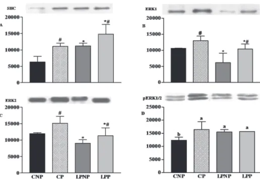
As shown in Figure 2A, the expression of SHC increased 1.5-fold in the islets from rats fed a low-protein diet in relation to rats fed the control diet (F1,8= 16.25, P<0.01). Similar results were observed
in the islets from pregnant rats in relation to those from non-pregnant rats (F1,8=15.63, P<0.01). The
protein levels of both ERK1 (Fig. 2B) and ERK2 (Fig. 2C) were decreased in the islets from rats fed a low-protein diet (F1,10=11.64, P<0.01 and F1,8=
11.97, P<0.01, respectively) compared with those
from rats maintained on the control diet. However, the expression of ERK1 and ERK2 was 1.4-fold (F1,10=10.18, P<0.01) and 1.3-fold (F1,8= 7.91, P<0.05) higher in the islets from pregnant rats compared with non-pregnant rats, respectively. The phosphoERK1/2 levels were similar in islets from the LPNP, LPP and CP groups and were
significantly higher in relation to those from the
CNP group (Fig. 2D).
The insulin content in the pancreatic islets did not differ among groups (CNP= 26±8 ng/islet, n=4; CP= 28±8 ng/islet, n=4; LNP= 34±8 ng/islet, n=3; LP= 44±18 ng/islet, n=4).
The islet area in LPP rats was lower in relation to that in LPNP rats, whereas the islet area in CP
rats was similar to that in CNP rats. No significant
differences were observed among the islet areas in LPNP, CNP and CP rats (CNP= 1869±2615 µm2, n=91; CP= 2613±4360 µm2, n=137; LPNP=2585
±3892 µm2, n=138; LPP=1502±2000 µm2, n=91).
Values represent the means ± SDs for the number of rats shown in parentheses. #Indicates differences in relation to non-pregnant rats (Two-way ANOVA, P<0.05). a,b Mean values with
unlike superscript letters were significantly different (LSD test; P<0.05). TABLE I
Body weight gain, final body weight, and serum albumin, 17β-estradiol, progesterone, prolactin and corticosterone levels in pregnant and non-pregnant rats maintained on control (CNP and CP) or low-protein diets (LPNP and LPP).
Variable Groups
CNP CP LPNP LPP
Body weight gain (g) 35±16
(17)
78±13# (22)
29±12 (20)
79±15# (28) Final body weight (g) 258±23
(17)
309±24# (22)
270±18 (20)
306±29# (28) Serum albumin (g/dl) 4.4±0.7
(6)
3.9±0.4 (7)
4.3±0.4 (10)
4.3±0.8 (6)
Serum 17β-estradiol (pmol/L) 89±15 (6) 198±48(7) # 81±18(6) 206±44(8) #
Serum progesterone (nmol/L) 51±22 (5)
232±35# (7)
54±19 (5)
188±60# (9) Serum prolactin (ng/ml) 5.5±1.1
b
(3)
4.0±0.6b (4)
5.6±1.3b (4)
12.6±6.6a (3) Serum corticosterone (µg/dl) 15.8±1.1
a
(7)
14.9±0.6a (6)
15.3±0.3a (5)
Figure 1 - Protein expression of IR-β (A), IRS-1 (B), IRS-2 (C), the p85 subunit of PI3K (D) and AKT1 (E), phospho-[Se473] AKT1 levels (F), phospho-[Se473]AKT1/AKT1 ratios (G), the protein expression of p70S6K (H), phospho-p70S6K levels (I) and phospho-p70S6K/p70S6K ratios (J) in the islets of CNP, CP, LPNP and LPP rats. Equal amounts of protein (60 µg) were subjected
to immunoprecipitation using antibodies anti-IRβ , anti-IRS1, anti-IRS2, anti-PI3K-p85, anti-p70S6K
, or anti-phospho p70S6.
Immunoprecipitates were analyzed by SDS-PAGE followed by immunoblotting with antibodies to IR-β, IRS-1 IRS-2, PI3K-p85,
Figure 2 - The levels of the SHC (A), ERK1 (B) and ERK2 proteins (C) and phospho-ERK1/2 levels (D) in the islets of CNP, CP, LPNP and LPP rats. Equal amounts of protein (50 µg) were run on SDS-PAGE and blotted with anti- SHC, anti- ERK1, anti-ERK2 and anti-phospho-ERK1/2. The columns represent the means ± SDs of 3-4 independent experiments. #Indicates differences in relation to non-pregnant rats (Two-way ANOVA, P<0.05). *Indicates differences in relation to rats maintained on a control diet (Two-way ANOVA, P<0.05). a,b Mean values with unlike superscript
letters were significantly different (LSD test; P<0.05).
DISCUSSION
After the short period of low-protein diet consumption (15 d) used here, the classical alterations observed in malnourished animals (weight loss and hypoalbuminemia) were not yet
present. We verified an increase in E2 and P in both
CP and LPP rats, which is normally observed during pregnancy (Nadal et al. 2009). In disagreement with the observation that the maternal plasma total and free corticosteroneconcentrations in rats are increased by protein restriction and pregnancy at 15 days (Dupouy et al. 1975, Martin et al. 1977, Herbert and Carrillo 1982), in the present study, the serum corticosterone levels did not increase in CP rats and were decreased in LPP rats. Interestingly, the unexpected decrease in the serum corticosterone levels in LPP rats was accompanied by an early increase in the serum prolactin concentration. This
increase is commonly observed in the later stages of pregnancy (Nadal et al. 2009) and suggests an inhibitory role for corticosterone in prolactin secretion (Nishino et al. 1992).Corticosterone and prolactin are involved in the structural adaptations of pancreatic islets to pregnancy.
Some studies have attempted to identify the molecular mechanisms underlying these adaptations in response to pregnancy and protein deprivation. Long-term exposure to a low-protein diet during the growth phase produces an increase in IR, IRS2 and PI3K levels and a reduction in IRS1 and p70S6K expression (Araujo et al. 2004, Filiputti
accompanied by the reduced expression of AKT1 and p70S6K, whereas normal pregnancy did result in an increase in the expression of p70S6K, but did not
alter AKT1 expression. Although similar in CNP and CP groups, the AKT1 expression tended to increase in CP rats compared with CNP rats. Neither condition resulted in changes in the expression of IR, IRS1/2 or PI3K-p85. Interestingly, normal pregnancy does not affect the protein levels of IRS1 and IRS2; however, it increases IRS1/2 phosphorylation and its association with p85, and also increases AKT1, p70S6Kexpression and phosphorylation in pancreatic islets (Amaral et al. 2004).
Several studies indicate that the structure
and function of pancreatic islets are influenced by
alterations in the expression and/or activity of IR, IRS-1, IRS-2, AKT1 and p70S6K. A reduced IR content in β-cells strongly impairs insulin secretion in
response to glucose (Kulkarni et al. 1999). In contrast,
IRS-1 deficiency produces hyperinsulinemia and increases of the β-cell mass, and the absence of
IRS-2 produces hyperinsulinemia followed by islet hypoplasia and early diabetes (Kulkarni et al. 1999, Withers et al. 1998). It has been demonstrated that AKT promotes protein synthesis by modulating mTOR and its downstream target, p70S6K (Hannan et al. 2003). Increased AKT1 activity is accompanied
by a marked increase in β-cell mass due to enhanced
cell survival and increased cell size (Tuttle et al. 2001). Additionally, p70S6K deficiency produces hypoinsulinemia, islet insensitivity to glucose, a low
insulin content and reduced β-cell size (Pende et al.
2000). In the present study, changes in the AKT1 and p70S6K levels were not followed by alterations in the insulin content of islets. However, changes in the levels of these two proteins were accompanied by a decrease in islet area in the LPP group. Because the LPP group had lower AKT1 and pAKT1 contents and a lower pAKT1Ser473/AKT1 ratio, which are
indirect indicatives of AKT1 activity, it is possible that the reduction in islet area resulted from elevated apoptosis and/or reduced AKT1-mediated cellular
proliferation and growth. Corroborating with the hypothesis of elevated apoptosis, we observed that islets from LPP group exhibited elevated levels of caspase 3 and caspase 9 (unpublished data).
In several signaling pathways, the expression and phosphorylation of members of the MAPK cascade activate several transcriptional regulators to control cellular growth. Although ERK1/2 modulate insulin secretion (Longuet et al. 2005), the most important role of these proteins in
pancreatic β-cells is the regulation of proliferation,
cell growth, and survival (Lawrence et al. 2008). In the present study, pregnancy increased the expression of SHC and ERK1/2 independent of diet and enhanced the level of pERK1/2 only in the islets of normal pregnant rats, as previously shown (Bordin et al. 2004, Amaral et al. 2004).A low-protein diet also elevated the levels of SHC and pERK1/2 and reduced the expression of ERK1 and ERK2. Pregnancy did not increase the pERK1/2 levels in the islets of rats exposed to a low-protein diet.
In pancreatic β-cells, nutrients, hormones
and growth factors modulate the expression of proteins in the SHC/ERK and IRS/PI3K/ AKT/ p70S6K pathways. Prolactin has been
considered an important growth factor. Prolactin is involved in the increase in islet mass and increased sensitivity to glucose that occur during pregnancy. Mechanistically, it activates proteins that are downstream of PI3K and MAPK cascades (Amaral et al. 2004). Dexamethasone, a synthetic glucocorticoid, increases the p70S6K expression and pAKT1 level in pancreatic islets (Rafacho et al. 2007). Curiously, in this study, LPP rats that exhibited an increase in serum prolactin levels and a decrease in serum corticosterone concentrations also had lower p70S6K and AKT1 levels. These results
Our results show that short-term exposure to a low-protein diet during pregnancy reduces pancreatic islet area that could be related to
deficiencies in the AKT1 and p70S6K levels. We
suggest that reduced circulating corticosterone levels altered the PI3K pathway and, consequently, cellular proliferation, growth, survival and death.
ACKNOWLEDGMENTS
This work was partly supported by the Fundação de Amparo à Pesquisa do Estado de Mato Grosso (FAPEMAT, grant number: 0786/2006) and
Conselho Nacional de Desenvolvimento Científico
e Tecnológico (CNPq, grant number: 305155/2004-0). Celso Roberto Afonso provided technical assistance for the study. This work is part of a dissertation that was presented by Cristiana dos Santos Barbosa Salvatierra as a partial requirement for her Master’s degree in Health Sciences at the School of Medical Sciences, UFMT.
RESUMO
As vias da fosfatidilinositol-3-cinase e da proteína cinase ativada por mitógeno medeiam crescimento, proliferação, sobrevivência e morte das células β. Investigamos se a restrição proteica durante a prenhez altera a morfometria da ilhota ou a expressão e a fosforilação de diversas proteínas envolvidas nas vias da fosfatidilinositol-3-cinase e da proteína cinase ativada por mitógeno. Como controles, ratas adultas não prenhes e prenhes foram alimentadas com uma dieta norpoproteica (17%). As ratas não prenhes e prenhes dos grupos experimentais foram alimentadas com dieta hipoproteica (6%) por 15 dias. A dieta hipoproteica durante a prenhez aumentou as concentrações séricas de prolactina, reduziu as concentrações séricas de corticosterona e a expressão da proteína cinase B/ AKT1 (AKT1) e da proteína p70 ribossomal S6 cinase (p70S6K
), bem como a área das ilhotas, mas não alterou o conteúdo de insulina nas ilhotas pancreáticas. A prenhez aumentou a expressão da proteína homóloga ao colágeno com domínio SH2 (SHC) e a proteína
cinase regulada por sinal extracelular 1/2 (ERK1/2), independente da dieta consumida. A fosforilação da ERK1/2 (pERK1/2) foi similar em ilhotas de ratas prenhes e não prenhes alimentadas com dieta hipoproteica, e foi maior em ilhotas de ratas prenhes do que não prenhes alimentadas com dieta normoproteica. Portanto, um curto período de dieta hipoproteica durante a prenhez foi suficiente para reduzir o conteúdo de proteínas da via da fosfatidilinositol-3-cinase e afetar a morfometria da ilhota.
Palavras-chave: via da MAP-cinase, ilhotas pan-creáticas, via da PI3-cinase, prenhez, restrição de proteína, ratos.
REFERENCES
AMARAL ME, CUNHA DA, ANHE GF, UENO M, CARNEIRO
EM, VELLOSO LA, BORDIN S AND BOSCHERO
AC. 2004. Participation of prolactin receptors and phosphatidylinositol 3-kinase and MAP kinase pathways in the increase in pancreatic islet mass and sensitivity to glucose during pregnancy. J Endocrinol 183: 469-476. AMARAL ME, UENO M, CARVALHEIRA JB, CARNEIRO EM,
VELLOSO LA, SAAD MJ AND BOSCHERO AC. 2003.
Prolactin-signal transduction in neonatal rat pancreatic islets and interaction with the insulin-signaling pathway. Horm Metab Res 35: 282-289.
ARAUJO EP, AMARAL ME, FILIPUTTI E, DE SOUZA CT,
LAURITO TL, AUGUSTO VD, SAAD MJ, BOSCHERO AC,
VELLOSO LA AND CARNEIRO EM. 2004. Restoration of
insulin secretion in pancreatic islets of protein-deficient
rats by reduced expression of insulin receptor substrate (IRS)-1 and IRS-2. J Endocrinol 181: 25-38.
BORDIN S, AMARAL ME, ANHE GF, DELGHINGARO-AUGUSTO
V, CUNHA DA, NICOLETTI-CARVALHO JE AND BOSCHERO
AC. 2004. Prolactin-modulated gene expression profiles
in pancreatic islets from adult female rats. Mol Cell Endocrinol 220: 41-50.
BRADFORD MM. 1976. A rapid and sensitive method for the quantitation of microgram quantities of protein utilizing the principle of protein-dye binding. Anal Biochem 72: 248-254. BRELJE TC, SCHARP DW, LACY PE, OGREN L, TALAMANTES
F, ROBERTSON M, FRIESEN HG AND SORENSON RL. 1993.
Effect of homologous placental lactogens, prolactins, and growth hormones on islet B-cell division and insulin secretion in rat, mouse, and human islets: implication for placental lactogen regulation of islet function during pregnancy. Endocrinology 132: 879-887.
BRELJE TC, SVENSSON AM, STOUT LE, BHAGROO NV AND
SORENSON RL. 2002. An immunohistochemical approach to monitor the prolactin-induced activation of the JAK2/STAT5 pathway in pancreatic islets of Langerhans. J Histochem Cytochem 50: 365-383.
COUSIN SP, HUGL SR, MYERS JR MG, WHITE MF, R
EIFEL-MILLER A AND RHODES CJ. 1999. Stimulation of pancreatic beta-cell proliferation by growth hormone is glucose-dependent: signal transduction via janus kinase 2 (JAK2)/signal transducer and activator of transcription 5 (STAT5) with no crosstalk to insulin receptor substrate-mediated mitogenic signalling. Biochem J 344 Pt 3: 649-658.
DICKSON LM, LINGOHR MK, MCCUAIG J, HUGL SR, SNOW
L, KAHN BB, MYERS Jr MG AND RHODES CJ. 2001.
Differential activation of protein kinase B and p70(S6)K by glucose and insulin-like growth factor 1 in pancreatic beta-cells (INS-1). J Biol Chem 276: 21110-21120. DOUMAS BT, WATSON WA AND BIGGS HG. 1971. Albumin
standards and the measurement of serum albumin with bromcresol green. Clin Chim Acta 31: 87-96.
DUPOUY JP, COFFIGNY H AND MAGRE S. 1975. Maternal and foetal corticosterone levels during late pregnancy in rats. J Endocrinol 65: 347-352.
FILIPUTTI E, FERREIRA F, SOUZA KL, STOPPIGLIA LF, ARANTES
VC, BOSCHERO AC AND CARNEIRO EM. 2008. Impaired insulin secretion and decreased expression of the nutritionally responsive ribosomal kinase protein S6K-1 in pancreatic islets from malnourished rats. Life Sci 82: 542-548.
GAHR S, MERGER M, BOLLHEIMER LC, HAMMERSCHMIED CG,
SCHOLMERICH J AND HUGL SR. 2002. Hepatocyte growth factor stimulates proliferation of pancreatic beta-cells particularly in the presence of subphysiological glucose concentrations. J Mol Endocrinol 28: 99-110.
GALSGAARD ED, NIELSEN JH AND MOLDRUP A. 1999.
Regulation of prolactin receptor (PRLR) gene expression in insulin-producing cells. Prolactin and growth hormone activate one of the rat prlr gene promoters via STAT5a and STAT5b. J Biol Chem 274: 18686-18692.
HANNAN KM, THOMAS G AND PEARSON RB. 2003. Activation of S6K1 (p70 ribosomal protein S6 kinase 1) requires an initial calcium-dependent priming event involving formation of a high-molecular-mass signalling complex. Biochem J 370: 469-477.
HEARD CR AND STEWART RJ. 1971. Protein-calorie deficiency
and disorders of the endocrine glands. Hormones 2: 40-64.
HERBERT DC AND CARRILLO AJ. 1982. The hypophyseal-adrenal axis in the protein-calorie malnourished rat. Horm Metab Res 14: 205-207.
HOGG J, HAN VK, CLEMMONS DR AND HILL DJ. 1993.
Interactions of nutrients, insulin-like growth factors (IGFs) and IGF-binding proteins in the regulation of DNA synthesis by isolated fetal rat islets of Langerhans. J Endocrinol 138: 401-412.
HUGL SR, WHITE MF AND RHODES CJ. 1998. Insulin-like growth factor I (IGF-I)-stimulated pancreatic beta-cell growth is glucose-dependent. Synergistic activation of insulin receptor substrate-mediated signal transduction pathways by glucose and IGF-I in INS-1 cells. J Biol Chem 273: 17771-17779.
IHLE JN, STRAVAPODIS D, PARGANAS E, THIERFELDER W,
FENG J, WANG D AND TEGLUND S. 1998. The roles of Jaks and Stats in cytokine signaling. Cancer J Sci Am 4 (Suppl. 1): S84-91.
KELLEY GG, ZAWALICH KC AND ZAWALICH WS. 1994.
Calcium and a mitochondrial signal interact to stimulate phosphoinositide hydrolysis and insulin secretion in rat islets. Endocrinology 134: 1648-1654.
KULKARNI RN, WINNAY JN, DANIELS M, BRUNING JC, FLIER
SN, HANAHAN D AND KAHN CR. 1999. Altered function
of insulin receptor substrate-1-deficient mouse islets and
cultured beta-cell lines. J Clin Invest 104: R69-75. LAWRENCE M, SHAO C, DUAN L, MCGLYNN K AND COBB
MH. 2008. The protein kinases ERK1/2 and their roles in pancreatic beta cells. Acta Physiol (Oxf) 192: 11-17. LECHNER J, WELTE T AND DOPPLER W. 1997. Mechanism of
interaction between the glucocorticoid receptor and Stat5: role of DNA-binding. Immunobiology 198: 112-123. LEVY DE AND DARNELL Jr JE. 2002. Stats: transcriptional
control and biological impact. Nat Rev Mol Cell Biol 3: 651-662.
LONGUET C, BROCA C, COSTES S, HANI EH, BATAILLE D AND
DALLE S. 2005. Extracellularly regulated kinases 1/2 (p44/42 mitogen-activated protein kinases) phosphorylate synapsin I and regulate insulin secretion in the MIN6 beta-cell line and islets of Langerhans. Endocrinology 146: 643-654. MAES M, UNDERWOOD LE AND KETELSLEGERS JM. 1984. Low
serum somatomedin-C in protein deficiency: relationship
with changes in liver somatogenic and lactogenic binding sites. Mol Cell Endocrinol 37: 301-309.
MARTIN CE, CAKE MH, HARTMANN PE AND COOK IF. 1977.
Relationship between foetal corticosteroids, maternal progesterone and parturition in the rat. Acta Endocrinol (Copenh) 84: 167-176.
MOLDRUP A, PETERSEN ED AND NIELSEN JH. 1993. Effects of sex and pregnancy hormones on growth hormone and prolactin receptor gene expression in insulin-producing cells. Endocrinology 133: 1165-1172.
NADAL A, ALONSO-MAGDALENA P, SORIANO S, ROPERO AB
AND QUESADA I. 2009. The role of oestrogens in the adaptation of islets to insulin resistance. J Physiol 587: 5031-5037.
NISHINO Y, MICHNA H, HASAN SH AND SCHNEIDER MR. 1992.
Involvement of the adrenal glands in the prolactin rise induced in the female rat by an antiprogestin, onapristone. J Steroid Biochem Mol Biol 41: 841-845.
PENDE M, KOZMA SC, JAQUET M, OORSCHOT V, BURCELIN R,
LE MARCHAND-BRUSTEL Y, KLUMPERMAN J, THORENS
B AND THOMAS G. 2000. Hypoinsulinaemia, glucose intolerance and diminished beta-cell size in
S6K1-deficient mice. Nature 408: 994-997.
PETRIK J, REUSENS B, ARANY E, REMACLE C, COELHO C, HOET
JJ AND HILL DJ. 1999. A low protein diet alters the balance of islet cell replication and apoptosis in the fetal and neonatal rat and is associated with a reduced pancreatic expression of insulin-like growth factor-II. Endocrinology 140: 4861-4873. PICARD F, WANATABE M, SCHOONJANS K, LYDON J, O'MALLEY
BW AND AUWERX J. 2002. Progesterone receptor knockout mice have an improved glucose homeostasis secondary to beta -cell proliferation. Proc Natl Acad Sci USA 99: 15644-15648.
PIMSTONE B. 1976. Endocrine function in protein-calorie malnutrition. Clin Endocrinol (Oxf) 5: 79-95.
PREWITT TE, D'ERCOLE AJ, SWITZER BR AND VAN WYK
JJ. 1982. Relationship of serum immunoreactive somatomedin-C to dietary protein and energy in growing rats. J Nutr 112: 144-150.
RAFACHO A, ROMA LP, TABOGA SR, BOSCHERO AC AND
BOSQUEIRO JR. 2007. Dexamethasone-induced insulin resistance is associated with increased connexin 36 mRNA and protein expression in pancreatic rat islets. Can J Physiol Pharmacol 85: 536-545.
RAO YP, BUCKLEY DJ AND BUCKLEY AR. 1995. Rapid activation of mitogen-activated protein kinase and p21ras by prolactin and interleukin 2 in rat Nb2 node lymphoma cells. Cell Growth Differ 6: 1235-1244.
SCHARFMANN R AND CZERNICHOW P. 1996. Differentiation and growth of pancreatic beta cells. Diabetes Metab 22: 223-228.
SOKAL RR AND ROHLF FJ. 1995. Assumptions of analysis of variance. In: SOKAL RR and ROHLF FJ (Eds), Biometry: the principles and practice of statistics in biological research: WH Freeman & Co, p. 392-450.
SORENSON RL AND BRELJE TC. 1997. Adaptation of islets of Langerhans to pregnancy: beta-cell growth, enhanced insulin secretion and the role of lactogenic hormones. Horm Metab Res 29: 301-307.
STOCKLIN E, WISSLER M, GOUILLEUX F AND GRONER B.
1996. Functional interactions between Stat5 and the glucocorticoid receptor. Nature 383: 726-728.
SWENNE I, BORG LA, CRACE CJ AND SCHNELL LANDSTROM
A. 1992. Persistent reduction of pancreatic beta-cell mass after a limited period of protein-energy malnutrition in the young rat. Diabetologia 35: 939-945.
TUTTLE RL, GILL NS, PUGH W, LEE JP, KOEBERLEIN B, FURTH
EE, POLONSKY KS, NAJI A AND BIRNBAUM MJ. 2001.
Regulation of pancreatic beta-cell growth and survival by the serine/threonine protein kinase Akt1/PKBalpha. Nat Med 7: 1133-1137.
WEINHAUS AJ, BHAGROO NV, BRELJE TC AND SORENSON RL.
2000. Dexamethasone counteracts the effect of prolactin on islet function: implications for islet regulation in late pregnancy. Endocrinology 141: 1384-1393.
WEINKOVE C, WEINKOVE E, TIMME A AND PIMSTONE B.
1977. Pancreatic islets of malnourished rats: quantitative
histologic and electron microscopic findings. Arch Pathol
Lab Med 101: 266-269.
WITHERS DJ ET AL. 1998. Disruption of IRS-2 causes type 2 diabetes in mice. Nature 391: 900-904.
WYSZOMIERSKI SL, YEH J AND ROSEN JM. 1999.
Glucocorticoid receptor/signal transducer and activator of transcription 5 (STAT5) interactions enhance STAT5 activation by prolonging STAT5 DNA binding and tyrosine phosphorylation. Mol Endocrinol 13: 330-343. YAMAUCHI T ET AL. 1998. Growth hormone and prolactin
![Figure 1 - Protein expression of IR-β (A), IRS-1 (B), IRS-2 (C), the p85 subunit of PI3K (D) and AKT1 (E), phospho- [Se473]](https://thumb-eu.123doks.com/thumbv2/123dok_br/15975217.688908/7.892.123.827.129.882/figure-protein-expression-irs-irs-subunit-akt-phospho.webp)