(Annals of the Brazilian Academy of Sciences)
Printed version ISSN 0001-3765 / Online version ISSN 1678-2690 www.scielo.br/aabc
Production of 5-hydroxy-7-methoxy-4-methylphthalide in a culture of Penicillium crustosum
ANGELA M.M.P. VALENTE1, ANTONIO G. FERREIRA2, CRISTINA DAOLIO2, EDSON RODRIGUES FILHO3, ELISANGELA F. BOFFO4, ANTONIA Q.L. SOUZA5, FERNANDA L.S. SEBASTIANES6 and ITAMAR S. MELO1
1
Laboratório de Microbiologia Ambiental, Embrapa Meio Ambiente (CNPMA), Via SP 340, Km 127,5, Caixa Postal 69, 13820-000 Jaguariúna, SP, Brasil 2
Laboratório de Ressonância Magnética Nuclear and Laboratório de Microbiologia Molecular e Espectroscopia de Massas, Departamento de Química, Universidade Federal de São Carlos (UFSCar),
Via Washington Luís, Km 235, Caixa Postal 676, 13565-905 São Carlos, SP, Brasil 3
Laboratório de Microbiologia Molecular e Espectroscopia de Massas, Departamento de Química, Universidade Federal de São Carlos (UFSCar), Via Washington Luís, Km 235,
Caixa Postal 676, 13565-905 São Carlos, SP, Brasil
4Departamento de Química Orgânica, Instituto de Química, Universidade Federal da Bahia (UFBA), Rua Barão de Geremoabo, 147, Campus Universitário de Ondina, 40170-115 Salvador, BA, Brasil
5
Laboratório de Produtos de Origem Microbiana, Instituto de Ciências Biológicas/ ICB-DFCA, Universidade Federal do Amazonas (UFAM), Av. General Rodrigo Octávio Jordão Ramos, 3000,
Campus Universitário, Coroado I, 69077-000 Manaus, AM, Brasil
6Departamento de Genética, Escola Superior de Agricultura ‘Luiz de Queiroz’, Universidade de São Paulo (USP), Avenida Pádua Dias, 11, Caixa Postal 83, 13400-970 Piracicaba, SP, Brasil
Manuscript received on February 12, 2012; accepted for publication on July 9, 2012
ABSTRACT
The chemical reactions carried out by microorganisms have been used as a tool in modern chemistry. This paper reports the production of mycophenolic acid and a new phthalide by the endophytic fungus
Penicillium crustosum obtained from coffee seeds. The fungus was cultivated in a liquid medium for a period of seven days and after that the culture medium was divided into four treatments: A, B, C and D, to which different organic substances were added. Treatment A was maintained as the control to evaluate the occurrence of biotransformation. Organic acids were added to the culture media of treatments B (ferulic and quinic acids) and C [cinnamic and 3,4-(methylenedioxy) cinnamic acids], and caffeine was added in the treatment D. All these organic compounds were dissolved in DMSO, and the fermentation was maintained for more 13 days, totalizing 20 days. Mycophenolic acid was isolated from the culture with no added acids (treatment A). Mycophenolic acid and a new phthalide, 5-hydroxy-7-methoxy-4-methylphthalide were isolated from treatments B and C, and mycophenolic acid and caffeine (added to the culture medium) were isolated from treatment D. The structures were determined by NMR techniques and confi rmed by MS and MS/MS techniques.
Key words: coffee, Endophytic fungus, Mycophenolic acid, Penicillium crustosum, 5-hydroxy-7-methoxy-4-methylphthalide.
INTRODUCTION
The plant kingdom is colonized by a diversity of endophytic microorganisms, resulting in benefi cial associations that can stimulate plant growth, and increased resistance to diseases and adverse environmental conditions. There are several studies showing that endophytes do not act alone or only at the level of the host, but rather in a complex network of interactions with the microbial fl ora and native plant metabolism (Silva et al. 2006).
The colonization of the host plant by endophytes may be mediated by interesting secondary metabolites of potential use to modern medicine, agriculture and industry (Strobel and Daisy 2003, Mitchell et al. 2008). Surveys of the endophytes have been carried out in dozens of plants of important agricultural commodities, but coffee has been practically unexplored (Vega et al. 2005). Various endophytic Penicillium species were isolated from Coffea arabica, including
Penicillium crustosum. The role that these endophytes play in the biology of the coffee plant remains enigmatic, but the fact that no Penicillium
species have been reported as pathogens of
Coffea spp implies that these endophytes are not latent pathogens, suggesting either commensal or mutualistic relationships (Vega and Posada 2006).
The Penicillium genus is noted for producing a variety of bioactive metabolites possessing diverse biological properties including plant growth regulators (Arnold 2007).
Mycophenolic acid is produced by
Byssochlamys nivea and mainly by several
Penicillium species, such as P. brevi-compactum,
P. roqueforti, P. roqueforti var. carneum, P.
bialowiezense, P. chrysogenum, P. raciborkii, P. stoloniferum, and other Penicillium spp. It presents several biological activities, amongst which antibiotic activity, and it has also been successfully applied as an immunosuppressant after organ transplantation, administered in the form of the
pro-drug Mycophenolate Mofetil (Grinyo 2001). Recently mycophenolic acid was obtained by the biotransformation of imbricatolic acid, obtained from a culture of Cunninghamella echinulata
(Schmeda-Hirschmann et al. 2007).
The chemical reactions carried out by microorganisms have been used as a tool in modern chemistry, but the use of microorganisms in chemistry is not a new issue. Bacteria and fungi have been used to produce chemicals, pharmaceuticals and perfumes for decades. However their use for modifying the chemical structures of natural products is more recent, it is very well documented (Boaventura et al. 2004).
This study aimed to use the endophytic fungus
Penicillium crustosum, isolated from coffee beans, to transform characteristic substances of the coffee, such as caffeine, cinnamic, ferulic and quinic acids, and 3,4-methylenedioxycinnamic acid, which is a semi-synthetic substance.
MATERIALS AND METHODS
SAMPLING
Apparently healthy coffee (Coffea arabicaCoffea arabica L.) fruits L.) fruits from ripe berries (crown, peduncle, pulp and seeds) were harvested from coffee plantations in Águas da Prata, Brazil and taken to the Laboratory of Molecular Microbiology and Mass Spectroscopy of the Chemistry Department at the Universidade Federal de São Carlos, where the fungus was isolated.
FUNGAL ISOLATION
sterilized by a series of immersions in the following order: 1 min in 70% ethanol, 4 min in 11% sodium hypochlorite, 0.5 min in 70% alcohol and 1 min in distilled water. The beans seeds were inoculated onto PDA contained in Petri dishes, and 0.1% stock antibiotic solution was then added (stock: 0.02 g streptomycin in 10 mL sterile distilled water, fi lter sterilized; from this 1 mL was added per liter of medium). The cultures were incubated at room temperature (25°C).
SCANNING ELECTRON MICROSCOPE
The method used to prepare the material for scanning electron microscopy (SEM) was similar to the one described by Melo and Faull (2004). The samples were dried in a critical point (Dryer) (Emitech), sputtered (Emitech plating) with gold for 3 minutes at 30 mA and observed under a Field Emission Scanning Microscope (Gemini Leo 982 Zeiss + Leica) at the Environmental Microbiology Laboratory of the Brazilian Agriculture Research Corporation – Embrapa.
STRAIN TAXONOMY
The genomic DNA from the P. crustosum strain, Catl2.3, was extracted according to the method described by Raeder and Broda (1985). The fungi were identifi ed by comparing the rDNA from the internal transcribed spacer (ITS) regions with sequences deposited in the GenBank. The ITS1-5.8S-ITS2 region was amplifi ed by the polymerase chain reaction (PCR) using the ITS1 primer (5'-TCCGTAGGTGAACCTGCGG-3') and the ITS4 primer (5'-TCCTCCGCTTATTGATATGC-3') in a Peltier Thermal Cycler 200, MJ Research. The conditions for DNA amplifi cation included an initial denaturation step of 5 min at 94°C, followed by 24 cycles of 30 s at 94°C (denaturation), 30 s at 55°C (annealing) and 30 s at 72°C (elongation), plus a fi nal elongation step of 7 min at 72°C. The amplicons were purifi ed using GFX columns (GE
Healthcare UK Ltd., Buckinghamshire, England), and then cycle-sequenced using the BigDye® Terminator Cycle Sequencing Kit (Applied Biosystems, Nieuwerkerk, The Netherlands). The PCR products were sequenced by using the same primers used for amplifi cation. Each sequence was obtained in duplicate from each of two separate PCR amplifi cations. The ITS sequence of the strain found in this study matched the ITS sequence of
Penicillium crustosum (GenBank Accession No. GU723443), and displayed 99% similarity. The ITS sequence of the Penicillium crustosum isolated was aligned with different Penicillium isolates available in the GenBank nucleotide data base, using the MEGA 4.0 program (Tamura et al. 2007). The phylogenetic analysis was carried out by a neighbor-joining method to infer the relationships between the Penicillium crustosum isolate and sequences available for Penicillium in the GenBank. For this analysis, 1,000 bootstrap replicates were carried out to assess the statistical support for each tree. The fungus was deposited and preserved in the collection of the Environmental Microbiology Laboratory of the Brazilian Agriculture Research Corporation - Embrapa.
CULTURE (FUNGUS CATL2.3)
After fi ve days of growth, 3 - 5 pieces of the PDA culture containing mycelium, each 5 mm in diameter, were inoculated into 40 Erlenmeyer fl asks (500 mL) containing 250 mL of the following medium: 3.0 g NaNO3, 1.0 g K2HPO4, 0.5 g MgSO4.7H2O, 0.5 g KCl, 0.01 g FeSO4.7H2O, 10.0 g glucose, and 8.0 g yeast extract in 1.0 liter of distilled water. This culture was autoclaved for 15 minutes at 120°C and incubated at room temperature (28°C) in the static mode for seven days.
(C) and 2 mL of caffeine added to the fi nal 10 (D). These solutions were sterilized using Millipore fi lters, and the cultivation continued for 13 days in the static mode.
EXTRACTION, PURIFICATION AND ISOLATION
After 20 days of fermentation, 350 mL of CH2Cl2/ MeOH (7:3 v/v) were added to each fl ask and the fl asks allowed to rest for 24 hours. The mycelium was then separated from the culture medium, the organic and aqueous phases separated from each other, and the aqueous phase partitioned twice with dichloromethane. The dried organic extracts [A (338.4 mg), B (524.3 mg), C (537.7 mg) and D (964.0 mg)] were fractionated by CC over common silica (with vacuum) using a gradient elution system [n-C6H14/EtOAc (10:0, 7:3; 5:5; 4:6, 0:10 v/v) and EtOAc/MeOH (9:1, 8:2, 6:4, 4:6, 2:8 and 0:10 v/v)].
Very pure mycophenolic acid was isolated from extract A (fraction 5, 100 % EtOAc).
Fractions 5 and 6 from extract B (100 % EtOAc and EtOAc/MeOH 9:1, respectively) were mixed together [154.5 mg], re-chromatographed on Sephadex LH-20 (2.5 cm - diameter x 64.0 cm - length) and eluted in MeOH/CH3OCH3 (8:2 and 10:0 v/v). Mycophenolic acid was isolated from subfractions 4, 5 and 6 (MeOH/CH3OCH3, 8:2 v/v), and 5-hydroxy-7-methoxy-4-methylphthalide was isolated from subfractions 7, 8 and 9, eluted in MeOH (100%).
Fractions 5, 6 and 7 from extract C (100 % EtOAc, EtOAc/MeOH - 9:1 and EtOAc/MeOH - 8:2, respectively) were mixed [270.6 mg] and chromatographed under the same conditions as extract B. Mycophenolic acid and 5-hydroxy-7-methoxy-4-methylphthalide were again isolated.
Fractions 4 and 5 from extract D (100 % EtOAc and EtOAc/MeOH - 9:1, respectively) were mixed [141.5 mg] and subjected to column chromatography on fl ash-type silica gel (3.0 cm x
24.5 cm), and eluted in a gradient system: EtOAc/ Hex 8:2; EtOAc/Hex 9:1; EtOAc 100%; EtOAc/ MeOH 9:1; EtOAc/MeOH 1:1 and MeOH 100%. All the fractions were analyzed by TLC, and the caffeine was isolated from the fractions eluted in EtOAc (100%). Fraction 6 from extract D was subjected to preparative-layer chromatography (PLC), eluted with CH2Cl2/CH3OCH3 (7:3), and six subfractions obtained (a - g). Mycophenolic acid was obtained in fraction 6a.
NMR DATA
All the NMR data were recorded at 298 K using a Bruker DRX400 spectrometer operating at 9.4 T (1H at 400.2 and 13C at 100.6 MHz). Tetramethylsilane (TMS) was employed as the internal reference for chemical shifts (δ 0.0 ppm). Mycophenolic acid was dissolved in 600 μL of CDCl3 and 5-hydroxy-7-methoxy-4-methylphthalide in 600 μL of DMSO-d6. 1H, 13C,
gHSQC and gHMBC NMR spectra were acquired using standard pulse sequences. The TopSpin software was used for data processing.
MASS SPECTROSCOPY DATA
Low-resolution ESI-MS and ESI-MS/MS data were acquired in the negative and positive ion modes, using a QuattroLC mass spectrometer (Micromass, triple-quadrupole, ESI/APCI). Mycophenolic acid and 5-hydroxy-7-methoxy-4-methylphthalide, dis-solved in methanol, were constantly introduced by direct infusion using the equipment syringe pump at 5 mL.min-1. The Masslynx 3.5 software was used for data processing.
RESULTS AND DISCUSSION
dried in the sun. This strain was identifi ed as belonging to the genus Penicillium using Scanning Electron Microscope (SEM) methodology, based on the morphological structures.
The fungus was taxonomically assigned as
Penicillium crustosum on the basis of the molecular techniques. The ITS sequence of the strain isolated in this study matched the ITS sequence of P. crustosum (GenBank Accession Nº GU723443), displaying 99% similarity.
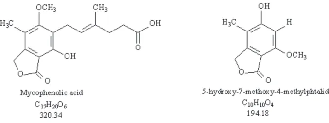
P. crustosum produced mycophenolic acid in the four treatments evaluated (A, B, C and D). However, in the treatments B and C in which acids were added, P. crustosum produced mycophenolic acid and 5-hydroxy-7-methoxy-4-methylphthalide (Figure 1).
In treatment A (with no additional substances added) this fungus produced only mycophenolic acid (102.0 mg) with a high degree of purity.
In the treatments in which organic acids were added (treatments B and C), the biotransformation of the acids (B - ferulic and quinic acids; C - cinnamic and 3,4-methylenedioxycinnamic acid) did not occur, as expected in this assay. The acids added to the culture medium were completely metabolized by the fungus, since these compounds were not detected in the fractions. However, two compounds were isolated, mycophenolic acid [B - 83.2 mg and C - 85.3 mg] and 5-hydroxy-7-methoxy-4-methylphthalide [B - 22.3 mg and C - 27.1 mg].
Mycophenolic acid was isolated in treatments A, B and C due to its production in the fermentation
Figure 1 - Structures of the compounds mycophenolic acid and 5-hydroxy-7-methoxy-4-methylphthalide.
medium before supplementation with the acids. These results were confi rmed by new cultures at several periods (Valente et al. 2007).
The synthesis of 5-hydroxy-7-methoxy-4-methylptalide only occurred after the addition of the organic acids, since it was not isolated in treatment A. In experiment D (supplemented with caffeine), P. crustosun only produced mycophenolic acid (3.25 mg), and the caffeine was completely recovered (70.1 mg). This change in the metabolite produced in these treatments is probably related to
the chemical composition of the culture medium, confi rming the data in the literature that reports that the production of mycophenolic acid is highly dependent on the chemical composition of the culture medium, since there is no specifi c enzyme involved in the process (Demain 1968).
The characterization of the compounds produced by P. crustosun is described and discussed.
methylene chloride. The structure of the compound was determined by 1D (1H and 13C) and 2D (1H-13C gggHMBC) NMR experiments (Table I) and HMBC) NMR experiments (Table I) and confi rmed by comparing the data with those from the literature (Rovirosa et al. 2006).
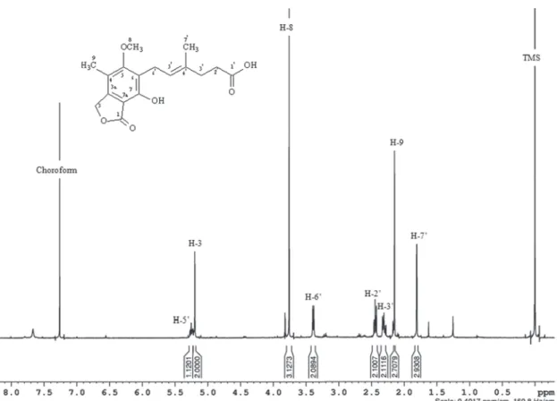
The 1H NMR spectrum of the mycophenolic acid (Figure 2) showed signals for a methyl group at δ 1.78 (H-7’, singlet, 3H) and a methyl group linked to aromatic ring at δ 2.13 (H-9, singlet, 3H), a signal for a methoxyl group at δ 3.74 (H-8, singlet, 3H), two signals corresponding to aliphatic methylenes in the region of δ 2.47 to δ 2.38 (H-3a’ and H-3b’, multiplet 2H), of δ 2.32 to δ 2.24 (H-2’, multiplet, 2H), a broad doublet for a methylene group at δ 3.37 (H-6’, 2H), a broad singlet at δ 5.18 (H-3, 2H) referring to an aliphatic
methylene bearing oxygen, and a multiplet for a methyne group of δ 5.23 to δ 5.28 (H-5’,1H).
In the MS/MS experiment (ESI - negative mode, with a collision energy of 5 eV) a fragment of m/zm/z 319.3 was obtained for the ion with a 319.3 was obtained for the ion with a m/zm/z of of 639.5. This fragment was exactly half of the mass, suggesting that the ion with a m/zm/z of 639.5 was of 639.5 was formed by two molecules of mycophenolic acid linked by hydrogen bonds (dimmer), with lose a hydrogen [H+] [M+M-H]-. The mass of 320.0 Da was coherent with the NMR results. The MS/MS experiment for the ion with a m/zm/z of 319.3 showed of 319.3 showed the following fragments: m/zm/z 191.1 (100%), 191.1 (100%), referring to the loss of a terpenic unit with a methyl arrangement; m/zm/z 275.3, referring to the loss of a 275.3, referring to the loss of a carbon dioxide unit.
Position
Mycophenolic acid 5-hydroxy-7methoxy-4methylphthalide
δ 13
C δ
1 H (multiplicity, JJ in Hz)
1 H-13
C gHMBC* δ 13
C δ
1 H (multiplicity, JJ in Hz)
1 H-13
C gHMBC*
1 173.0 - 173.0 -
-3 70.0 5.18 (2H, br s) 1; 3a 68.0 5.15 (2H, br s) 1; 3a; 4; 5; 7; 7a
3a 122.1 - - 104.0 -
-4 116.7 - - 110.0 -
-5 163.7 - - 163.0 -
-6 106.4 - - 98.0 6.49 (1H, s) 1; 4; 5; 7; 7a
7 153.7 - - 158.0 -
-7a 144.0 - - 151.0 -
-8 61.0 3.74 (3H, s) 5 57.0 3.79 (3H, s) 7
9 11.5 2.13 (3H, s) 3; 3a; 4; 5 11.0 1.95 (3H, s) 3a; 4; 5
OH - 7.67 (1H, br s) - - 10.59 (1H, br s) 4; 6; 5
1’ 179.1 - - - -
-2’ 32.7 2.38-2.47 (2H, m) 1’; 3’; 4’ - -
-3’ 34.2 2.24-2.32 (2H, m) 1’; 2’; 4’; 5’; 7’ - -
-4’ 133.9 - - - -
-5’ 123.0 5.23-5.28 (1H, m) 3’; 7’ - -
-6’ 22.3 3.37 (2H, br dbr d; 6.8) 5; 7; 5’ - -
-7’ 16.1 1.78 (3H, s) 3’; 4’; 5’ -
-Abbreviations: s – singlet, br s – broad singlet, br dbr d – broad doublet, m – multiplet. *gggHMBC data set: the numbers correspond to the correlated carbons.
TABLE I 1
H, 13
Figure 2 -1H NMR spectrum of mycophenolic acid (400 MHz, CDCl3).
5-hydroxy-7-methoxy-4-methylphthalide is a solid white powder – chemical formula: C10H10O4, molecular weight (M.W.): 194.0, soluble in methanol and DMSO. The structure of the compound was determined by 1D and 2D NMR techniques (Table I) and confi rmed by comparing the data with that of the NMR data for mycophenolic acid, especially using the gggHMBC experiment.
The 1H NMR spectrum of 5-hydroxy-7-methoxy-4-methylphthalide (Figure 3) showed signals for methyl group at δ 1.95 (H-9, singlet, 3H), a signal for the methoxyl group at δ 3.79 (H-8, singlet, 3H), a signal corresponding to one methylene group at δ 5.15 (H-3, singlet, 2H), and a signal for aromatic hydrogen at δ 6.49 (H-6, singlet, 1H).
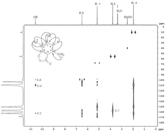
In the 3JCH gggHMBC experiment (Figure 4), HMBC experiment (Figure 4), H-3 correlated with C3a (δ 104.0), C4 (δ 110.0), C5 (δ 163.0), C7 (δ 158.0), C7a (δ151.0) and C1
(δ 173.0); H-6 correlated with C1, C4, C5, C7 and C7a; H-8 correlated with C3a, C4 and C5 and H-9 correlated with C7.
The mass spectrum (ESI - negative ion mode) of 5-hydroxy-7-methoxy-4-methylphthalide showed the [M-H]–– ion at ion at m/zm/z 193.0 and the chloride adduct 193.0 and the chloride adduct [M+Cl]–– at at m/zm/z 229.0. The mass spectrum (ESI - 229.0. The mass spectrum (ESI - positive ion mode) showed the [M+H]+ ion at m/z 195.0. The MS data suggested that the molecular mass of the compound was 194 Da. In the MS/ MS experiment (ESI - negative mode) for the ion m/z 193.0 was obtained the fragment of m/z 178.0 [M-CH3]–.
Figure 3 -1H NMR spectrum of 5-hydroxy-7-methoxy-4-methylphthalide (400 MHz, DMSO).
compound could be used as an intermediate in the process of synthesizing some signifi cant products, such as 5,7-dimethoxy-4-methylphthalide and 5,7-dihydroxy-4-methylphthaIide (Zuo et al. 2008), or mycophenolic acid and its analogs (Lee et al. 2001).
From the results obtained, it was concluded that the production of mycophenolic acid was related to the acidic or basic nature of the substances added to the culture medium.
ACKNOWLEDGMENTS
The authors are thankful to the Coordenação de Aperfeiçoamento de Pessoal de Nível Superior (CAPES), the Conselho Nacional de Desenvolvimento Científi co e Tecnológico (CNPq), the Fundação de Amparo à Pesquisa do Estado de São Paulo (FAPESP) and to the Financiadora de Estudos e Projetos (FINEP) for their fi nancial support.
RESUMO
As reações químicas realizadas por microorganismos têm sido utilizadas como uma ferramenta na química moderna. Este artigo relata a produção de ácido micofenólico e uma nova ftalida pelo fungo endofítico
Penicillium crustosum obtido a partir de grãos de café. O fungo foi cultivado em meio líquido durante um período de sete dias, e depois disso, o meio de cultura foi dividido em quatro lotes: A, B, C e D, nos quais diferentes substâncias orgânicas foram adicionadas. O lote A foi mantido como controle para avaliar a ocorrência de biotransformação. Os ácidos orgânicos foram adicionados ao meio de cultura dos lotes B (ácidos ferúlico e quínico) e C [ácido cinâmico e 3,4-(metilenodioxi) cinâmico], e cafeína foi adicionada ao lote D. Todos estes compostos orgânicos foram dissolvidos em DMSO, e a fermentação foi mantida por mais 13 dias, totalizando 20 dias. O ácido micofenólico foi isolado a partir da cultura sem adição de ácidos (lote A). O ácido micofenólico e uma nova ftalida, 5-hidroxi-7-metoxi-4-metilftalida, foram isolados a partir dos lotes B e C, e ácido micofenólico e cafeína
(adicionada ao meio de cultura) foram isolados a partir do lote D. As estruturas foram determinadas pela técnica de RMN e confi rmadas pelas técnicas de MS e MS/MS.
Palavras-chaves: café, fungo endofítico, ácido micofenólico, Penicillium crustosum, 5-hidroxi-7-metoxi- 4-metilftalida.
REFERENCES
ARNOLD AE. 2007. Understanding the diversity of foliar endophytic fungi: progress, challenges, and frontiers. Fungal Biol Rev 21: 51-66.
BOAVENTURA MAD, LOPES RFAP AND TAKAHASHI JA. 2004. Microorganisms as tools in modern chemistry: the biotransformation of 3-indolylacetonitrile and tryptamine by fungi. Braz J Microbiol 35: 345-347.
DEMAIN AL. 1968. Regulatory mechanisms and the industrial production of microbial metabolites. Lloydia 31: 395-418. FUJIMOTO H, FUJIMAKI T, OKUYAMA E AND YAMAZAKI M.
1999. Immunomodulatory constituents from an ascomycete, Microascus tardifaciens. Chem Pharm Bull 47: 1426-1432.
GRINYO JM. 2001. Place of mycophenolate mofetil in renal transplantation. Transplant Proc 33: 997-999.
HABIB E, LEO´N F, BAUER JD, HILL RA, CARVALHO P, CUTLER GH AND CUTLER SJ. 2008. Mycophenolic Derivatives from Eupenicillium parvum. J Nat Prod 71: 1915-1918.
LEE Y, FUJIWARA Y, UJITA K, NAGATOMO M, OHATA H AND SHIMIZU I. 2001. Syntheses of mycophenolic acid and its analogs by palladium methodology. Bull Chem Soc Jpn 74: 1437-1443.
MELO IS AND FAULL JL. 2004. Scanning electron microscopy of conidia of Trichoderma stromaticum, a biocontrol agent of witches' broom disease of cocoa. Braz J Microbiol 35: 330-332.
MITCHELL AM, STROBEL GA, HESS WM, VARGAS PN AND EZRA D. 2008. Muscodor crispans, a novel endophyte from Ananas ananassoides in the Bolivian Amazon. Fung Divers 31: 37-44.
PUEL O, TADRIST S, GALTIER P, OSWALD IP AND DELAFORGE M. 2005.Byssochlamys nivea as a source of mycophenolic acid. Appl Environ Microbiol 71: 550-553.
RAEDER R
R U AND BRODA P. 1985. Rapid preparation of DNA from fi lamentous fungi. Lett Appl Microbiol 1: 17-20. ROVIROSA J, DIAZ-MARRERO A, DARIAS J, PAINEMAL KK AND AND
SAN-MARTIN A. 2006. Secondary metabolites from marine
Penicillium brevicompactum. J Chil Chem Soc 51(1): 775-778.
SILVA HSA, BETTIOL W, TERRASAN CRF, TOZZI JPL, MELO IS AND NUNES FV. 2006. Microrganismos endofíticos: potencial de uso como agentes de biocontrole da ferrugem do cafeeiro. Boletim de Pesquisa e Desenvolvimento 38, EMBRAPA-CNPMA. http://ainfo.cnptia.embrapa.br/digital/ bitstream/CNPMA/7434/1/boletim_38.pdf
STROBEL G AND DAISY B. 2003. Bioprospecting for Microbial Endophytes and Their Natural Products. Mol Biol Rev 67: 491-502.
SUN MX, LI X, LIU WY AND XIAO K. 2009. 5,7-Dimethoxy-isobenzofuran-1(3H)-one. Acta Cryst E65: o2146. TAMURA K, DUDLEY J, NEI M AND KKKUMAR SM. 2007.
Molecular Evolutionary Genetics Analysis. Mol Biol Evol 24: 1596-1599.
VALENTE AMMP, BOFFO EF, FERREIRA AG, RODRIGUES FILHO E, NASCIMENTO RS, SILVA JL, VILELA ESD AND MELO IS. 2007. Processo para produção e isolamento de ácido micofenólico e seus sais. UFSCAR; EMBRAPA. No: PI0704700-2. DEDF/INPI.
VEGA FE, PAVA-RIPOLL M, POSADA F AND BUYER JS. 2005. Endophytic bacteria in Coffea arabica L. J Basic Microbiol 45: 371-380.
VEGA FE AND POSADA F. 2006.Penicillium species endophytic in coffee plants and ochratoxin A production. Mycologia 98: 31-42.