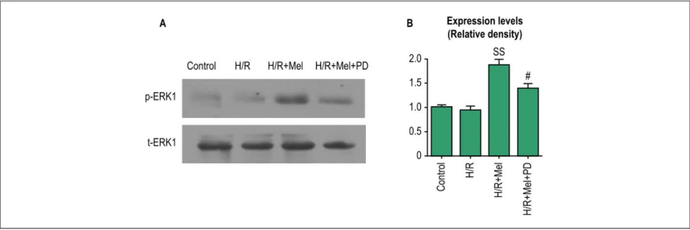
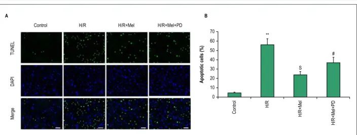
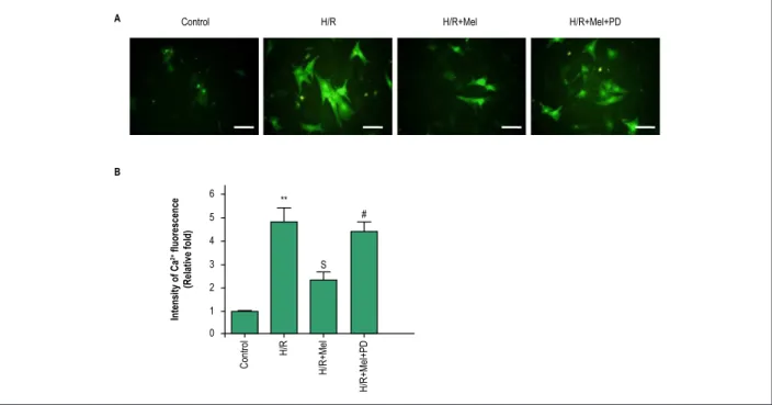
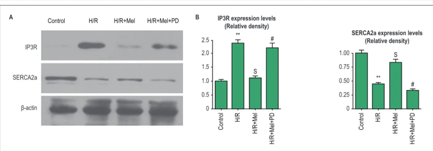
Melatonin-Induced Protective Effects on Cardiomyocytes Against Reperfusion Injury Partly Through Modulation of IP3R and SERCA2a Via Activation of ERK1
Texto
Imagem
Documentos relacionados
Endothelial kinin B(1)-receptors are induced by myocardial ischaemia-reperfusion in the rab- bit. Amelioration of ischemia-reperfusion injury with cyclic peptide blockade of
membro posterior de rato. Acta Cir Bras. Saita Y, Yokoyama K, Nakamura, Itoman M. Protective effect of ischaemic preconditioning against ischaemia-induced reperfusion injury
CPU0213, a novel endothelin type A and type B receptor antagonist, protects against myocardial ischemia/reperfusion injury in
Superoxide dismutase activity was significantly higher (p<0.029) and creatinine (p<0.001) and urea (p<0.001) concentrations were significantly lower in the
There were also significant differences in terms of the last evaluated parameter, caspase-3 immunohis- tochemistry levels (p: 0.002; p < 0.05).. Figure 1 A) I/R group, Left
Here, we demonstrate that miR-145 protects against the activation of mitochondria apoptotic pathway in cardiomyocytes under oxidative stress through directly targeting
H9C2 cells were subjected to H/R with or without pre-treatment with 20 U/ml EPO for 24 hrs, H/R induced cells showed increase in green fluorescence (A), more fluorescence intensity
Administration of fasudil, a Rho-kinase inhibitor, significantly reduced the I/R-induced expression of the proinflammatory cytokines interleukin (IL)-6, C-C motif chemoattractant
