https://doi.org/10.1590/0004-282X20180059
ARTICLE
Association between blood pressure and
acute phase stroke case fatality rate: a
prospective cohort study
Associação entre níveis de pressão arterial e letalidade na fase aguda do acidente
vascular cerebral: estudo de coorte prospectivo
Natalia Eduarda Furlan1,2, Silméia Garcia Zanati Bazan1, Gabriel Pereira Braga1, Meire Cristina Novelli e Castro1, Roberto Jorge da Silva Franco1, Ana Lúcia Gut1, Rodrigo Bazan1, Luis Cuadrado Martin1
Stroke is one of the leading causes of death and disability among adults in Brazil and worldwide1,2,3. Fifteen million
peo-ple suffer from a stroke annually, of whom five million die and another five million become disabled1,2,3. Arterial hypertension
is the major modifiable cause of stroke4,5. In addition, blood
pressure (BP) control is a priority in stroke prevention6,7,8,9.
Nevertheless, few studies have evaluated the relationship between arterial hypertension in the first 48 hours of stroke
onset and the case fatality rate. In the acute phase of stroke, 80% of patients present with elevated BP10. High BP can
pro-duce hematoma enlargement in hemorrhagic stroke (HS). On the other hand, in ischemic stroke (IS), high BP during this phase can induce cerebral edemaandhemorrhagic transfor-mation10. Alternatively, low BP can lead to low perfusion of
the ischemic penumbra zone, which leads to enlargement of the stroke area11,12.
1Universidade Estadual Paulista Julio de Mesquita Filho, Faculdade de Medicina, Botucatu SP, Brasil; 2Serviço Nacional de Aprendizagem Comercial, Botucatu SP, Brasil.
Correspondence: Luis Cuadrado Martin; Rua Rubião Jr, s/n; 18618-970 Botucatu SP, Brasil; E-mail: cuadrado@fmb.unesp.br
Clinical Trial registration number: NCT01195350.
Conflict of interest: There is no conflict of interest to declare.
Received 28 June 2017; Received in final form 21 February 2018; Accepted 04 April 2018.
ABSTRACT
Objective: ed to investigate the association between blood pressure and acute phase stroke lethality in a Brazilian intensive care unit. Methods: This was an observational, prospective cohort study of hemorrhagic and ischemic stroke intensive care patients. The primary outcome was all-cause mortality during the first seven days. Results: There were 146 patients, aged 66 ± 13.4 years, 56% men, 89% Caucasian, 69% had ischemic stroke, and 80% were hypertensive. The median of the National Institutes of Health Stroke Scale score was 16. There were 101 ischemic stroke patients and 45 hemorrhagic stroke patients. In the ischemic stroke patients, logistic regression analysis identified low systolic blood pressure as an independent ominous prognostic factor and the optimal cut off was a mean of systolic blood pressure ≤ 131 mmHg during the first 48 hours from admission for prediction of death. No association was found for hemorrhagic stroke. Conclusions: There was a negative association between systolic blood pressure and case fatality ratio of acute phase stroke in ischemic stroke intensive care patients.
Keywords: stroke; blood pressure, hypertension.
RESUMO
Objetivo: Investigar a associação entre pressão arterial e letalidade do acidente vascular cerebral (AVC) em uma unidade de terapia intensiva brasileira. Métodos: estudo de coorte prospectivo de pacientes com AVC hemorrágico (AVC-H) ou isquêmico (AVC-I) internados em terapia intensiva. O desfecho primário foi a letalidade por todas as causas nos primeiros sete dias. Resultados: Avaliados 146 pacientes, idade: 66 ± 13,4 anos, 56% homens, 89% brancos, 69% AVC-I e 80% hipertensos. A mediana do NIH foi de 16. Os pacientes com AVC-I foram 101 e 45 com AVC-H. Para AVC-I, a análise de regressão logística identificou baixa pressão arterial sistólica como um fator prognóstico negativo e o melhor corte foi uma média da pressão arterial sistólica nas primeiras 48 h de admissão ≤ 131 mmHg para a predição da morte. Para o AVC-H, nenhuma correlação foi encontrada. Conclusões: houve associação negativa entre a pressão arterial sistólica e a letalidade do AVC-I em fase aguda em pacientes de terapia intensiva.
Through these mechanisms, the relationship between BP and death during the acute phase of stroke depicts a “U” shape in many studies, i.e. both low and high BP extremes are associated with a higher chance of death11,13,14,15. Other
studies have demonstrated a direct relationship16,17. For
critical care stroke patients, these mechanisms may play a significant role in survival too. However, information is limited on the impact of BP on the case fatality rate of strokes in the intensive care unit (ICU). Some studies have evaluated other prognostic factors of stroke in the ICU18,19,20,21,22,23,24,. However, we identified only one study
performed in the ICU evaluating BP as a prognostic fac-tor; but the evaluation was performed only for the first 12 hours after onset and did not mention any association with death25. Therefore, the aim of the present study was
to evaluate the relationship between BP and fatality dur-ing the acute phase of stroke in critical care patients.
METHODS
We conducted an observational, prospective cohort study in the ICU of a tertiary hospital of Botucatu Medical School - São Paulo State University (UNESP) in Brazil. The participants included were patients over 18 years old, who were admitted to the ICU with a stroke from March 2012 to April 2016 during the study time period. The criteria for stroke patients for the ICU admission were: vasopressor drug use due to hemodynamic instability and/or depressed consciousness and/or invasive ventilation. All survivors of the initial stroke event stayed in the ICU for at least seven days. We excluded patients with subarachnoid hemorrhage or incomplete records. All patients underwent computed tomography of the head.
Age, gender, ethnic group, stroke type, occurrence of infection or myocardial infarction during ICU hospitaliza-tion, and previous diagnosis of hypertension or diabetes were recorded. Stroke evaluation and BP measurements were performed on admission and every two hours there-after, throughout the patient’s first week in the ICU. Stroke was evaluated with the National Institutes of Health Stroke Scale (NIHSS)26. Noninvasive automatic BP measurements
were taken with a multiparameter monitor (Dixtal®) with
the cuff positioned on the patient’s arm. The first BP was analyzed on admission, with a mean of 48 hours and the variation was quantified by the coefficient of variation (standard deviation divided by mean) of the first 48 hours. Body temperature was measured every two hours and cap-illary blood glucose every six hours throughout the first week. On admission, laboratory tests were performed for glucose and creatinine.
This study was approved by the local ethics committee, protocol number: 3429 01/February/2010, and was regis-tered in the Clinical Trials database (NCT01195350). The few
patients who were conscious, alert and oriented gave their written informed consent. Legal representatives signed con-sent agreements for unconscious patients.
Statistical Analysis
All patients were analyzed in two sets separately accord-ing to the stroke subtypes. The sample size was calculated to detect a 30% difference in the case fatality ratio with an alpha error of 0.05 and statistical power of 0.9; this resulted in an estimated required sample size of 132 patients.
Variables were described as mean ± standard deviation. Patients were divided into two groups: patients alive within seven days and patients deceased during this period. All eval-uated data from these two groups were compared to each other. Univariate analysis was performed by the Chi square test to compare categorical variables and the T-test to com-pare continuous variables.
After this univariate analysis, a backward stepwise multiple logistic regression was performed. The depen-dent variable of logistic regression was death during the first seven days of stroke. The association between BP and death was adjusted for variables that differed at p < 0.1 in univariate analysis (confounders). After backward analy-sis, variables with p < 0.1 were preserved in the final model. Only in these phases of analysis, the different p value used was set to identify variables that, by themselves, were not statistically associated with the outcome, but could con-tribute to the model together with other variables27. The
collinearities were tested and, if present, the more rep-resentative variable was selected. Statistical significance was set at p < 0.05.
An ROC curve was performed to verify the discrimina-tory power of BP to case fatality. The optimal cut off was determined by the higher sum of sensibility and specificity (Youden Index). Sensibility and specificity for this cut off was calculated. All analysis was performed using SPSS20.0 soft-ware (Illinois Chicago).
RESULTS
During the study period, there were 1,432 admissions for stroke to our hospital. Of these patients, 260 required treat-ment in the ICU, 48 patients refused to give informed con-sent or there were difficulties obtaining informed concon-sent in an interview with the patient’s family. Therefore, a total of 212 patients were screened. There were 66 patients who were excluded due to incomplete records, and of these, 13 died in the seven first days (19%), which is a frequency similar of the mortality of the analyzed patients. The final sample included 146 patients; 101 with IS and 45 with HS (Figure 1).
was 16 ± 6.5 points. Initial mean systolic BP (SBP) was 161 ± 40.9 mm Hg. The mean SBP in the first 48 hours after admission was 138 ± 16.7 mm Hg.
During the first week of admission, 31 patients (21%) died. The causes of death were intracranial hypertension in 17 patients (55%), endocarditis in one patient (3%), car-diogenic shock in four patients (13%), septic shock in seven patients (23%), and indefinite causes in two patients (6%).
Among the patients with HS (Table 1) when compar-ing those who died against those who survived durcompar-ing the first week, the groups were similar except for sex and body temperature where differences were statistically sig-nificant. Infection was marginally associated with out-come. No BP parameter was associated with outout-come. We took into account the Glasgow Coma Scale and hema-toma volume, and ran a multiple regression with BP and these variables. Even so, BP was not correlated with out-come. The baseline SBP adjusted odds ratio (OR) and 95% confidence interval (95% CI) was: 1.026 (0.986-1.067); p = 0.209. Baseline diastolic BP (DBP) OR and CI 95% was: 1.054 (0.984-1.130); p = 0.132. The 48-hour SBP average OR and CI 95% was: 1.003 (0.935-1.077); p = 0.926. The 48-hour DBP average OR and CI 95% was: 1.031 (0.913-1.164); p = 0.620. No patient was on anticoagulation treatment prior to the hemorrhagic stroke.
Table 2 shows comparisons between the deceased and surviving patients among those with IS, during the first week. The groups differed statistically in age, NIHSS score on admission, initial SBP, SBP in the first 48 hours, initial DBP, DBP in the first 48 hours, and occurrence of myo-cardial infarction. In the first 48 hours, the average of the capillary blood glucose, the coefficient of variation of SBP, and serum creatinine had a p value < 0.10. The variables of age, NIHSS score on admission, mean SBP during the first
48 hours, myocardial infarction, average of the first 48 hours of capillary blood glucose, the coefficient of variation of SBP in the first 48 hours and serum creatinine were selected as predictors in a multiple logistic regression analysis. Baseline SBP and DBP, and 48-hour DBP were excluded by collinear-ity with the SBP mean during the 48 hours. Some antihyper-tensive drug classes were more commonly used among sur-vivors than the deceased patients (angiotensin converting enzyme inhibitors: 47% vs. 9%; p < 0.01; beta-blockers: 34% vs. 6%; p < 0.01). The frequency of use of other antihyperten-sive drug classes were homogeneous between survivors and deceased patients.
Multiple logistic regression analysis for IS
For the subset of IS patients, the association between SBP in the first 48 hours from stroke onset and outcome was adjusted for confounders. This model is shown in Table 3. The SBP showed a statistically significant association with all-cause mortality. The final model, including age and NIHSS score demonstrated an OR of 0.960 (CI 95%: 0.922-0.999; p = 0.047).
A post hoc logistic regression analysis was performed.
This analysis included the mean BP in first 48 hours after stroke, myocardial infarction and occurrence of any infec-tion (including septic shock) as confounders. In this analysis, low BP in the first 48 hours after stroke was associated with death after adjusting for these confounders (Table 4). Among the 23 deaths of IS patients, 13 were caused by intracranial hypertension, six were caused by septic shock and four by cardiogenic shock. The Hosmer-Lemeshow test was not sig-nificant for all logistic regressions (p range from 0.22 to 0.40), demonstrating the goodness-of-fit.
ROC curve for IS
Figure 2 shows the ROC curve. The area under the ROC curve between the 48-hour-average SBP and mortality was 0.74 (CI 95%: 0.63-0.84; p < 0.001). The optimal cut off was ≤ 131 mm Hg. This BP cut off demonstrated a sensibility of 0.65 and a specificity of 0.67 to predict one-week mortal-ity in the ICU.
DISCUSSION
Of the patients studied, 80% had hypertension, dem-onstrating the high prevalence of this risk factor in criti-cal care stroke patients, as observed in other studies9,10.
Nevertheless, few studies have evaluated the association between BP and the case fatality ratio in acute stroke. Some international studies have identified a “U” curve relation-ship between BP and in-hospital mortality, where both high and low BP were associated with an increased early fatal-ity ratio11. In our study, the association between BP and
the case fatality ratio in the acute phase of stroke in ICU
All 1,432 strokes in study period
260 ICU admission
212 patients
146 patients
1,172 excluded (no need of ICU treatment)
48 not assessed (refused consent)
66 excluded (due to incomplete records)
101 ischemic stroke 45 hemorrhagic stroke
patients did not follow this expected relationship; rather there was an increase in the probability of death in the first week only among patients with low SBP (≤ 131 mm Hg) in the first 48 hours after admission among IS patients, and no relationship for HS. In the current study, we analyzed BP and variation of BP during the first 48 hours of admission to exclude pre-mortem phenomena as a cause of BP abnormal-ities. This procedure was performed to exclude epiphenom-ena, such as septic or cardiogenic shock, as an explanation for the associations observed.
There were no differences in the case fatality ratio between patients with HS (18%) and IS (23%) in this study. This data demonstrated the profile of severity of patients in our study that evaluated only ICU patients. Alonso et al.28 found similar
results in ICU stroke patients. In the current study, the NIHSS score in IS was similar to that of HS (16 ± 6.7 vs. 17 ± 5.8).
The lack of relationship between BP and fatality in HS in the present work may be explained by the low average BP (141 mm Hg) that was obtained in ICU care; also, higher
SBP levels, sufficient to increase fatality, were not found in this series of patients. The fact that low BP was not associ-ated with the case fatality ratio is in consonance with the INTERACT-229 study, and corroborates the safety of intensive
BP intervention in HS.
The IS patients with SBP values below 147 mm Hg at admission and 131 mm Hg in the first 48 hours of hospitaliza-tion had a higher risk of death. These values were obtained by ROC curve analysis. The different results for IS and HS may be explained by the different pathophysiology models. In IS, the ischemic penumbra zone30, mainly in the early hours
from onset of acute stroke, is relevant. On the other hand, perihematoma and vasogenic edema prevails in HS, leading to elevation of intracranial pressure.
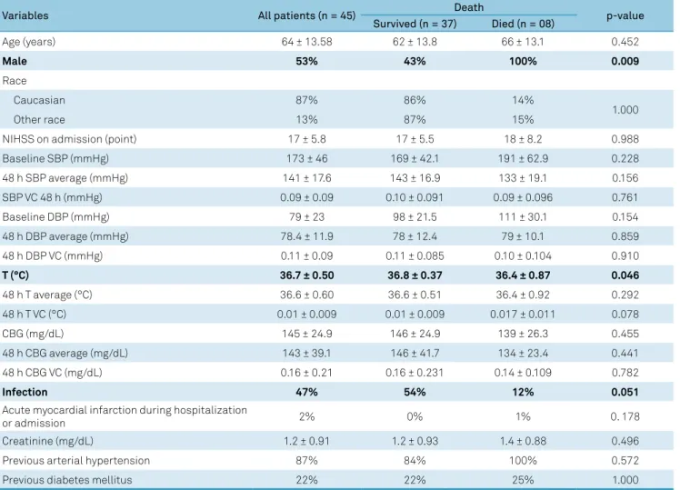
A post hoc logistic regression analysis of our data (Table 4) showed that in IS patients, the relationship between low BP and the case fatality ratio was maintained even after adjust-ment for myocardial infarction and infection. This finding indi-cates that the effect of low BP was independent of myocardial Table 1. Comparison of prognostic variables by outcome during the acute phase of hemorrhagic stroke.
Variables All patients (n = 45) Death p-value
Survived (n = 37) Died (n = 08)
Age (years) 64 ± 13.58 62 ± 13.8 66 ± 13.1 0.452
Male 53% 43% 100% 0.009
Race
Caucasian 87% 86% 14%
1.000
Other race 13% 87% 15%
NIHSS on admission (point) 17 ± 5.8 17 ± 5.5 18 ± 8.2 0.988
Baseline SBP (mmHg) 173 ± 46 169 ± 42.1 191 ± 62.9 0.228
48 h SBP average (mmHg) 141 ± 17.6 143 ± 16.9 133 ± 19.1 0.156
SBP VC 48 h (mmHg) 0.09 ± 0.09 0.10 ± 0.091 0.09 ± 0.096 0.761
Baseline DBP (mmHg) 79 ± 23 98 ± 21.5 111 ± 30.1 0.154
48 h DBP average (mmHg) 78.4 ± 11.9 78 ± 12.4 79 ± 10.1 0.859
48 h DBP VC (mmHg) 0.11 ± 0.09 0.11 ± 0.085 0.10 ± 0.104 0.910
T (°C) 36.7 ± 0.50 36.8 ± 0.37 36.4 ± 0.87 0.046
48 h T average (°C) 36.6 ± 0.60 36.6 ± 0.51 36.4 ± 0.92 0.292
48 h T VC (°C) 0.01 ± 0.009 0.01 ± 0.009 0.017 ± 0.011 0.078
CBG (mg/dL) 145 ± 24.9 146 ± 24.9 139 ± 26.3 0.455
48 h CBG average (mg/dL) 143 ± 39.1 146 ± 41.7 134 ± 23.4 0.441
48 h CBG VC (mg/dL) 0.16 ± 0.21 0.16 ± 0.231 0.14 ± 0.109 0.782
Infection 47% 54% 12% 0.051
Acute myocardial infarction during hospitalization
or admission 2% 0% 1% 0. 178
Creatinine (mg/dL) 1.2 ± 0.91 1.2 ± 0.93 1.4 ± 0.88 0.496
Previous arterial hypertension 87% 84% 100% 0.572
Previous diabetes mellitus 22% 22% 25% 1.000
Table 2. Comparison of prognostic variables by outcome during the acute phase of ischemic stroke.
Variables All patients (n =
101)
Death
p-value Survived (n = 78) Died (n = 23)
Age (years) 68 ± 13.1 66 ± 12.5 75 ± 12.8 0.003
Male 55% 60% 39% 0.120
Race
Caucasian 87% 85% 96%
0.301
Other race 13% 66% 23%
NIHSS on admission (point) 16 ± 6.7 15 ± 6.2 20 ± 7.4 0.006
APACHE score 17 ± 7.5 16 ± 6.1 24 ±8.6 0.004
Baseline SBP (mmHg) 156 ± 37 164 ± 36.2 130 ± 27.4 <0.001
48 h SBP average (mmHg) 136 ± 16.1 138 ± 14.9 127 ±16.9 0.003
48 h SBP VC (mmHg) 0.09 ± 0.07 0.08 ± 0.060 0.12 ± 0.107 0.051
Baseline DBP (mmHg) 91 ± 21 95 ± 20.7 79 ± 19.4 0.001
48 h DBP average (mmHg) 73.9 ± 10.9 75 ±11.1 70 ± 9.2 0.049
48 h DBP VC (mmHg) 0.09 ± 0.08 0.08 ± 0.082 0.10 ± 0.089 0.376
T (°C) 36.8 ± 0.80 36.8 ± 0.71 36.8 ± 1.07 0.944
48 h T average (°C) 36.8 ± 1.74 36.8 ± 1.91 36.7 ± 0.88 0.747
48 h T VC (°C) 0.002 ± 0.04 0.02 ± 0.049 0.02 ± 0.014 0.738
CBG (mg/dL) 150 ± 28.3 150 ± 27.6 149 ± 30.8 0.880
48 h CBG average (mg/dL) 147 ± 36.3 144.4 ± 32.1 159 ± 46.2 0.075
48 h CBG VC (mg/dL) 0.16 ± 0.20 0.15 ± 0.210 0.18 ± 0.154 0.509
Infection 50% 54% 39% 0.316
Previous myocardial infarction 12% 6% 30% 0.006
Acute myocardial infarction (during ICU stay) 4% 1% 13% 0.053
Thrombolysis 12% 13% 9% 0.613
Hemorrhagic transformation 77% 79% 70% 0.324
Creatinine (mg/dL) 1.3 ± 1.08 1.17 ± 0.93 1.68 ± 1.42 0.056
Previous arterial hypertension 77% 79% 70% 0.324
Previous diabetes mellitus 34% 33% 35% 0.898
Data presented as the mean ± standard deviation or percent (%).IS: ischemic stroke; NIHSS: National Institute of Health Stroke Scale; SBP: systolic BP; 48 h SBP average: average of systolic BP during the 48 hours after admission; 48 h SBP VC: systolic BP variation coefficient at 48 hours after admission; DBP: diastolic BP; 48 h DBP average: average of diastolic BP during 48 hours after admission; 48 h DBP VC: diastolic BP variation coefficient at 48 hours after admission; T: temperature at admission; 48 h T average: average of body temperature at 48 hours after admission; 48 h T VC: average of body temperature variation coefficient at 48 hours after admission; CBG: capillary blood glucose; 48 h CBG VC: average of capillary blood glucose variation coefficient at 48 hours after admission; Infection: infection during the first 7 days of hospitalization. Bold indicates p < 0.10, therefore inclusion criteria for multiple logistic analysis.
Table 3. Adjusted odds ratios by backward stepwise multiple logistic regression analysis for the association of SBP in the first 48 hours of stroke onset with death in ischemic stroke patients.
Variables OR 95%iIC for OR p-value
Lower Upper
Step 1
Age (year) 1.033 0.985 1.085 0.181
NIHSS (point) 1.074 0.983 1.172 0.113
48 h SBP VC (mmHg) 0.245 0.011 145.206 0.666
Acute myocardial infarction during hospitalization or admission 2.292 0.455 11.547 0.315
Creatinine (mg/dL) 1.470 0.936 2.307 0.094
48 h CBG average (mg/dL) 1.007 0.991 1.023 0.400
48 h SBP average (mmHg) 0.960 0.923 0.999 0.043
Thrombolysis 4.038 1.245 13.096 0.020
Step 5
Age (year) 1.090 1.041 0.994 0.086
NIHSS (point) 1.087 1.001 1.180 0.046
48 h SBP average (mmHg) 0.960 0.926 0.996 0.030
Creatinine (mg/dL) 1.516 0.987 2.327 0.057
Thrombolysis 4.204 1.345 13.138 0.014
infarction and infection. Moreover, even excluding patients deceased from infection or cardiogenic shock (post hoc analysis not shown in the results) BP was lower among the 20 patients deceased from purely neurologic complications (131 ± 17.6 mm Hg) than among the survivors (140 ± 15.7 mm Hg; p = 0.02). Conversely, myocardial infarction and cardiogenic shock may contribute to fatality among these patients independent of neurological factors. Therefore, these factors, in addition to low BP, may potentiate deleterious effects on the penumbra zone during cerebral ischemia. A seminal study has shown that the restoration of blood flow in acute IS cases could improve the outcome in these patients31. In an experimental study, an
artifi-cial increase in BP promoted an improvement in the ischemic core and penumbra zone32.
During hospitalization, no patient presented with SBP > 180 mm Hg, making it challenging to confirm if there was a bimodal distribution for BP and risk of death from stroke from our study results. Antihypertensive treatment was
more common in survivors than in deceased patients, so antihypertensive treatment cannot be attributed as a cause of mortality. On the other hand, we cannot rule out that the lack of antihypertensive cardioprotective drugs among the patients who died could have influenced, at least partially, the lethality ratio.
In the present study, SBP was assessed over DBP because it has a more accurate prognostic significance in the predom-inant age of the patients studied. In this age group, the rigid-ity of large arteries may explain this phenomenon32.
We observed that the case fatality ratio of stroke in the present study was 21% over the total period of seven days. In the published literature, lethality in the first 30 days after IS is approximately 10%33. The high lethality in our patient
population may be explained by the fact that we studied ICU patients, who often present with a more severe stroke. The high NIHSS scores observed corroborate this idea; patients with an NIHSS score less than 5 points are often discharged quickly, and patients with scores above 14 frequently need long-term ICU care34.
It is important to emphasize that the sample in the cur-rent study comprised stroke patients admitted in ICU. The NIHSS scores of our patients were very high, and there were patients with compromised consciousness and many comor-bidities. These characteristics make the management of BP a very difficult challenge in these patients, and these facts must be taken into account in the interpretation of our find-ings. On the other hand, this study contributes to the knowl-edge of acute BP management in ICU stroke patients that is more relevant nowadays because of the possibility of throm-bolysis and thrombectomy. It is of note that BP control must be initiated during prehospital assistance and be maintained not only through the hospital/ICU phase, but for the whole life of the patients.
Limitations
The greatest limitation of this study was the relatively small sample size. However, patients were recruited from the ICU, which provided information on a specific high-risk patient group. It would have been better to take into account the confounding variables—the creatinine clear-ance and cardiac output—instead of myocardial infarction and creatinine. But cardiac output and creatinine clear-ance are measurements performed in only a few patients in the ICU. Therefore, as a substitute to cardiac and renal involvement, we used infarction and creatinine. Another limitation, due to the use of conventional brain comput-erized tomography, in this study, we did not evaluate the penumbra zone for IS or the edema zone in HS. Finally, we did not assess atrial fibrillation (persistent or paroxysmal). Additional study strengths include the prospective study design and addressing a topical subject—prognostic factors of stroke in the acute phase in Brazilian ICU patients, which has a dearth of published research.
0.0 0.2 0.4 0.6 0.8 1.0
1.0 0.8
0.6 0.4
0.2 0.0
Sensitivity
1-Specificity
AUC/SBPadmission: 0.75 ic95% (0.65-0.86) P<0.01 Cutoff< 147 mm Hg; sens: 0.7; spec.: 0.67
AUC/SBPmean24h: 0.74 ic95% (0.63-0.84) P<0.01
Cutoff< 131 mm Hg; sens: 0.65; spec.: 0.67
Figure 2. ROC curve between systolic blood pressure and outcome in ischemic stroke.
Table 4. Post hoc logistic regression analysis including confounders associated with low BP.
Variable OR 95% CI for OR p
Lower Upper
48 h SBP average (mmHg) 0.961 0.929 0.995 0.024
Infection 0.607 0.217 1.692 0.339
In conclusion, there was a negative association between BP and the case fatality ratio of acute phase IS stroke patients in ICU, even after adjustment for cardio-genic shock and infection. This result indicates an inde-pendent effect of low BP. Values of SBP below 147 mm Hg
on admission or 131 mm Hg within the first 48 hours were associated with the highest risk of death. There was no relationship between BP and outcome in HS. Our results are a contribution to the study of prognostic factors in the acute phase of stroke in ICU patients.
References
1. Grysiewicz RA, Thomas K, Pandey DK. Epidemiology of ischemic and hemorrhagic stroke: incidence, prevalence, mortality, and risk factors. Neurol Clin. 2008 Nov;26(4):871-95. https://doi.org/10.1016/j.ncl.2008.07.003
2. Garritano CR, Luz PM, Pires ML, Barbosa MT, Batista KM. Analysis of the mortality trend due to cerebrovascular accident in Brazil in the XXI century. Arq Bras Cardiol. 2012;98(6):519-27. https://doi.org/10.1590/S0066-782X2012005000041
3. Instituto Brasileiro de Geografia e Estatística. Sistema de Informação Hospitalar do SUS (SIH/SUS). Rio de Janeiro, RJ: IBGE; 2009 [cited 2013 Oct 10]. Available on: http://ces.ibge.gov.br/pt/ base-de-dados/metadados/ministerio-da-saude/sistema-de-informacoes-hospitalares-do-sus-sih-sus
4. O’Donnell MJ, Xavier D, Liu L, Zhang H, Chin SL, Rao-Melacini P et al. Risk factors for ischaemic and intracerebral haemorrhagic stroke in 22 countries (the Interstroke study): a case-control study. Lancet. 2010 Jul;376(9735):112-23. https://doi.org/10.1016/S0140-6736(10)60834-3
5. Dunbabin DW, Sandercock PA. Preventing stroke by the modification of risk factors. Stroke. 1990 Dec;21(12 Suppl):IV36-9.
6. Nogueira D, Faerstein E, Coeli CM, Chor D, Lopes CS, Werneck GL. Awareness, treatment, and control of arterial hypertension: Pró-Saúde Study, Brazil. Rev Panam Salud Publica. 2010 Feb;27(2):103-9. Portuguese. https://doi.org/10.1590/S1020-49892010000200003
7. Gagliardi RJ. Hipertensão arterial e AVC. ComCiência.2009;(109).
8. Adams HP Jr, Bendixen BH, Kappelle LJ, Biller J, Love BB, Gordon DL et al. Classification of subtype of acute ischemic stroke: definitions for use in a multicenter clinical trial. TOAST. Trial of Org 10172 in Acute Stroke Treatment. Stroke. 1993 Jan;24(1):35-41. https://doi.org/10.1161/01.STR.24.1.35
9. Dahlöf B, Devereux RB, Kjeldsen SE, Julius S, Beevers G, Faire U et al. Cardiovascular morbidity and mortality in the Losartan Intervention for Endpoint reduction in hypertension study (LIFE): a randomized trial against atenolol. Lancet. 2002 Mar;359(9311):996-1003. https://doi.org/10.1016/S0140-6736(02)08089-3
10. Spence JD. Treating hypertension in acute ischemic stroke. Hypertension. 2009 Oct;54(4):702-3.
https://doi.org/10.1161/HYPERTENSIONAHA.109.134486
11. Geeganage CM, Bath PM. Relationship between therapeutic changes in blood pressure and outcomes in acute stroke: a metaregression. Hypertension. 2009 Oct;54(4):775-81. https://doi.org/10.1161/HYPERTENSIONAHA.109.133538
12. Massaro AR. Hipertensão arterial e acidente vascular cerebral: as bases científicas para a decisão terapêutica. Rev Soc Cardiol Estado de São Paulo. 2008;18(2):182-92.
13. Leonardi-Bee J, Bath PM, Phillips SJ, Sandercock PA; IST Collaborative Group. Blood pressure and clinical outcomes in the International Stroke Trial. Stroke. 2002 May;33(5):1315-20. https://doi.org/10.1161/01.STR.0000014509.11540.66
14. Tikhonoff V, Zhang H, Richart T, Staessen JA. Blood pressure as a prognostic factor after acute stroke. Lancet Neurol. 2009 Oct;8(10):938-48. https://doi.org/10.1016/S1474-4422(09)70184-X
15. Vemmos KN, Tsivgoulis G, Spengos K, Zakopoulos N, Synetos A, Manios E et al. U-shaped relationship between
mortality and admission blood pressure in patients with acute stroke. J Intern Med. 2004 Feb;255(2):257-65. https://doi.org/10.1046/j.1365-2796.2003.01291.x
16. Boreas AM, Lodder J, Kessels F, de Leeuw PW, Troost J. Prognostic value of blood pressure in acute stroke. J Hum Hypertens. 2002 Feb;16(2):111-6. https://doi.org/10.1038/sj.jhh.1001304
17. Carlberg B, Asplund K, Hägg E. The prognostic value of admission blood pressure in patients with acute stroke. Stroke. 1993 Sep;24(9):1372-5. https://doi.org/10.1161/01.STR.24.9.1372
18. Milhaud D, Popp J, Thouvenot E, Heroum C, Bonafé A. Mechanical ventilation in ischemic stroke. J Stroke Cerebrovasc Dis. 2004 Jul-Aug;13(4):183-8. https://doi.org/10.1016/j.jstrokecerebrovasdis.2004.06.007
19. Golestanian E, Liou JI, Smith MA. Long-term survival in older critically ill patients with acute ischemic stroke. Crit Care Med. 2009 Dec;37(12):3107-13. https://doi.org/10.1097/CCM.0b013e3181b079b2
20. Lan MY, Wu SJ, Chang YY, Chen WH, Lai SL, Liu JS. Neurologic and non-neurologic predictors of mortality in ischemic stroke patients admitted to the intensive care unit. J Formos Med Assoc. 2006 Aug;105(8):653-8. https://doi.org/10.1016/S0929-6646(09)60164-9
21. Lahiri S, Mayer SA, Fink ME, Lord AS, Rosengart A, Mangat HS et al. Mechanical Ventilation for Acute Stroke: A Multi-state Population-Based Study. Neurocrit Care. 2015 Aug;23(1):28-32. https://doi.org/10.1007/s12028-014-0082-9
22. Moon BH, Park SK, Jang DK, Jang KS, Kim JT, Han YM. Use of APACHE II and SAPS II to predict mortality for hemorrhagic and ischemic stroke patients. J Clin Neurosci. 2015 Jan;22(1):111-5. https://doi.org/10.1016/j.jocn.2014.05.031
23. Handschu R, Haslbeck M, Hartmann A, Fellgiebel A, Kolominsky-Rabas P, Schneider D et al. Mortality prediction in critical care for acute stroke: severity of illness-score or coma-scale? J Neurol. 2005 Oct;252(10):1249-54. https://doi.org/10.1007/s00415-005-0853-5
24. Riachy M, Sfeir F, Sleilaty G, Hage-Chahine S, Dabar G, Bazerbachi T et al. Prediction of the survival and functional ability of severe stroke patients after ICU therapeutic intervention. BMC Neurol. 2008 Jun;8:24. https://doi.org/10.1186/1471-2377-8-24
25. Jeng JS, Huang SJ, Tang SC, Yip PK. Predictors of survival and functional outcome in acute stroke patients admitted to the stroke intensive care unit. J Neurol Sci. 2008 Jul;270(1-2):60-6. https://doi.org/10.1016/j.jns.2008.01.015
26. Cincura C, Pontes-Neto OM, Neville IS, Mendes HF, Menezes DF, Mariano DC et al. Validation of the National Institutes of Health Stroke Scale, modified Rankin Scale and Barthel Index in Brazil: the role of cultural adaptation and structured interviewing. Cerebrovasc Dis. 2009;27(2):119-22. https://doi.org/10.1159/000177918
27. Bursac Z, Gauss CH, Williams DK, Hosmer DW. Purposeful selection of variables in logistic regression. Source Code Biol Med. 2008 Dec;3(12):17-25. https://doi.org/10.1186/1751-0473-3-17
intensive care treatment. Cerebrovasc Dis. 2015;40(1-2):10-7. https://doi.org/10.1159/000430871
29. Anderson CS, Heeley E, Huang Y, Wang J, Stapf C, Delcourt C et al. Rapid blood-pressure lowering in patients with acute intracerebral hemorrhage. N Engl J Med. 2013 Jun;368(25):2355-65. https://doi.org/10.1056/NEJMoa1214609
30. Olsen TS, Larsen B, Herning M, Skriver EB, Lassen NA. Blood flow and vascular reactivity in collaterally perfused brain tissue. Evidence of an ischemic penumbra in patients with acute stroke. Stroke. 1983 May-Jun;14(3):332-41. https://doi.org/10.1161/01.STR.14.3.332
31. Shin HK, Nishimura M, Jones PB, Ay H, Boas DA, Moskowitz MA et al. Mild induced hypertension improves blood flow and oxygen metabolism in transient focal
cerebral ischemia. Stroke. 2008 May;39(5):1548-55. https://doi.org/10.1161/STROKEAHA.107.499483
32. Agabiti-Rosei E, Muiesan ML. Carotid atherosclerosis, arterial stiffness and stroke events. Adv Cardiol. 2007;44(10):173-86. https://doi.org/10.1159/000096729
33. Bamford J, Dennis M, Sandercock P, Burn J, Warlow C. The frequency, causes and timing of death within 30 days of a first stroke: the Oxfordshire Community Stroke Project. J Neurol Neurosurg Psychiatry. 1990 Oct;53(10):824-9. https://doi.org/10.1136/jnnp.53.10.824