Lopes Cardoso et al. European Journal of Biomedical and Pharmaceutical Sciences
BIOCHEMICAL MECHANISMS OF THE EFFECT OF ENVIRONMENTAL FACTORS
(DIET, FLUORINE, ALCOHOL AND TOBACCO) ON ORAL HEALTH
Lopes Cardoso I.*, Leal M.F.C., Santos D.R. and Azevedo W.S.
Health Sciences Faculty, University Fernando Pessoa, Porto, Portugal.
Article Received on 31/01/2020 Article Revised on 20/02/2020 Article Accepted on 11/03/2020
1. INTRODUCTION
Oral diseases such as dental caries affect most of the world's population. Dental caries is a biofilm-related oral disease that results from the interaction of specific bacteria and salivary constituents with dietary carbohydrates in the oral cavity. The appearance of biofilms on the tooth surface is the first clinical evidence of diet-bacteria interaction. Sucrose is considered the “arch enemy” from the dietary point of view, as it is fermentable, serving as a substrate for the synthesis of extracellular polysaccharides (EPS) in oral biofilm. In modern societies, starches are an important source of fermentable carbohydrate and are usually consumed simultaneously with sucrose. The combination of starch and sucrose is highly cariogenic and may increase the pathogenicity of biofilms in humans.[1]
Although the prevalence and severity of dental caries have decreased substantially over the past two decades, this widely preventable disease is still common. This disease increases significantly with age, remaining a public health problem. Sugars are recognized as the most important dietary factor in the development of dental caries and there is a clear understanding of the biology of the enamel dissolution process induced by acid products fermented from sugars by the action of bacteria. Several
factors modify this effect such as saliva, the frequency of sugar intake, the individual tooth chosen for examination, as well as the post-eruptive age of the tooth and whether prolonged fluorine use changed the enamel to make it more resistant to acid attack.
One of the biggest advantages of fluorine is its efficacy in prevention and treatment of dental caries, leading to the decrease in the demineralization capacity, promoting enamel remineralization. In this way, breast-feeding helps to reduce the index of dental caries due to the presence of fluorine. However, despite the use of fluorine and improvements in preventive dentistry, dental caries levels remain unacceptably high worldwide, especially when, in addition to the traditional focus on childhood caries, the level of caries in adults is considered.[2]
Since it has high solubility in water, fluorine is present in the form of fluoride (F-). Fluorine high electronegativity allows it to combine with other elements, so mostly this element is not present in the free form (in its pure state) but instead associated with other positively charged elements such as calcium, aluminium and sodium, and it can also be present in silicates, forming more stable compounds. The most abundant combined forms in
European Journal of Biomedical
AND Pharmaceutical sciences
http://www.ejbps.com
Volume: 7 Issue: 4
52-63 Year: 2020
*Corresponding Author: I. Lopes Cardoso
Health Sciences Faculty, University Fernando Pessoa, Porto, Portugal.
ABSTRACT
Nowadays, oral diseases are prevalent chronic diseases present in worldwide population. Dental caries results from a complex interaction between acid-producing bacteria, fermentable carbohydrates and host factors, including teeth and saliva. Risk factors for caries development include physical, biological, environmental, behavioral factors such as high numbers of cariogenic bacteria, inadequate salivary flow, poor oral hygiene and poor eating habits. Moreover, due to its efficacy in caries prevention, fluorine has been added to toothpaste, supplements and water. However, despite of its beneficial effect, excessive intake can lead to acute or chronic toxic effects, that depend on ingestion period and ingested amount, age, cardiovascular or renal problems and genetic changes. The most common adverse effects of excessive fluorine intake are dental and skeletal fluorosis. Another oral health problem is oral cancer, one of the most common cancers in the world and a highly relevant problem of global public health, mainly affecting lips and oral cavity. It is a preventable disease, where tobacco and alcohol, considered major risk factors, are present with a synergic effect in 90% of cases. Alcohol can act as a risk factor both locally and systemically, being causally associated with oropharyngeal and larynx cancer, esophageal cancer, among others.
nature are calcium fluoride (CaF2) and sodium and
aluminium fluoride (NaAlF6). Fluorides are associated
with calcified/mineralized tissues like bones and teeth.[3]
Nowadays, as a measure of community oral health, it is done the fluoridation of water, consisting of the addition of fluoride compounds to the water used for human consumption. This measure is quite advantageous since it allows access to fluoride compounds of a big part of the population, in an easy and economic fashion. However, although it is known that fluorine use in adequate amounts is beneficial to the organism and to oral health, its excessive use in paediatric ages can lead to the development of dental fluorosis.[4] For this reason, the use of fluoridated toothpastes by children under 6 years of age has been questioned because this practice has been associated with the increase in dental fluorosis prevalence in children.
Besides the development of dental fluorosis, the excessive exposure to fluorine can also lead to severe health problems, such as disturbances in thyroid function, increased risk of neurological problems, or even the appearance of bone fluorosis that in an advanced stage causes pain and damage to bones and joints.
Another oral health problem is oral cancer. Oral cancer is one of the most common cancers in the world, with more than 300,000 cases diagnosed per year and annual mortality of around 145,000 people. It is considered a highly relevant health problem not only because of the number of deaths it causes, but also because of its impact on the quality of life of patients with some type of this tumor. In many countries around the world there has been an increase in the incidence of oral cancer, especially in the oropharynx region.[5-7]
In 90% of oral cancer cases, the main risk factors, smoking and alcohol, are present, both having a synergistic effect. Regular consumption of alcoholic beverages increases the risk of developing oral cancer. The association between tobacco and alcoholic beverages greatly increases the risk for oral cancer. These factors are strongly associated with behavior, so oral cancer is a preventable disease.[8,9]
In fact, alcoholism is a public health problem worldwide, increasing the morbidity and mortality of oral cancer cases.[10] Regular and high-dose alcohol consumption has often been associated with the development of oral cancer, especially in men between 50 and 70 years of age.[11,12]
1.1. MATERIALS AND METHODS
A bibliographic search was made in the PUBMED and SciELO databases, having as criterion articles published in the last 10 years, in English and Portuguese, but considering the information of the articles, even if older, since related to the theme.
The articles available in full were included, with the keywords searched in the title or abstract of the paper. The keywords searched were: Caries; diet; fluorine; oral cancer; alcohol drinking.
2. DENTAL CARIES AND ORAL CANCER AS ORAL HEALTH DISEASES
2.1. Aetiology of dental caries
The oral cavity is a complex environment where high humidity, moderate temperature and abundance of nutrients promote the proliferation of different microorganisms and microbial biofilms. All surfaces exposed to the oral environment are constantly covered by a film derived from the adsorption of organic and inorganic molecules in saliva, giving rise to the biofilm. Floating bacterial cells bind to salivary film receptors and form microcolonies. Over time, bacteria aggregate and multiply giving rise to a mature biofilm firmly adhering to these surfaces. Thus, bacterial cells present in the biofilm do not exist as independent entities, but as a coordinated and metabolically integrated microbial community.[13]
Caries is now known to have multiple aetiologies, but
Staphyloccus mutans (S. mutans) often makes a critical
contribution.[14] The importance of this bacterium in the aetiology and pathogenesis of dental caries is certainly controversial, in part because too much attention has been paid to the amount of this bacterium present in biofilm and acid production, while the dental plaque matrix has been neglected. S. mutans does not always predominate in plaque. Many other microorganisms are equally acidogenic and aciduric.
Moreover, it is also recognized that S. mutans glycosyltransferases (Gtfs) play a critical role in the development of virulent dental plaque. Gtfs absorb enamel by synthesizing glucans in situ, creating sites for avid colonization by microorganisms and an insoluble matrix for plaque. Gtfs also absorb surfaces from other oral microorganisms, making them glucan producers. S.
mutans expresses three genetically distinct Gtfs, playing
a different but overlapping role in virulent plaque formation. Gtf C is adsorbed by the enamel on the film, while Gtf B avidly binds to bacteria, promoting tight cell clustering and improving plaque cohesion. On the other hand, Gtf D forms a soluble and easily metabolizable polysaccharide and acts as an initiator of Gtf B action.[15]
In addition, the polysaccharide structure of the matrix changes over time as a result of the action of plaque mutanases and dextranases. Gtfs in different locations offer chemotherapeutic targets for caries prevention. However, agents that inhibit Gtfs in solution often have little or no effect on adsorbed enzymes.[15]
Gtfs synthesized glucan present on the tooth surface and embedded in the film from sucrose can provide enhanced binding sites for various oral microorganisms, especially
glucans, or adsorb Gtf and become glucan producers, form highly stable and persistent microcolonies. In situ produced exopolymers provide a coherent, adherent and mechanically stable matrix and offer a continuous diverse range of binding sites. The structure of glucan is altered over time by the presence of mutanases and dextranases. In addition, Gtfs can bind to glucan already formed in dental plaque. Glucan increases plaque volume leading to a dense concentration of microorganisms at the enclosed sites of tooth surfaces protected from enemy influences.[15]
On the other hand, saliva, with its buffering capacity and antimicrobial properties, can play a key role in maintaining ecological balance. Saliva contains calcium and phosphate ions, which are part of the mineral structure of the teeth, and the concentration of these ions is above the mineral solubility product. Therefore, in addition to reducing tooth dissolution, saliva tends to repair mineral losses that occur in the oral environment (remineralizing property) whenever sugar is ingested (demineralization process). This remineralizing capacity of saliva is increased by the salivary flow and the effect of the fluoride ion, since in the presence of this ion, in addition to the tendency of formation of the hydroxyapatite mineral, fluoroapatite will also be formed, thus improving the process of remineralization.[16]
The neutralizing and cleaning effect of saliva has been associated with the immediate effect of remineralization of enamel or dentin, relative to the critical pH for dissolution. However, maintaining acidic pH in the biofilm also selects aciduric and acidogenic bacteria such as S. mutans and lactobacilli.[16]
During periods of hunger, salivary flow neutralizes acids, contributing to pH recovery. The night rest period acts as a natural phase of recovery and growth of oral microbial communities, with saliva being the main source of nutrients. The difference between composition stability and daily functional variability suggests that the oral ecosystem has powerful mechanisms for maintaining its stability. Zaura et al. [17] observed specific differences in microbiome and salivary metabolites between genders that were associated with salivary pH and dietary protein intake.
The buffering capacity of saliva is catalysed by the carbonic anhydrase enzyme and results in the release of water and carbon dioxide and consequent pH increase.[18] The critical pH is the pH value at which a solution is saturated with respect to a mineral, such as tooth enamel. Saliva and plaque fluid, for example, are usually supersaturated in relation to enamel because the pH is higher than the critical value, so teeth do not dissolve in these fluids.[19]
2.2. General considerations and epidemiology of oral cancer
Cancer is the name given to a group of more than 100 diseases that have in common the disordered growth of cells, which invade tissues and organs. These cells multiply quickly, uncontrolled and aggressive, giving rise to neoplasms (tumors) that can spread to other regions of the body.[20] According to the International Classification of Diseases (ICD-10), oral cancer consists of a set of malignant neoplasms that affect anywhere in the oral cavity, from the lips to the oropharynx.[21]
The term “oral cavity” generally refers to the lips, two anterior thirds of the tongue, buccal and labial mucosa, gums, hard palate, retromolar region and buccal floor. The pharynx consists of nasopharynx, hypopharynx and oropharynx with the term "oropharynx" referring generally to the posterior third of the tongue, palatine and lingual tonsils, soft palate and posterior pharyngeal wall.[22]
More than 90% of cancers of the oral cavity and oropharynx are oral squamous cell carcinomas (OSCC), an extremely aggressive malignant neoplasm with a high capacity for expansion, and mainly affects the tongue, lips and oropharynx.[23]
Global estimates made by the International Agency for Research on Cancer of the World Health Organization (IARC - WHO) indicated that 302,500 cases of cancer of the oral cavity, including cases of oropharyngeal cancer, were diagnosed in the year 2012. The rate of oral cancer for that year was 2.7 cases per 100,000 inhabitants, with the highest proportion (48.7%) detected in central-south Asia, affecting mainly men.[6]
For 2018, estimates of the incidence of new cancer cases and cancer deaths for Europe indicate more than 3.9 million new cases, of which 53% occur in men and 47% in women. Of these cases, 3.1% are cancer of the lip, oral cavity and pharynx for both sexes, with an estimated mortality rate of 2.8% for both sexes. For Portugal, in 2018, the standardized rates for age (SRAs) per 100,000 inhabitants, of incidence of cancer of the lip, oral cavity and pharynx were 24.9% for men and 3.5% for women.[24]
3. THE ROLE OF DIET IN ORAL DISEASES
Diet plays an important role in the development of oral diseases, including tooth decay, dental erosion, developmental defects, oral mucosal disease and, to a lesser extent, periodontal disease. Evidence suggests that soft drinks, an important source of dietary acids in developed countries, are a significant causal factor. Compelling evidence from many studies shows that sugars are the main dietary factor associated with tooth decay.[25]
Despite the undisputed role of fluorine in caries prevention, tooth decay has not eliminated, and many
communities are not exposed to optimal amounts of fluorine. Thus, control of sugar intake remains crucial for caries prevention.[25]
Although few studies suggest that some starchy foods and fruits are cariogenic, this has not yet been confirmed by epidemiological data showing that high intakes of starchy foods, fruits and vegetables are associated with low levels of tooth decay.[26]
Epidemiological evidence shows that starchy staple foods present a low risk to oral health. People who eat high starch/low sugar diets usually have low levels of caries. On the other hand, people who eat low starch/high sugar diets have high levels of caries. Studies have shown that raw starch is low cariogenesis and cooked starch is about one third to half the cariogenic value of sucrose. However, starch and sucrose mixtures are potentially more cariogenic than just sugars.[26]
There is no clear evidence that, except for lactose (which is less cariogenic), the cariogenesis of different sugars varies. Several studies [26] found no significant difference in caries development between those who consumed sucrose and those who consumed fructose. Based on this finding, the term “free sugars” is used to refer to all monosaccharides and disaccharides added to foods in addition to those naturally present in honey, fruit juices and syrups.[27]
Limited evidence suggests that glucose polymers are potentially cariogenic. Isomaltose and glucose oligosaccharides may be less acidogenic than sucrose, but fructose oligosaccharides may be as acidogenic as sucrose.[28]
3.1. CARBOHYDRATES 3.1.1. Sucrose
Refined sugarcane or beet sucrose is the most common food sugar. A wide range of other common foods, such as most breakfast cereals, many dairy products and some meat and fish products, also contain sucrose. This sugar is also present in fruits. It is from sucrose that most oral bacteria can synthesize soluble and insoluble EPS, which increase plaque volume and facilitate the fixation of bacteria, especially S. mutans. Unlike other sugars, sucrose can directly serve as a glycosyl group donor in EPS synthesis.[29]
All sugars in the diet diffuse rapidly into plaque, being fermented in acids or stored as polysaccharides by bacteria. Thus, sucrose favours colonization of oral microorganisms and increases plaque viscosity, allowing greater adhesion to teeth. This property, coupled with the high specificity of the enzymes involved in EPS synthesis, has led some researchers to consider sucrose as having a unique role in caries. Therefore, sucrose can be expected to be slightly more cariogenic than other sugars.[30]
There is evidence to show that tooth decay is lower when the intake of free sugars is < 10%. Data analysis suggests that there may be benefits in limiting sugar consumption to < 5% to minimize the risk of tooth decay over a lifetime. The risk of caries is higher if sugars are consumed with high frequency and are kept in the mouth for long periods.[31]
Glucan, formed from sucrose, allows firm bacterial adhesion to the teeth and limits the diffusion of acid and buffer solutions into plaque. There is an emerging interest in the effects of glucan on dental development and its role in the future risk of dental caries in children.[25]
Biochemical analysis showed that the EPS matrix of biofilms formed in the presence of sucrose and starch contained large amounts of highly branched insoluble glucans (with branching points 3,4-, 3,6- and 3,4,6-) and consequently with higher biomass than biofilms formed in the presence of sucrose alone or in the presence of sucrose and glucose.[1] Modified polysaccharide synthesis can be partially explained by the presence of oligosaccharides resulting from starch digestion by the adsorbed amylase in surface. High amounts of highly branched insoluble glucans can increase the physical integrity and stability of the biofilm, influencing diffusion properties and providing greater protection against enemy influences of antimicrobials and other environmental aggressions. In addition, structural differences between starch hydrolysate-produced glucan and starch-free glucan may also provide distinct bacterial binding sites.[1]
3.1.2. Starch
Although the causal relationship between sucrose and dental caries development is unquestionable, the relationship between food starch and dental caries remains under debate.
The current view of the aetiology of dental caries suggests that in-depth evaluation of the starch-caries relationship requires consideration of several critical cariogenic determinants: (1) the intensity (the amount and frequency) of dental surface exposure to sugars and starches, (2) the bioavailability of starch, (3) the nature of the microbial flora present in dental plaque, (4) the ability to reduce the pH of dental plaque and (5) the saliva flow rate. Animal studies on human plaque pH response and enamel/dentin demineralization leave no doubt that the processed starches present in the modern human diet have significant cariogenic potential.[32] However, studies available with humans do not provide unambiguous data on their actual cariogenicity. Although food starches do not appear to be particularly caries-inducing, they cannot be ruled out that they may contribute significantly to caries activity in modern human populations.
The commonly used term "dietary starch content" is misleading as it represents a wide variety of foods manufactured and processed with varied composition and potential cariogenicity. Therefore, a greater focus is needed on the cariogenesis of starchy foods.
Other aspects of consumption of starchy foods that deserve attention include the bioavailability of starch in processed foods, its properties as co-cariogenics, the frequency of consumption, the effect of hyposalivation on their cariogenesis, and its impact on caries. The issue of caries resulting from starch ingestion is a complex problem and much remains uncertain, and further studies are needed.[32]
3.2. Lipids
Biofilms exposed to sucrose followed by fatty acids showed less demineralization than when exposed to sucrose alone. Biomass, S. mutans colonies and insoluble EPS production decreased in biofilms treated with oleic and linoleic unsaturated fatty acids. No differences were observed when stearic acid, a saturated fatty acid, was used.[33] Poly and monounsaturated fatty acids appear to reduce enamel demineralization and cariogenesis of S.
mutans biofilms.[33] A study by Huang et al.[34] demonstrated that fatty acids and their esters exhibited strong antimicrobial activity against oral microorganisms, having shown some specificity for certain microbial species.
When examining the antibacterial activity of various saturated and unsaturated fatty acids against different oral pathogens involved in dental caries, stomatitis, gingivitis and periodontitis, it was observed that saturated (palmitic acid and stearic acid) and unsaturated ω7 and ω9 had low antimicrobial activity against 12 pathogenic strains usually present in the oral cavity. On the other hand, the polyunsaturated ω3 and ω6 showed considerable antimicrobial activity.[35]
3.3. Proteins
Calcium binding to proteins is not unique to macromolecules in teeth and bones. Extensive calcium binding occurs with the milk protein casein, the yolk protein fosvitine, and various other proteins, including proteins from the bone matrix, dentin and enamel. These proteins have much in common, especially the high polarity regions created by acidic amino acid groups and the presence of a significant percentage of phosphorylated amino acids.
Glimcher et al.[36] studied embryonic and bovine enamel proteins and found that both contained relatively high amounts of organic phosphorus. Apparently, only fosvitine contains significantly more phosphorus than enamel proteins. As with other phosphoproteins, most organic phosphorus appears to be present in the form of serine phosphate. The isolation of a serine phosphate-rich dentin phosphopeptide has been reported.[37]
An anti-cariogenic diet containing probiotics may be effective in preventing tooth decay. An animal study compared the effects of milk and yogurt on
Streptococcus sobrinus counts and the presence of
caries.[38] This study observed that microbial counts decreased in the yogurt group compared to the milk group and the control group. Both milk and yogurt were also found to significantly reduce the presence of caries on cracked surfaces compared to the control.[38]
3.4. Food with protective effect
Commonly consumed beverages, such as milk, coffee and tea, have a preventive action against caries. The effect of milk is still controversial due to its effect on caries development using bottle feeding, however its caries protective action, resulting from its bioactive components, cannot be refuted.
The preventive action of cheese on caries development has been reported in several studies. Cow's milk contains calcium, phosphorus and casein, which inhibits caries and controls plaque pH. Recent epidemiological studies have indicated a positive result or neutral effect of caries milk consumption. Moreover, breastfeeding is associated with low levels of tooth decay. Breast milk was insignificant in the development of caries in early childhood compared to the nightly bottle that can lead to caries in early childhood. Foods that stimulate salivary flow, including whole foods, peanuts, cheeses and chewing gum also protect against caries.[39]
Coffee is a rich source of antioxidants with antibacterial activity. It also inhibits the adhesion of S. mutans to the tooth surface.[39]
The anti-cariogenic effects of tea are attributed to its content in fluorine, tannins and polyphenolic compounds. Oral tea solutions are being investigated extensively. Since these drinks are part of the daily routine of many people, they can be recommended as caries to prevent caries, changing the pattern of consumption of cariogenic sweets, carbonated drinks and juices.[39]
One study showed that apple polyphenols markedly inhibited Gtf activity.[15] However, these polyphenols showed no significant effect on reducing growth of cariogenic bacteria.[15]
4. FLUORINE EFFECT ON DENTAL HEALTH
Fluorine, particularly in toothpastes, is an important preventive agent for tooth decay. The fluoride ion alters the teeth 'resistance to acid attack and interacts with plaque sugars. This ion affects tooth structure during development and after eruption, reducing tooth decay by inhibiting enamel dissolution. Fluoride has strong affinity for apatite. Due to its small size and strong electronegativity, the fluoride ion is incorporated into the enamel structure and/or bonded to the crystal surface. By converting apatite to fluorapatite, which is more stable
than hydroxyapatite, fluoride reduces the rate of enamel dissolution by replacing hydroxyl groups in hydroxyapatite to form fluorapatite.[40]
However, excessive fluorine intake can lead to intoxication and severe adverse effects in several systems of the organism. Children, diabetics, kidney patients, individuals with osteoporosis, pregnant or breast-feeding women and individuals with deficiency in specific micronutrients (calcium, magnesium, iodine and selenium) are the most susceptible to the adverse effects of fluorine.[41]
Although the human body has the capacity of metabolizing fluorine, in cases of ingestion of low but constant doses (≥ 2µmol/L), its metabolization does not occur. In these circumstances, its effect is cumulative. In this way, chronic toxic secondary effects start to appear.[42-45]
4.1. Dental fluorosis
Dental fluorosis is one of the several chronic manifestations of excessive ingestion of fluorine and is related with exposure to high concentrations of fluorine during the period of enamel formation (amelogenesis).[46] The consequence of this exposure is deficit in enamel mineralization, with a severity proportional to the ingested amount.[47] Typical characteristic of fluorosis is the development of a brownish pigmentation with white spots and superficial hypomineralization of teeth, that can worsen until enamel becomes extremely porous and highly pigmented.[48,49] This increase in enamel porosity makes it fragile and susceptible to fractures and dental sensitivity (with pain).[50] Dental fluorosis is the first alert of fluorine toxicity.[51]
Children below 8 years of age are more susceptible to fluorosis development since it is in this stage that enamel maturation is completed, and eruption of permanent dentition occurs.[52,53] From this age the susceptibility to dental fluorosis decreases. However, recent studies alert to dental fluorosis increase in adolescents.[54,55] Stages with higher fluorine absorption are during low body weight, skeletal growth and periods of bone remodelling. The nutritional state, stature and changes in kidney function and in calcium homeostasis are also relevant factors. In this way, this disease affects mainly teeth with late mineralization (permanent dentition) in children with low body weight, with precarious nutritional state or with chronic renal insufficiency.[47]
It is estimated that, in all continents, exists endemic fluorosis affecting millions of people. As an example, in China around 38 million of people can have dental fluorosis.[56]
Diverse studies confirm association between dental fluorosis and high fluorine levels in drinking water.[57-60] Therefore, it is important to evaluate if supplementation
of consumed water with fluorine should continue to be done.
4.2. Interaction with enzymes
According with Domingos et al.[61], although most fluorine is incorporated in mineralized tissues, a low concentration of fluorine can alter the metabolism and enzymatic activity of soft tissues, functioning as a potent inhibitor of several enzymes.
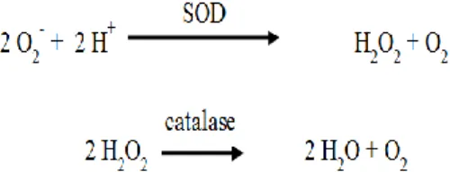
The effects of fluorine in human cell functions have been a target of study, showing that it can interact with cell processes dependent on enzymes and corresponding genes modulated by fluorine, including processes related with stress response, metabolic enzymes, cell cycle, communication between cells and signal transduction.[51] Several studies show that exposure to fluorine can be related with the inhibition of activity of several enzymes and with the formation of free radicals that, consequently, interfere with the antioxidant defence mechanisms in living systems.[62] In one of these studies[62], rats were injected with a unique dose of NaF (15 mg/kg of weight) and were killed in several time periods till 24 hours after fluorine exposure. The goal was to evaluate the effects of low doses of fluorine on antioxidant enzymatic activity of enzymes like superoxide dismutase (SOD) and catalase (Fig. 1), and the levels of lipidic peroxidation in salivary glands (parotid and submandibular) of rats.
Fig. 1: Reactions catalysed by superoxide dismutase (SOD) and catalase.
Results showed that exposure to NaF did not induce significative changes in the activities of SOD and catalase, or in the levels of lipidic peroxidation in the parotid gland when compared with the control group. However, submandibular glands showed an increase in SOD activity after 3 hours and decrease of that activity after 1, 12 and 24 hours. Lipidic peroxidation suffered an increase after 6, 12 and 24 hours of NaF exposure. Concerning catalase activity, no significative change occurred when compared with controls. These results showed that NaF exposure triggered oxidative stress in salivary glands few hours after NaF intake. These effects were more pronounced in the submandibular gland than in the parotid gland.[62]
4.3. Citotoxicity
The action of fluorine in cell metabolism varies with its concentration, exposure period and type of cell.[63] In teeth and bones, micromolar concentrations are beneficial to cells by promoting cell proliferation and
growth. However, millimolar concentrations supress cell division and induce apoptosis. Studies have demonstrated that exposure to high concentrations of fluorine induce apoptosis of ameloblasts[64,65], odontoblasts[66] and osteoblasts.[67]
Toxic effects include induction of inflammatory reactions, inhibition of protein synthesis and the progression of cell cycle, oxidative stress and DNA damage. NaF can induce apoptosis of cells of different organs and tissues.[68,69]
5. ALCOHOL AS AN INDUCER OF ORAL CANCER
The International Agency for Research on Cancer (IARC) identified alcohol (ethanol) as a human carcinogen and considered it as an individual risk factor.[23]
Based on the sum total of research evidence on mechanistic studies of alcohol carcinogenesis and epidemiological evidence relating alcohol to an increased risk of multiple forms of cancer, primarily oral cancer, the American Institute for Cancer Research (AICR) classified alcohol as a group 1 carcinogen because it causes cancer in humans. The AICR concluded that there is enough evidence in humans for the carcinogenicity not only of alcohol consumption, but also for the carcinogenicity of acetaldehyde associated with the consumption of alcoholic beverages.[70]
After smoking and the infectious agents responsible for chronic infections, alcohol is the most important known cause of cancer in humans. According to epidemiological studies, which consider that alcohol consumption is responsible for approximately 5.2% of deaths of men and 1.7% of women from cancer in the world. High cancer risks are observed for all organs with direct contact with non-metabolized alcohol, such as the oral cavity, pharynx, larynx and esophagus.[71]
Scientific evidence from recent years shows that the consumption of alcoholic beverages increases the risk of tumors of the oral cavity, both in quantity and in duration. Some researchers reported that consuming 50 g of alcohol daily increases the risk of these tumors two or three times when compared to non-consumers of alcoholic beverages.[72] In another study, individuals who consumed 30 g or more of alcohol per day were 6.39% more likely to develop oral cavity cancer and were 3.52% more likely to develop oropharyngeal or hypopharyngeal cancer than non-ethanol consumers.[73]
A study that associated oral and pharyngeal cancer with alcohol consumption, showed that the risk of these cancers increases in direct proportion to alcohol consumption. The risk increases 1.7 times with the consumption of 1 to 2 drinks per day, 2.3 times with the consumption of 3 to 4 drinks per day, and 5.5 times when the individual consumes 5 or more drinks per day.[74]
A systematic review by Mello et al.[75] observed an increased probability of occurrence of OSCC in individuals who consume alcohol and in those who consume fermented beverages such as wine, cider, spirits and beer. The isolated consumption of beer and other types of alcoholic beverages was not associated with OSCC.
Alcohol has genotoxicity and mutagenic effects, causing reduced saliva flow and affecting the liver's ability to metabolize toxic and potentially carcinogenic compounds. Its chronic use is associated with a considerable impairment of innate and acquired immunity, resulting in an increased susceptibility to infections and neoplasms.[76]
Ethanol is eliminated from the body by its oxidation to acetaldehyde by the enzyme alcohol dehydrogenase (ADH). Ethanol alone is not mutagenic, but acetaldehyde is carcinogenic and mutagenic, as it binds to deoxyribonucleic acid (DNA) and proteins.[77]
Most of the acetaldehyde resulting from the degradation of alcohol is readily converted to acetate by the enzyme aldehyde dehydrogenase (ALDH). However, a genetic variant of ALDH that encodes a protein without enzymatic activity has been identified, which potentiates the mutagenic action and the harmful effects of alcohol. The administration of ethanol or acetaldehyde to drinking water led to an increased incidence of various tumors in rats.[78]
A study carried out on rats[71] examined the effect of alcohol consumption on immune regulation and the damage induced by dibenzo[a, l]pyrene (DB[a, l]P), an environmental polycyclic aromatic hydrocarbon. The levels of glutathione (GSH) found in the oral mucosa after alcohol intake (6.6% v/v in the diet) for 24 days, decreased by 28%, suggesting a possible reduction in the detoxification of diol epoxides derived from DB[a, l]P in oral tissues. In addition, analysis of the levels of DNA adducts (a segment of DNA linked to a chemical carcinogen) induced by DB[a, l]P in oral tissues revealed that alcohol consumption increased (-)anti-trans-DB[a,
l]P adduct levels by 25%. These researchers also found
an increase in the expression of several genes (coding for the interleukin 22-binding protein, lymphotoxin α and arginase) associated with the negative regulation of the immune response. This study allowed to conclude that chronic alcohol consumption increases the genotoxicity induced by DB[a, l]P and promotes alterations in the expression of genes that can suppress the defense mechanisms of the immune system in the oral cavity.
According to this model, several genes are modulated by the presence of alcohol. Changes in chemostatic signals and arginase production lead to a reduction in the recruitment activity of T and NK cells. Decreased signaling of type I interferon or innate detection by toll-like receptors may allow suppressive myeloid cells to
produce interleukin 23 (IL-23) to amplify the response of the innate lymphoid cell 3, leading to IL-17A and IL-22 induced inflammation of the epithelium. This state of inflammation can be further amplified by the increased production of α-lymphotoxin and the consequent increased activation of the respective receptor that positively regulates the production of IL-23. Alcohol leads to the production of high levels of the soluble IL-22ra2 receptor, perhaps by local or accumulated dendritic cells, which can moderate tissue damage, as well as obstruct tissue repair.[71]
In another study[79], it was found that individuals who abuse alcohol chronically and chronically are more likely to develop various diseases, including cancer. Chronic alcohol consumption leads to depletion of GSH levels and inhibition of cell transsulfurization/transmethylation, key pathogenic events involved in alcohol-related tissue damage and carcinogenesis.
Some studies supported the hypothesis that chronic exposure of tissues in the oral cavity to acetaldehyde can lead to DNA damage. Balbo et al.[80] quantified the presence of N2-ethylidenodesoxyguanosine (N2 -ethyl-dG), the main product of the reaction of acetaldehyde with DNA, in human oral exfoliating cells, at various time points after consuming increasing doses of alcohol. The superficial oral exfoliating cells represent the first cell layer of lining of the oral cavity, so they are those that are directly exposed to alcohol. During the study, participants consumed one drink/week over 3 weeks. The results demonstrated a 100-fold increase in N2-ethyl-dG levels compared to baseline values within four hours after each dose. Despite the small number of participants, the results were significant even after the first dose and indicated a direct link between alcohol consumption and DNA damage.
Acetaldehyde can react with tissue DNA to form adducts that can serve as biomarkers of carcinogenic exposure and potentially cancer risk. In order to clarify the mechanism of alcohol-induced carcinogenesis, Balbo et al. [81] investigated the use of N2-ethyl-dG as a biomarker of alcohol-induced DNA damage in oral and esophageal tissues of Rhesus monkeys exposed to daily alcohol consumption (2.3 ± 0.8 g of ethanol/kg/day, corresponding to approximately 9 drinks/day, with 1 drink equivalent to 15 g of alcohol) over 12 months. This study observed higher levels of N2-ethyl-dG in the DNA of the oral mucosa of the exposed animals compared to the control animals. In the esophageal mucosa, the levels of N2-ethyl-dG were not significantly different from the control. These results reinforce the role of acetaldehyde in inducing DNA damage in the oral mucosa exposed to alcohol, playing a role in the carcinogenic process induced by alcohol.
6. SYNERGISM BETWEEN ALCOHOL AND TOBACCO IN ORAL CANCER
A fact to be considered in the etiology of oral cancer is
the magnitude of the risk due to the interaction between alcohol consumption and smoking, whose adverse effects are not simply additive, but rather multiplicative, reaching quite high risks in individuals with simultaneous high consumption of alcohol and tobacco.[82]
Scientific literature points out that alcohol enhances the carcinogenic role of tobacco. Alcohol, particularly ethanol, can act both as a risk factor locally and systemically, increasing the permeability of the oral mucosa, dissolving the lipid components of the epithelium, generating epithelial atrophy, promoting the penetration and solubilization of the carcinogenic metabolites of tobacco and causing interference in DNA synthesis and repair.[76,83] In one study[84] it was stated that the synergism between alcohol and tobacco consumption increases 10 to 15 times the risk of developing oral cancer.
A systematic review[75] evaluated the available evidence on the association between the simultaneous consumption of alcohol and tobacco and the occurrence of OSCC. This study showed that the synergistic consumption of alcohol and tobacco increased 2.44 to 23.7 times the probability of occurrence of OSCC when compared to none or minimum consumption of alcohol and tobacco. The simultaneous consumption of alcohol and tobacco increased the probability of developing OSCC from 4.21 to 24.28 times. When the individual simultaneously consumes alcohol, smoked and non-smoked tobacco, the probability of developing OSCC varied from 4.8 to 16.34 times.
A study[85] of 132 subjects diagnosed with OSCC and 320 control subjects without oral cancer, evaluated different potential risk factors for OSCC and its influence on the recurrence or occurrence of a new second primary tumor (SPT) of OSCC. The study showed that all individuals with OSCC consumed more alcohol and tobacco than the control subjects. The consumption of alcohol (> 350 g/week) and tobacco (11 to 20 cigarettes/day) were dose-dependent risk factors. In addition, high alcohol consumption was associated with a high relative rate of recurrence or occurrence of SPT.
It is speculated that alcohol acts as a solvent for carcinogenic tobacco smoke, allowing better absorption through the mucosa.[23,73,76] Very high-level alcoholics who smoke more than 20 cigarettes a day are more likely to develop some form of head and neck cancer than non-smoking alcoholics.[73] It is estimated that 75% of oropharyngeal cancers can be prevented by eliminating tobacco and reducing alcohol consumption.[86]
7. CONCLUSIONS
Dental caries stands out as a highly prevalent oral condition that affects oral health in different age groups and socioeconomic strata. Dental caries is a dynamic process involving the interaction between susceptible
dental surfaces, cariogenic bacteria such as S. mutans or
Lactobacillus and a source of fermentable carbohydrates.
Fermentable carbohydrates are primarily sucrose; however, all carbohydrates are generally considered cariogenic. Other factors such as salivary gland hypofunction, lack of motivation to maintain oral hygiene, socioeconomic and cultural differences, parenting practices, genetics, among other factors, play an important role in the development of dental diseases. Frequent consumption of carbohydrates in the form of simple sugars increases the risk of tooth decay.
There are also foods like some milk proteins (casein) and fluorine that are protective, reducing the enamel demineralization. Further studies on oral health protective foods are needed, as well as more information on their populations, aiming at a more balanced and healthy diet.
This study also allows to conclude that alcohol consumption is associated with the development of several types of oral cancer, being a relevant risk factor for this disease, with a synergistic effect when associated with any form of smoking. Alcohol (ethanol) alone is not associated with the development of cancer, but the products of its metabolism, especially acetaldehyde, are directly related to the occurrence of oral cancer, producing toxic and mutagenic effects. The appearance of oral cancer is related to the amount and duration of alcohol consumption. As it is a modifiable risk factor, since it results from behavior, prevention and education measures on this relationship must be adopted by all health professionals.
8. REFERENCES
1. Klein MI, Duarte S, Xiao J, Mitra S, Foster TH, Koo H. Structural and molecular basis of the role of starch and sucrose in Streptococcus mutans biofilm development. Appl Environ Microbiol, 2009; 75(3): 837-841.
2. Sheiham A, James WPT. A reappraisal of the quantitative relationship between sugar intake and dental caries: the need for new criteria for developing goals for sugar intake. BMC Public Health, 2014; 14(1): 1-8.
3. Tu CL, He LL, Cui LF, Zhang QH, Zeng QB, Pan XL, et al. Environmental and geochemical behaviors of fluorine and its impact on ecological environment. Ying Yong Sheng Tai Xue Bao., 2019; 30(1): 21-29.
4. Ullah R, Zafar M, Shahani N. Potential fluoride toxicity from oral medicaments: a review. Iran J Basic Med Sci., 2017; 20(8): 841-848.
5. Denson L, Janitz AE, Brame LS, Campbell JE. Oral cavity and oropharyngeal cancer: changing trends in incidence in the United States and Oklahoma. J Okla State Med Assoc, 2016; 109(7-8): 339-345.
6. Shield KD, Ferlay J, Jemal A, Sankaranarayanan R, Chaturvedi AK, Bray F, et al. The global incidence
of lip, oral cavity, and pharyngeal cancers by subsite in 2012. CA Cancer J Clin., 2017; 67(1): 51-64. 7. Weatherspoon DJ, Chattopadhyay A, Boroumand S,
Garcia I. Oral cavity and oropharyngeal cancer incidence trends and disparities in the United States: 2000-2010. Cancer Epidemiol, 2015; 39(4): 497-504.
8. Rivera C. Essentials of oral cancer. Int J Clin Exp Pathol, 2015; 8(9): 11884-11894.
9. Sankaranarayanan R, Ramadas K, Amarasinghe H, Subramanian S, Johnson N. Oral cancer: prevention, early detection, and treatment. In: Gelband, H, Jha P, Sankaranarayanan R, Horton S, ed. Cancer:
disease control priorities. 3rd ed. Washington (DC): The International Bank for Reconstruction and Development/The World Bank, 2015.
10. Bezerra NV, Leite KL, de Medeiros MM, Martins ML, Cardoso AM, Alves PM, et al. Impact of the anatomical location, alcoholism and smoking on the prevalence of advanced oral cancer in Brazil. Med Oral Patol Oral Cir Bucal, 2018; 23(3): e295-e301. 11. Albuquerque R, López-López J, Mari-Roig A,
Jané-Salas E, Roselló-Llabrés X, Santos JR. Oral tongue squamous cell carcinoma (OTSCC): alcohol and tobacco consumption versus non-consumption. A study in a Portuguese population. Braz Dent J., 2011; 22(6): 517-521.
12. Kruse AL, Bredell M, Grätz, KW. Oral cancer in men and women: are there differences? Oral Maxillofac Surg, 2011; 15(1): 51-55.
13. Hao Y, Huang X, Zhou X, Li M, Ren B, Peng X, et al. Influence of dental prosthesis and restorative materials interface on oral biofilms. Int J Mol Sci., 2018; 19(10): E3157.
14. Banas JA, Drake DR. Are the mutans streptococci still considered relevant to understanding the microbial etiology of dental caries?. BMC Oral Health, 2018; 18(1): 1-8.
15. Bowen WH, Koo H. Biology of Streptococcus mutans-derived glucosyltransferases: role in extracellular matrix formation of cariogenic biofilms. Caries Res., 2011; 45(1): 69-86.
16. Ferreira-Nóbilo NP, Tabchoury CPM, Sousa MLR, Cury JA. Knowledge of dental caries and salivary factors related to the disease: influence of the teaching-learning process. Braz Oral Res., 2015; 29(1): 1-7.
17. Zaura E, Brandt BW, Prodan A, Teixeira de Mattos MJ, Imangaliyev S, Kool J, et al. On the ecosystemic network of saliva in healthy young adults. ISME J., 2017; 11(5): 1218-1231.
18. Lenander-Lumikari M, Loimaranta V. Saliva and dental caries. Adv Dent Res., 2000; 14(1): 40-47. 19. Dawes C. What is the critical pH and why does a
tooth dissolve in acid?. J Can Dent Assoc, 2003; 69(11): 722-724.
20. INCA (2019). Instituto Nacional de Câncer. Available at https://www.inca.gov.br/o-que-e-cancer.
21. World Health Organization. International Statistical Classification of Diseases and Related Health Problems 10th Revision (ICD-10)-WHO Version for 2016. Available at: https://icd.who.int/browse10/2016/en#/II.
22. Cleveland JL, Junger ML, Saraiva M, Markowitz LE, Dunne EF, Epstein JB. The connection between human papillomavirus and oropharyngeal squamous cell carcinomas in the United States: implications for dentistry. J Am Dent Assoc, 2011; 142(8): 915-924.
23. Lambert R, Sauvaget C, de Camargo Cancela M, Sankaranarayanan R. Epidemiology of cancer from the oral cavity and oropharynx. Eur J Gastroenterol Hepatol, 2011; 23(8): 633-641.
24. Ferlay J, Colombet M, Soerjomataram I, Dyba T, Randi G, Bettio M, et al. Cancer incidence and mortality patterns in Europe: estimates for 40 countries and 25 major cancers in 2018. Eur J Cancer, 2018; 103: 356-387.
25. Doichinova L, Bakardjiev P, Peneva M. Assessment of food habits in children aged 6 12 years and the risk of caries. Biotechnol Biotechnol Equip, 2015; 29(1): 200-204.
26. Moynihan PJ. The role of diet and nutrition in the etiology and prevention of oral diseases. Bull WHO, 2005; 83(9): 694-699.
27. Feldens CA, Kramer PF, Vargas-Ferreira F. The role of diet and oral hygiene in dental caries. In: Coelho Leal, S., Takeshita E. ed. Pediatric Restorative
Dentistry. Springer, Cham., 2019.
28. Bica I, Cunha M, Reis M, Costa J, Costa P, Bica A. Food consumption, body mass index and risk for oral health in adolescents. Aten Primaria, 2014; 46(S5): 154-159.
29. Gupta M. Sugar substitutes: mechanism, availability, current use and safety concerns - an update. Open Access Maced J Med Sci., 2018; 6(10): 1888-1894. 30. Díaz-Garrido N, Lozano C, Giacaman RA.
Frequency of sucrose exposure on the cariogenicity of a biofilm-caries model. Eur J Dent, 2016; 10(3): 345-350.
31. Moynihan PJ, Kelly SAM. Effect on caries of restricting sugars intake: systematic review to inform WHO guidelines. J Dent Res., 2014; 93(1): 8-18.
32. Lingstrom P, van Houte J, Kashket S. Food starches and dental caries. Crit Rev Oral Biol Med., 2000; 11(3): 366-380.
33. Giacaman RA, Jobet-Vila P, Muñoz-Sandoval C. Fatty acid effect on sucrose-induced enamel demineralization and cariogenicity of an experimental biofilm–caries model. Odontology, 2015; 103(2): 169-176.
34. Huang CB, George B, Ebersole JL. Antimicrobial activity of n-6, n-7 and n-9 fatty acids and their esters for oral microorganisms. Arch Oral Biol., 2010; 55(8): 555-560.
35. Voytsekhovskaya IV, Axenov-Gribanov DV, Murzina SA, Pekkoeva SN, Protasov ES,
Gamaiunov SV, et al. Estimation of antimicrobial activities and fatty acid composition of actinobacteria isolated from water surface of underground lakes from Badzheyskaya and Okhotnichya caves in Siberia. Peer J., 2018; 6: e5832.
36. Glimcher MJ, Friberg UA, Levine PT. The isolation and amino acid composition of the enamel proteins of erupted bovine teeth. Biochem J., 1964; 93: 202. 37. Kreitzman SN. Enzymes and dietary factors in
caries. J Dent Res., 1974; 53(2): 218-225.
38. Ghasempour M, Rajabnia R, Ashrafpour M, Ehsani A, Moghadamnia AA, Gharekhani S, et al. Effect of milk and yogurt on Streptococcus sobrinus counts and caries score in rats. Dent Res J., 2015; 12(6): 569-573.
39. Smith DJ. Dental caries vaccines: prospects and concerns. Crit Rev Oral Biol Med., 2002; 13(4): 335-349.
40. Sheiham A. Dietary effects on dental diseases. Public Health Nutr, 2001; 4(2b): 569-591.
41. Krul L, Kremer BHA, Luijckx NBL, Leeman WR. Quantifiable risk-benefit assessment of micronutrients: from theory to practice. Crit Rev Food Sci Nutr, 2017; 57(17): 3729-3746.
42. Aoba T, Fejerskov O. Dental fluorosis: chemistry and biology. Crit Rev Oral Biol Med., 2002; 13(2): 155-170.
43. Tenuta LM, Cury JA. Fluoride: its role in dentistry. Braz Oral Res., 2010; 24(1): 9-17.
44. McGrady MG, Ellwood RP, Maquire A, Goodwin M, Boothman N, Pretty IA. The association between social deprivation and the prevalence and severity of dental caries and fluorosis in populations with and without water fluoridation. BMC Public Health, 2012; 12: 1122.
45. Bronckers AL, Lyaruu DM, DenBesten PK. The impact of fluoride on ameloblasts and the mechanisms of enamel fluorosis. J Dent Res., 2009; 88(10): 877-893.
46. Verkerk R. The paradox of overlapping micronutrient risks and benefits obligates risk/benefit analysis. Toxicology, 2010; 278(1): 27-38.
47. Cangussu MC, Narvai PC, Castellanos Fernandez R, Djehizian V. A fluorose dentária no Brasil: uma revisão crítica. Cad Saúde Pública, 2002; 18(1): 7-15.
48. Warren JJ, Kanellis MJ, Levy SM. Fluorosis of the primary dentition: what does it mean for permanent teeth?. J Am Dent Assoc, 1999; 130(3): 347-356. 49. Ulu Güzel KG, Özay Ertürk MS, Kirzioglu Z,
Özkorucuklu S. Evaluation of dentin permeability of fluorotic permanent teeth. Acta Odontol Scand, 2018; 76(6): 415-421.
50. Jimenez-Farfán MD, Hernandez-Guerrero JC, Juárez-López LA, Jacinto-Alemán LF, de la Fuente-Hernandéz J. Fluoride consumption and its impact on oral health. Int J Environ Res Public Health, 2011; 8(1): 48-60.
51. Peckham S, Awofeso N. Water fluoridation: a critical review of the physiological effects of ingested fluoride as a public health intervention. Sci World J., 2014; 2014: 293019.
52. Moimaz SA, Saliba O, Marques LB, Garbin CA, Saliba NA. Dental fluorosis and its influence on children’s life. Braz Oral Res., 2015; 29: 0014. 53. Molina-Frechero N, Pierdant-Rodríguez AI,
Oropeza-Oropeza A, Bologna-Molina R. Fluorosis and dental caries: an assessment of risk factors in Mexican children. Rev Invest Clin., 2012; 64(1): 67-73.
54. Verma A, Shetty BK, Guddattu V, Chourasia MK, Pundir P. High prevalence of dental fluorosis among adolescents is a growing concern: a school based cross-sectional study from Southern India. Environ Health Prev Med., 2017; 22(1): 17.
55. Kumar S, Chauhan A, Kumar A, Kumar S, Gupta A, Roy S, et al. Dental fluorosis and associated risk factors in early adolescents in India. Int J Adolesc Med Health, 2018. doi: https://doi.org/10.1515/ijamh-2017-0200. 56. Fawell J, Bailey K, Chilton J, Dahi E, Fewtrell L,
Magara Y. Fluoride in drinking-water. World Health Organization (WHO), IWA Publishing, London, UK, 2006.
57. Firempong CK, Nsiah K, Awunyo-Vitor D, Dongsogo J. Soluble fluoride levels in drinking water – a major risk factor of dental fluorosis among children in Bongo community of Ghana. Ghana Med J., 2013; 47(1): 16-23.
58. Chandrashekar J, Anuradha KP. Prevalence of dental fluorosis in rural areas of Davangere, India. Int Dent J., 2004; 54(5): 235-239.
59. Sarvaiya BU, Bhayya D, Arora R, Mehta DN. Prevalence of dental fluorosis in relation with different fluoride levels in drinking water among school going children in Sarada tehsil of Udaipur district, Rajasthan. J Indian Soc Pedod Prev Dent, 2012; 30(4): 317-322.
60. Rozier RG. Oral health in North Carolina: innovations, opportunities and challenges. N C Med J., 2012; 73(2): 100-107.
61. Domingos P, Ricci-Donato H, Russi A. Riscos do uso do fluor sistémico - Revisão de Literatura. J Res Dent, 2018; 6(4): 86-90.
62. Yamaguti PM, Simões A, Ganzerla E, Sousa DN, Nogueira FN, Nicolau J. Effects of single exposure of sodium fluoride on lipid peroxidation and antioxidant enzymes in salivary glands of rats. Oxid Med Cell Longev, 2013; 2013: 674593.
63. Barbier O, Arreola-Mendoza L, Del Razo LM. Molecular mechanisms of fluoride toxicity. Chem Biol Interact, 2010; 188(2): 319-333.
64. Kubota K, Lee DH, Tsuchiya M, Young CS, Everett ET, Martinez-Mier EA, et al. Fluoride induces endoplasmic reticulum stress in ameloblasts responsible for dental enamel formation. J Biol Chem., 2005; 280(24): 23194-23202.
65. Jacinto-Alemán LF, Hernández-Guerrero JC, Trejo-Solís C, Jiménez-Farfán MD, Fernández-Presas AM. In vitro effect of sodium fluoride on antioxidative enzymes and apoptosis during murine odontogenesis. J Oral Pathol Med., 2010; 39(9): 709-714.
66. Karube H, Nishitai G, Inageda K, Kurosu H, Matsuoka M. NaF activates MAPKs and induces apoptosis in odontoblast-like cells. J Dent Res., 2009; 88(5): 461-465.
67. Yang S, Wang Z, Farquharson C, Alkasir R, Zahra M, Ren G, et al. Sodium fluoride induces apoptosis and alters bcl-2 family protein expression in MC3T3-E1 osteoblastic cells. Biochem Biophys Res Commun, 2011; 410(4): 910-915.
68. Thrane EV, Refsnes M, Thoresen GH, Låg M, Schwarze PE. Fluoride-induced apoptosis in epithelial lung cells involves activation of MAP kinases p38 and possibly JNK. Toxicol Sci., 2001; 61(1): 83-91.
69. Refsnes M, Schwarze PE, Holme JA, Låg M. Fluoride-induced apoptosis in human epithelial lung cells (A549 cells): role of different G protein-linked signal systems. Hum Exp Toxicol, 2003; 22(3): 111-123.
70. van’t Veer P, Kampman E. Food, nutrition, physical activity, and the prevention of cancer: a global perspective. Washington DC, World Cancer Research Fund/American Institute for Cancer Research, 2007.
71. Boffetta P, Hashibe M, La Vecchia C, Zatonski W, Rehm J. The burden of cancer attributable to alcohol drinking. Int J Cancer, 2006; 119(4): 884-887. 72. Baan R, Straif K, Grosse Y, Secretan B, El Ghissassi
F, Bouvard V, et al. Carcinogenicity of alcoholic beverages. Lancet Oncol, 2007; 8(4): 292-293. 73. Maasland DH, van den Brandt PA, Kremer B,
Goldbohm RA, Schouten LJ. Alcohol consumption, cigarette smoking and the risk of subtypes of head-neck cancer: results from the Netherlands Cohort Study. BMC Cancer, 2014; 14(187): 1-14.
74. Blot WJ, McLaughlin JK, Winn DM, Austin DF, Greenberg RS, Preston-Martin S, et al. Smoking and drinking in relation to oral and pharyngeal cancer. Cancer Res., 1988; 48(11): 3282-3287.
75. Mello FW, Melo G, Pasetto JJ, Silva CAB, Warnakulasuriya S, Rivero ERC. The synergistic effect of tobacco and alcohol consumption on oral squamous cell carcinoma: a systematic review and meta-analysis. Clin Oral Investig, 2019; 23(7): 2849-2859.
76. Reidy J, McHugh E, Stassen LFA. A review of the relationship between alcohol and oral cancer. Surgeon, 2011; 9(5): 278-283.
77. Poschl G, Seitz HK. Alcohol and cancer. Alcohol Alcohol, 2004; 39(3): 155-165.
78. Chen K-M, Schell TD, Richie JP Jr, Sun Y-W, Zhang S-M, Calcagnotto A, et al. Effects of chronic alcohol consumption on DNA damage and immune regulation induced by the environmental pollutant
dibenzo[a,l]pyrene in oral tissues of mice. J Environ Sci Health C Environ Carcinog Ecotoxicol Rev., 2017; 35(4): 213-222.
79. Chen Y, Han M, Matsumoto A, Wang Y, Thompson DC, Vasiliou V. Glutathione and transsulfuration in alcohol-associated tissue injury and carcinogenesis. Adv Exp Med Biol., 2018; 1032: 37-53.
80. Balbo S, Meng L, Bliss RL, Jensen JA, Hatsukami DK, Hecht SS. Kinetics of DNA adduct formation in the oral cavity after drinking alcohol. Cancer Epidemiol Biomarkers Prev, 2012; 21(4): 601-608. 81. Balbo S, Juanes RC, Khariwala S, Baker EJ,
Daunais JB, Grant KA. Increased levels of the acetaldehyde-derived DNA adduct N 2-ethyldeoxyguanosine in oral mucosa DNA from Rhesus monkeys exposed to alcohol. Mutagenesis, 2016; 31(5): 553-558.
82. Szymanska K, Hung RJ, Wunsch-Filho V, Eluf-Neto J, Curado MP, Koifman S, et al. Alcohol and tobacco, and the risk of cancers of the upper aerodigestive tract in Latin America: a case-control study. Cancer Causes Control, 2011; 22(77): 1037-1046.
83. Paré A, Joly A. Oral cancer: risk factors and management. Presse Med, 2017; 46(3): 320-330. 84. Mummudi N, Agarwal JP, Chatterjee S, Mallick I,
Ghost-Laskar S. Oral cavity cancer in the Indian subcontinent: challenges and opportunities. Clin Oncol (R Coll Radiol), 2019; 31(8): 520-528. 85. Rosenquist K. Risk factors in oral and
oropharyngeal squamous cell carcinoma: a population-based case-control study in southern Sweden. Swed Dent J Suppl, 2005; 179: 1-66. 86. Marron M, Boffetta P, Zhang ZF, Zaridze D,
Wunsch-Filho V, Winn DM, et al. Cessation of alcohol drinking, tobacco smoking and the reversal of head and neck cancer risk. Int J Epidemiol, 2010; 39(1): 182-196.