Universidade do Minho
Escola de Engenharia
Cátia Sofia Almeida Cardoso
Improvement of oral health:
biofilm formation, oral implants and probiotics
11
Cátia Sof
ia Almeida Car
doso
Impro
vement of oral healt
h: biofilm formation, oral implants and probio
Dissertação de Mestrado
Ciclo de Estudos Integrados Conducentes
ao Grau de Mestre em Engenharia Biomédica
Trabalho realizado sob a orientação da
Professora Doutora Mariana Contente
Rangel Henriques
Universidade do Minho
e do
Professor Doutor Wim Teughels
KULeuven
Universidade do Minho
Escola de Engenharia
Cátia Sofia Almeida Cardoso
Improvement of oral health:
DECLARAÇÃO
Nome: Cátia Sofia Almeida Cardoso
Endereço electrónico: a49990@alunos.uminho.pt Telefone: +351 917999736 Número do Bilhete de Identidade: 13359006
Título dissertação:
Improvement of oral health: biofilm formation, oral implants and probiotics Ano de conclusão: 2011
Orientador:
Professora Doutora Mariana Contente Rangel Henriques Professor Doutor Wim Teughels
Designação do Mestrado:
Ciclo de Estudos Integrados Conducentes ao Grau de Mestre em Engenharia Biomédica Área de Especialização: Engenharia Clínica
Escola: de Engenharia
Departamento: de Engenharia Biológica
DE ACORDO COM A LEGISLAÇÃO EM VIGOR, NÃO É PERMITIDA A REPRODUÇÃO DE QUALQUER PARTE DESTA TESE/TRABALHO.
Braga, ____/____/________
iii
Acknowledgments
First of all, I would like to express my gratitude to my supervisors Dr. Mariana Henriques and Dr. Wim Teughels, whose expertise, attention and understanding contributed considerably to my dissertation work.
I am especially grateful to Dr. Mariana Henriques for the support, dedication and time put on my dissertation. All the tips, suggestions and advices were very important in the project and also all the enthusiasm that she gave me. Throughout my dissertation-writing period, she provided encouragement, good teaching, advising and a panoply of good ideas. This work would not have been possible without her.
To Dr. Wim Teughels, I want express my thankfulness for being so welcoming and for make me at ease in my work and for introducing me to the research made the Department of Periodontology of the Catholic University Leuven.
Martine Pauwels and Gitte Loozen deserve a special thank for the great support, time, patience, affection and friendship that they devoted to me. Since the first day they were fantastic making me feel at home. For all the teaching, knowledge and sharing experiences I am very grateful. Additionally, all the colleagues in the laboratory deserve my recognition for their encouragement and hospitality.
I have to thank all my Erasmus friends that spend a fantastic time with me and to my closest friends who accompanied me during all these years. They always provided me a stimulating and fun environment in which I learned and grew. They were always there in good and bad moments to give me a word of appreciation and courage, thank you for all.
Special thanks to my godfather of course for his motivation and encouragement, for the support during dissertation-writing and for all friendship.
The last words must be addressed to my beloved family, my parents, my sister and my grandparents. They are extremely important for me, for all the help that they give to get through the difficult times and to achieve my goals, for all the emotional support, love, affection and constant encouragement I am deeply grateful. They are the reason why I am here right now, without them this could not be possible. They are always my safe haven, my anchor and to them I dedicate this work.
v
Abstract
Oral health problems affect a large part of the world population and despite the numerous developments on technologies and products, there is still a need to know and understand those diseases. The recurring demands from society led to the development of new medical treatments and new materials used in dental implants. So, in this context, it became very important to evaluate the microbial colonization of implant materials because these materials can be important for future dental applications being necessary know the microbial adhesion. Therefore, the present dissertation aimed to improve the knowledge on oral microbial colonization of oral cavity.
The first goal was the evaluation of microbial colonization of different titanium surfaces (anodized and etched) that are normally used in implant applications. The bacteria studied (Prevotella intermedia, Porphyromonas gingivalis, Fusobacterium nucleatum) were able to form biofilms on both surfaces, although biofilm formation on anodized samples result in a higher amount of biomass than in the etched samples, although with a similar number of viable cells, indicating the higher presence of extracelular matrix in the former, which could induce lower mechanical friction on these samples.
In addition this study also evaluated the influence of fluoride and probiotic bacteria (Streptococcus salivarius) on biofilm formation. The presence of fluoride showed to inhibit biofilm formation on these biomaterials. The effect of probiotic bacteria has been evaluated on biofilm formation and S. salivarius had a direct influence on reducing the growth of pathogenic bacteria, such as F. nucleatum. However, these interactions are still unclear and there is a need to study these in greater detail.
Moreover, dental implants infection can also be reduced by controlling the presence of pathogenic species in oral environment, so another aim was the evaluation of the use of a sugar (C7) as a prebiotic agent. The different sources of energy (C7 sugar and glucose) had different influences on growth of pathogenic and probiotic bacteria. So, the sugar, C7, can be used to favour the growth of the beneficial oral bacteria.
In conclusion it can be pointed out that Ti anodized samples may be a good material for the production of dental implants due to their topography and also that the use of different energy sources allied with probiotics may be a start point for the development of new therapies.
vii
Resumo
Os problemas de saúde oral afectam grande parte da população mundial e apesar do grande desenvolvimento nas tecnologias e nos produtos aplicados a esta, existe ainda a necessidade de conhecer e compreender melhor as doenças orais de origem bacteriana. Assim, a presente dissertação teve como objectivo geral contribuir para melhorar o conhecimento sobre a interação bacteriana na cavidade oral.
O primeiro objectivo deste trabalho foi a avaliar a formação de biofilme em diferentes superfícies de titânio (anodizadas e com tratamento químico) normalmente utlizadas em implantes dentários. As bactérias estudadas (Prevotella intermedia, Porphyromonas gingivalis, Fusobacterium nucleatum) foram capazes de formar biofilme em ambas as superfícies, embora nas amostras anodizadas a quantidade de biomassa formada tenha sido maior que nas com tratamento quimico. No entanto, o número de células viáveis nos biofilmes formados em ambas as superfícies foi semelhante, indicando maior presença de matriz nas amostras anodizadas, o que pode induzir menor fricção nestas amostras.
Além deste estudo foi também avaliada a influência da adição flúor e a presença de uma bactéria probiotica (Streptococcus salivarius) nos biofilmes formados nas mesma amostras. A presença de flúor no meio de cultura mostrou ter uma influencia negativa na formação de biofilme. Quanto à presença de bactéria probiotica esta foi avaliada em relação a formação de biofilme pelas mesmas bactérias patogénicas. A bactéria probiotica teve influência direta na redução da proliferação das bactérias patogénicas em especifico da F. nucleatum.
As infecções orais podem também ser controladas alterando o equilíbrio entre a flora patogénica e probiótica. Assim sendo, foi também avaliada a influência de um açúcar (C7) neste equilíbrio, funcionado este como um agente prebiótico. As diferentes fontes de energia (glucose - controlo - e C7), tiveram diferentes influências sobre o crescimento das bactérias patogénicas e probioticas. A fonte energia, C7, poderá ser assim usada para favorecer o crescimento da bactéria probiotica e assim contribuir para uma melhoria da saúde oral.
Portanto, neste trabalho concluiu-se que as amostras de Ti anodizado poderão ter maior potencial na produção de implantes dentários devido à sua topografia e além disso o uso de diferentes fontes de energia aliada ao uso de bactérias probióticas pode ser um início de um desenvolvimento de uma nova terapia.
ix
Table of Contents
Acknowledgments ... iii Abstract ... v Resumo ... vii Table of Contents ... ix Abbreviations ... xiList of Figures ... xiii
List of Tables ... xv
1. Chapter I ... 1
1.1. Motivation and mains objectives ... 3
1.2. Introduction ... 4
1.2.1. Periodontal microorganisms ... 6
1.2.2. Biofilm and bacterial interactions ... 10
1.2.3. Relation between oral health and other diseases ... 16
2. Chapter II ... 19
2.1. Introduction ... 21
2.1.1. Formation of biofilm on dental implants ... 21
2.1.2. Titanium in dental implants ... 23
2.1.3. Influence of Fluoride in biofilm formation ... 23
2.1.4. Treatment of dental implant-associated infections ... 24
2.1.5. Probiotics as improvement for oral health ... 25
2.2. Materials and Methods ... 27
2.2.1. Sample preparation ... 27
2.2.2. Bacterial Culture ... 27
2.2.3. Culture media and solutions ... 28
2.2.4. Biofilm formation ... 29
2.2.5. Biofilm Analysis ... 29
2.2.6. Crystal violet ... 30
2.2.7. Microbial culturing ... 30
2.2.8. Scanning electron microscopy ... 30
2.3. Results ... 32
x
2.3.2. Evaluation of the presence of fluoride in biofilm formation ... 35
2.3.3. Analyses of the effect of the presence a probiotic bacteria on mixed biofilm formation by pathogenic bacteria ... 36
2.4. Discussion ... 38
2.4.1. Analysis of mixed biofilms formed on etched and anodized Ti samples ... 38
2.4.2. Evaluation of the presence of fluoride in biofilm formation ... 40
2.4.3. Analyses of the effect of the presence a probiotic bacteria on mixed biofilm formation by pathogenic bacteria ... 40
2.5. Conclusion ... 42
3. Chapter III ... 45
3.1. Introduction ... 47
3.2. Materials and Methods ... 49
3.2.1. Growth curves ... 49
3.3. Results and discussion ... 51
3.4. Conclusion ... 55
4. Chapter IV ... 57
4.1. Conclusions and future perspectives ... 59
xi
Abbreviations
BAKV: BAP: BHI: CFU: CV: CVE: DNA: FN: OD: PAA: PBS: PI: PG: QPCR: SEM: SS: Ti:Blood Agar plates enriched Kanamycin Vancomycin Blood Agar Plate for selective grow for P. intermedia Brain Heart Infusion
Colony-Forming Unit Crystal violet
Crystal Violet Erythromycin agar plates Deoxyribonucleic acid
Fusobacterium nucleatum Optical Density
Phenylethyl Alcohol Agar Phosphate Buffered Saline Prevotella intermedia Porphyromonas gingivalis
Quantitative polymerase chain reaction Scanning Electron Microscopy
Streptococcus salivarius Titanium
xiii
List of Figures
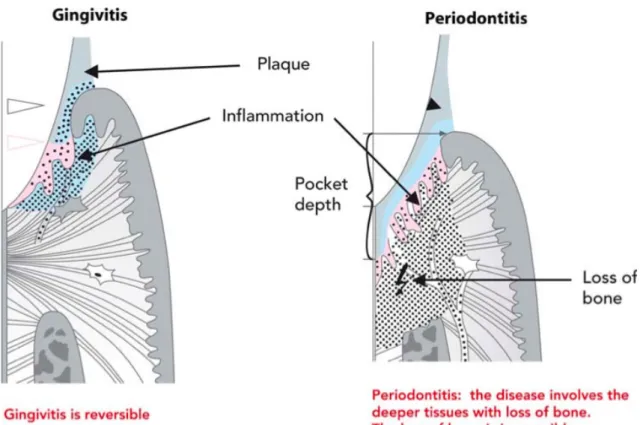
Figure 1. Representation of a chronic inflammatory infiltrate below the gingival margin (Gingivitis) and a chronic inflammatory condition affecting deeper periodontal tissues (connective tissue attachment and bone), a Periodontitis (6). ... 5
Figure 2. Ideal properties of a probiotic intended for use in disorders of the mouth (19). ... 9
Figure 3. Representation of bonding of oral bacteria on the tooth surface. The complementary sets of adhesin-receptor symbols (an example is shown at the top). Identical symbols are not intended to indicate identical molecules, but they are related functionally (45). ... 12
Figure 4. Diagrammatic representation of the effect of therapy on colonizing bacteria, the host and the habitat. (3). ... 17
The staining with CV, for quantification of biofilm biomass, includes both cells and exopolimeric matrix. As it is possible to observe in Figure 5, biofilm formation varied according to the surface topography, as the resultant staining of the anodized samples was stronger than for etched samples. ... 32
Figure 6. Crystal violet absorbance of mixed biofilm biomass formed on different titanium surfaces (Etched and Anodized). * represents the statistical differences between the two different samples. ... 32
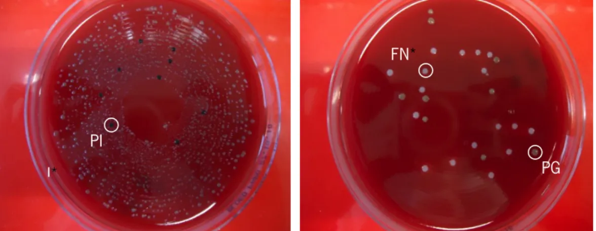
Figure 7. Blood agar plates supplemented with horse blood used to determine CFU of the 3 bacteria (PI - black, PG - green and FN - white). ... 33
Figure 8. Average values of the colony-forming units of multi-species biofilms of FN, PG and PI that grew during 8 days in anaerobic environment. The error bars represent the standard deviation and # represents the statistical differences between the colony-forming units formed for each bacteria. ... 33
Figure 9. Micrographs of anodized (a, c and e) and etched (b, c and f) Ti samples, obtained by SEM witch c, d, e and f are covered with a mixed biofilm of PI, PG and FN, c and d with a scale of 20 µm and e and f with 10 µm. ... 34
Figure 10. Blood agar plate supplemented with horse blood used to determine CFU of a biofilm formed in TI sample in the absence (left) and presence of fluoride (right). ... 35
Figure 11. Crystal violet absorbance of mixed biofilm biomass formed on different titanium surfaces (Etched and Anodized) after 8 for control and in the presence of S. salivarius. * represents the statistical differences between the two samples, # represents the statistical differences between two conditions in same type of sample. ... 36
xiv
Figure 12. Average values of the colony-forming units of multi-species biofilms of FN, PG, PI and SS, which grew during 8 days in anaerobic environment. The error bars represent the standard deviation and # represents the statistical differences between two conditions in same type of sample. ... 37
Figure 13. Growth curve of A. actinomycetemcomitans for six conditions based on different energy sources (BHI medium supplemented with glucose or the sugar under study - C7). ... 51
Figure 14. Growth curve of S. mitis, S. mutans and S. sobrinus, respectively, for six conditions based on different energy sources (BHI medium supplemented with glucose or the sugar under study - C7). ... 52
Figure 15. Growth curve of P. intermedia (left) and F. nucleatum (right), for six conditions based on different energy sources. ... 53
xv
List of Tables
1. Chapter I
General introduction
1.1. Motivation and mains objectives
Nowadays, oral health problems affect a large part of the world population (1). Oral diseases, such as dental caries and periodontal diseases are considered a major problem in our society. These diseases are the most common bacterial diseases occurring in man and greatly contribute towards the decrease oral health. Despite the numerous developments of oral products and technologies, there is still a need to know and understand how these bacterial diseases affect general health.
The use of dental implants has been increasing exponentially over the last few decades, making it essential to understand how the bacteria react and grow in these implants. Biofilm formation on oral implants can cause inflammation of peri-implant tissues (peri-implantitis), which endangers the long-term success of osseo-integrated implants. Other oral pathologies, such as dental caries and periodontal diseases, are also related to these biofilms. Therefore, the first goal of this work was to evaluate two different types of Titanium surfaces in microbial colonization, namely biofilm formation. It was also assessed the influence of the presence of fluoride and probiotic bacteria on biofilm formation.
Moreover, the influence of a specific sugar on the growth of probiotics and pathogenic bacteria on dental implants infection can also be reduced by controlling the presence of pathogenic species in oral environment, so, another aim was the evaluation of the use of a sugar (C7) as a prebiotic agent.
Therefore, the specific aims of this work were to:
- Study the biofilm formation on titanium surfaces (anodized and etched); - Evaluate biofilm formation in the presence of fluoride;
- Analyse biofilm formation by pathogenic bacteria in the presence of a probiotic bacteria; - Test the effect of surface topography on biofilm formation;
- Study of the effect of different sugars (Glucose and C7) on the growth of several oral bacteria.
This study intends to complement the general knowledge about oral health in specific in biofilm formation, oral implants and probiotics.
1.2. Introduction
Numerous oral pathologies, such as dental caries and periodontal diseases are plaque-related. These diseases are probably the most common bacterial diseases occurring in man and greatly contribute towards the decrease oral health. Dental caries and periodontal disease have historically been considered some of the most important global oral health burdens.
In the case of dental caries, this is still a major health problem in most industrialized countries affecting 60–90% of school-aged children and the vast majority of adults. Poor oral health may have a profound effect on general health. The experience of pain, problems with eating, chewing, smiling and communication due to missing, discoloured or damaged teeth have a major impact on people’s daily lives and well being. Furthermore, oral diseases restrict activities at home, at school and at work, causing millions of school and work hours to be lost each year throughout the world (2) (1).
Dental caries is considered a destructive condition of the dental hard tissues (teeth) that can progress to inflammation and death of vital pulp tissue, with eventual spread of infection to the periapical area of the tooth and beyond. This condition, if not treated, can lead to periodontal diseases and may even, in extreme cases, lead to tooth loss (3) (4).
Periodontal diseases are initiated by components of the plaque that develops on the tooth surface adjacent to the soft tissues of the supporting periodontium and may be confined to the gingiva (gingivitis) or extend to the deeper supporting structures with destruction of the periodontal ligament and the alveolar bone that supports the teeth (periodontitis). Periodontitis infections are characterized by the increased destruction of the periodontal ligament associated with detachment of collagen and consequent deepening of the pockets formed between the infected tissue and the teeth. Such infections associated with periodontal pocket formation, may ultimately lead to loosening and loss of the affected teeth (5). Depending on the immune response of the susceptible host, the presence of pathogenic bacterial species and the absence of beneficial bacteria, periodontitis can develop with more or less severity (6) (7).
Gingivitis, the most common form of gingival inflammation is a reversible inflammatory reaction of the dentogingival tissues (3). In contrast to gingivitis, periodontitis is greatly part a chronic inflammatory reaction of the same oral area but involving not only superficial gingival tissues but also periodontal ligament and the alveolar bone (Figure 1).
Figure 1. Representation of a chronic inflammatory infiltrate below the gingival margin (Gingivitis) and a chronic inflammatory condition affecting deeper periodontal tissues (connective tissue attachment and bone), a
Periodontitis (3).
The most common reported symptoms of these diseases are gingival bleeding and swelling. Other symptoms like gingival recession, drifting of teeth, mobility, and suppuration are also associated with periodontal diseases. These are signs of an advanced form of periodontitis due to progressive destruction of the dental supporting tissues. If left untreated, periodontitis results in a progressive deepening of the gingival sulcus associated to alveolar bone destruction up to the apex of the tooth, which eventually ends with its loss (8) (3) (9).
Moreover periodontitis is usually associated to polymicrobial infections of oral tissues, that result in chronic inflammation of the gingiva and surrounding connective tissue, in response to accumulations of bacteria on teeth (10).
The development of destructive periodontitis seems to be the result of a specific infection that normally starts with the formation of biofilms by specific microorganisms on tooth surfaces. The onset of these diseases is usually delayed for prolonged periods of time after initial colonization by the pathogen(s). The course of these diseases typically runs for years.
The responsible agents for colonization of oral areas, in most instances, appear to be members of the indigenous microbiota and, thus, the infections might be thought of as endogenous (3). Dental plaque biofilm formed by these endogenous bacteria on the tooth surface causes an immune response leading to the destruction of host tissues (11).
The difference between oral diseases, such gingivitis and periodontitis, and dental caries is that dental caries occurs supragingivally (on teeth above the gum line) and periodontal disease occurs subgingivally (below the gum line), attacking the tooth supporting tissues (12).
The microorganisms responsible for these disorders exhibit unique properties, conferred by their site of colonization and the nature of the environment in which they reside. They are capable to survive and to expand to other parts of the body. The microorganisms once in the blood system persist for long periods of time and they are able to adapt easily in a new environment and have high influence in other dangerous diseases (5).
1.2.1. Periodontal microorganisms
The knowledge of the complex interactions between the resident microbial communities and the human host is of extreme importance to understand the development and pathogenesis of a variety of diseases, not just the typical infectious diseases.
In the healthy oral cavity, the bacterial flora is different from that of diseased oral cavities and, often, certain indigenous bacterial species and their products are useful for a healthy periodontium (13). The commensal oral microbiota is a component of a complex homeostasis mechanism that interferes with the activity of pathogenic microorganisms (14). Commensal bacteria can affect the pathogenic species through different mechanisms, modifying the disease process by occupying a niche that could otherwise be colonized by pathogens, by actively limiting the capacity of pathogen to adhere to tissue surfaces, by affecting in a negative way the vitality or growth of a pathogen, by influencing the ability of a pathogen to produce virulence factors, or by degrading them (9). Additionally, beneficial bacteria are skilled to supply essential nutrients, regulate epithelial development, and contribute to the maturation and maintenance of the immune system (14). In other hand there are the periodontal pathogens that are the causative agents of
several periodontal diseases. For over 100 years, periodontal microbiologists have been searching for the causative agents of periodontal diseases (5).
Research indicates that there are several important species involved in the disease process, such as Treponema denticola, Prevotella intermedia, Fusobacterium nucleatum, Prevotella nigrescens, Campylobacter rectus, Capnocytophoga spp, Peptostreptococcus micros, Eikenella corrodens and several species of oral spirochaetes (15) (16) (17). Actinobacillus actinomycetem comitans, Porphyromonas gingivalis and Bacteroides forsythus, were also strongly associated with periodontal disease status (11). These bacterial species behave in a cooperative or synergistic fashion to initiate periodontitis, a mixed anaerobic infection (18). Although pathogenic bacteria are the main factor in the aetiology of periodontitis, tissue damage is also a consequence of the host response.
Periodontal pathogens have several virulence factors that allow bacteria to colonize the host and replicate, to avoid destruction or neutralization by the defence system of the host, and to finally cause tissue damage. During an infection, important virulence factors such as adhesins, lipopolysaccharides, hemolysins, proteinases and outer membrane vesicles may act alone or in combination (18). The microorganisms can cause disease directly, by invasion of the tissues, or indirectly through production of bacterial enzymes and toxins.
Besides commensal and pathogenic oral bacteria there are an important role represented by probiotic bacteria in oral health. The term probiotic is a relatively new word meaning “for life” and is presently defined as living microorganisms, principally bacteria that are safe for human consumption and, when ingested in sufficient quantities, have beneficial effects on human health (19) (20) (21). So, in the oral cavity you have pathogenic, commensal and beneficial species, probiotics can be indigenous beneficial species or can be beneficial species that are not present in the oral cavity like commensal. Probiotics have already been successfully used to control gastro-intestinal diseases, some systemic diseases, infectious diseases such as acute diarrhea and Crohn diseases and appear to performance through colonisation resistance and/or immune modulation (19) (21) (22) (23). The oral administration of probiotics has also been explored in the control of periodontal disease (24).
Given the widespread emergence of bacterial resistance to antibiotics, the concept of probiotic therapy has been considered for application in oral health. Dental caries, periodontal disease and halitosis are among the oral disorders that have been targeted. An essential condition
for a microorganism to represent a probiotic of interest for oral health is its capacity to adhere to and colonize various surfaces of the oral cavity.
The complex environment of the oral cavity that varies for each patient, and the nutrients present in their diet are important factors to be considered. The different types of sugars present in daily diet can also influence the growth of different bacteria. The possibility to control the amount of probiotic bacteria in oral environments using the different nutrients may provide beneficial effects in oral health. The human body lives in a highly contaminated bacterial environment, and symbiosis with these microorganisms seems to be a circumstance for survival (25). Although the recent availability and widespread use of effective and cheap antibiotics has encouraged the treatment of many diseases and reduction of the death rate in many countries, it has also led to the development of resistance to a range of antibiotics (21). As well as pharmacological therapy, probiotics may be a useful adjunct to conventional therapy, not a completely alternative.
Mechanisms of probiotic action within the oral cavity can possibly be suggested from gastrointestinal studies whereby the introduction of microorganisms as a therapeutic tool for the prevention and treatment of dental caries and periodontal disease could possibly act in the same way within the oral environment (26). Mechanisms could possibly include the disruption of plaque biofilm formation through competition for binding sites on host tissues and other bacteria, and competition for nutrients.
Several mechanisms have been proposed to explain how probiotics work (Figure 2). For example these bacteria secrete various antimicrobial substances such as organic acids, hydrogen peroxide and bacteriocins. The production of antimicrobial compounds inhibits some oral bacteria growth and the amount of probiotic bacteria to compete with the pathogenic agents permits a higher adhesion of probiotics on the sites of mucosa than for pathogenic agents. Probiotics can also modify the surrounding environment by modulating the pH and/or the oxidation-reduction potential, which may compromise the ability of pathogens to become established. Finally, probiotics may provide beneficial effects by stimulating nonspecific immunity and modulating the humoral and cellular immune response (20) (27).
Figure 2. Ideal properties of a probiotic intended for use in disorders of the mouth (20).
Probiotics can, not only suppress the emergence of endogenous pathogens or prevent the infection with exogenous pathogens they may also protect the host through the promotion of a beneficial host response (21). The production of hydrogen peroxide by members of the Sanguis group of Streptococci induces the reduction of organisms associated with periodontitis. These properties are consistent with the inverse proportions of oral Streptococci relative to Gram-negative anaerobes found in dental plaque. Therefore, the implantation of specific oral Streptococci or the
encouragement of their growth in dental plaque can be considered a probiotic approach for promoting the shift from a pathogenic to a less pathogenic biofilm (28).
Moreover, there are studies (29) (30) (31) (32) that examined the potential beneficial effect of some oral bacteria selected for their ability to inhibit the growth of pathogens, to down regulate fimbrial expression or biosurfactant production, for the nonappearance of co-aggregation or because of their high prevalence in periodontal health. Streptococcus sanguinis, Streptococcus salivarius and Streptococcus mitis appeared to be the bacterial species most effective in inhibiting periodonto-pathogen colonization in vitro. The inhibition is partially caused by environmental conditioning, bacterial interactions, and interaction with epithelial cells (21). S. salivarius, is known to produce bacteriocins, which could contribute towards decreasing the number of pathogenic bacteria (33) (34). The existence of probiotics in the indigenous oral microflora of humans permits exploration because these bacteria offer the advantage of being perfectly adapted to the human oral ecosystem (20).
So, the use of probiotics is an interesting emerging field in general and specifically in oral healthcare. Although various “statistically significant” improvements have been reported, but the knowledge of pathogen-host interactions and the role of benificial bacteria in preventing the emergence of pathogenic species and oral health remains obscure. There are great needs to elucidate the role of the oral beneficial microbiota and how the growth can be controlled, which requires making studies on the usefulness of probiotics to maintain or improve oral health (21).
1.2.2. Biofilm and bacterial interactions
Biofilm present on the tooth surface may be among the most complex biofilmthat exist in nature. This is due, in part, to the non-shedding surface of the tooth, which allows for the development of persistent colonization and very complex ecosystems. There is a dynamic co-existence between commensal and pathogenic bacteria and beneficial bacteria, which are protected from the natural physical and chemical antibacterial host defences in these communities (5) (35). There are several areas in oral environment that can be covered by a complex microbial community embedded in an extracellular matrix composed of polysaccharides, nucleic acids, proteins, and water, generally known as oral biofilm (36). By definition biofilm is considered a complex assemblage of microbial cells that are irreversibly attached to a surface and enclosed within a self produced protective polymeric matrix. Biofilms can form on diverse surfaces and can involve single or multiple microbial species. Usually a biofilm is highly resistant to conventional
antibiotics comparing with planktonic from of growth (37) (16).. The oral communities and consequently these biofilms can tolerate antimicrobial concentrations of 10-1000 times that the ones needed to kill planktonic counterparts and displays an inherent resistance to phagocytosis (38) (39).
The precise mechanism for antibiotic resistance remains unclear, however it is likely to be a manifestation of multiple factors. Firstly, the exopolymeric matrix secreted by biofilm bacteria plays a vital role in restricting the penetration of antimicrobials and antibodies (40) (41). Furthermore, negatively charged molecules within the matrix are capable of binding to antimicrobial agents (42). Secondly, bacteria deeply embedded within a biofilm exhibit a reduced growth and metabolic rate and thus are less permeable to antibiotics. Thirdly, inactivation of antibiotics can occur either on the biofilm surface or within the matrix itself (43) with a drug-inactivating of enzymes, such as β-lactamase that causes the degradation of β-lactam antibiotics, and its retention in the dental biofilm amplifies its barrier function (44). Moreover, there may be subpopulation of drug resistant, phenotypically and genetically different bacteria within the biofilm, as the close-knit community provides the ideal niche for the exchange of extra-chromosomal DNA (42).
Biofilm formation is the result of a succession of events, which are very well organized and complex, including the adhesion and the multiplication of the bacteria (45) (46) (47). Plaque or biofilm formation in oral cavity is described as one of highest ordered sequence of events in biofilm formation.
The process starts with acquired pellicle formation and reversible adhesion involving weak long-range physicochemical interactions between the cell and surfaces. With the formation of the pellicle, eventually, the interaction leads to stronger adhesion receptors, that mediate attachment and a succession of co-adhesions which can occur resulting in attachment of secondary colonizers to the bacteria that already are attached. Consequentially, there is bacterial proliferation and biofilm formation and eventually some detachment can happen (35) (16).
The development of a microbial community is initiated by a pioneer microbial population present on oral habitat. These microbial populations have the ability to modify the habitat and thus, new populations may develop (15). These Early colonizers, such as many oral Streptococci and Actinomyces, have the capability to bind to proteins named adhesins such as alpha-amylase, proline rich proteins, and proline rich glycoproteins that bind to receptors present on glycoproteins (e.g. mucin) in the conditioning film at oral surfaces. Steptococcus species, such as S. sanguinis, S. oralis, S. gordonii, S. mitis and S. sobrinus represent 60 to 80% of all primary colonizers, which
also include 5-30% species of Actinomyces naeslundii, Fusobacterium nucleatum, Capnocytophaga ochraceae. Different adhesins are present in the adherence of Streptococcus species and acquired pellicle. S. sanguinis and S. oralis possess adhesins similar to lectine cellular membranes, which are called lectins. Additionally, S. gordonii presents more than one adhesin that binds at least to three receptors, namely proline-rich proteins, salivary agglutinins and saliva amylase (48) (49).
Several diagrams show the different “congregate” pairings between the bacteria found in the construction of dental plaque. In Figure 3 it is possible see a simplified diagram about this bacteria aggregation (46) (50).
Figure 3. Representation of bonding of oral bacteria on the tooth surface. The complementary sets of adhesin-receptor symbols (an example is shown at the top). Identical symbols are not intended to indicate identical molecules,
but they are related functionally (46).
The partnerships between dental plaque bacteria are highly specific and primary colonizers can interact and connect with each other but not usually with secondary colonizers. However, the major periodontal pathogen P. gingivalis, a secondary colonizer, can connect with primary
colonizers. One of these early colonizers is F. nucleatum that is proposed to be a bridge organism because it can bond with both primary and secondary colonizers (51). In the absence of F. nucleatum many other secondary colonizers cannot become part of dental plaque community (52). Additionally, anaerobic secondary colonizers cannot survive in planktonic plaque unless congregated to F. nucleatum. Thus, the multiplicity of its congregation interactions and it role as a bridging organism could make F. nucleatum an essential organism in the development of dental plaque (46).
Successful colonization of new environments requires several important factors including nutrient supply, an environment conducive to proliferation and an environment with limited potential hazards. Biofilms develop in a vast array of differing environments and thus the structural composition of the biofilm and the extracellular polymeric substances will vary accordingly. The ability to incorporate hydrogen bonding makes the exopolymeric matrix a high hydrated structure. In addition to polysaccharides and water, a wide variety of proteins, glycoproteins, glycolipids and extra-cellular DNA are also present (53).
Bacteria establish interactions between individual cells within one population or between different bacterial populations forming a diversified and complex community, and between them positive and negative interactions can occur. The balance of these interactions is responsible for maintaining the ecological homeostasis within the community. The positive interactions include the relationship between two species where both will benefit from the association (mutualism), when only one of the species benefits whereas the other one obtains nothing from the association (commensalism), and when the interaction between the two microbial species have a greater effect than the sum of the effect of both species taken individually (synergism - mutual adhesion, nutrient cross feeding, complementation in macromolecule hydrolysis, defences against host antibacterial factors). On the other hand the negative interactions include competition, characterized by two populations competing for multiplication and survival and they try to occupy a particular site or obtaining specific nutrients and antagonism, as example when a bacterial population secretes products (hydrogen peroxide, bacteriocin, organic acid) that inhibit other populations or negatively alter environmental conditions (pH, oxidation-reduction potential) (28).
According to what was detailed previously, biofilm microorganisms are held together and protected by a complex matrix of excreted polymeric compounds, the exopolymeric matrix. This matrix functions mainly to protect the microorganisms within, as well as to facilitate intercellular communication (37). This means, microorganisms within a biofilm community actively
communicate through a cell-to-cell signalling system. Therefore, a chemical communication process among them, known as quorum sensing, represents an important bacterial function. Quorum sensing is defined as gene regulation in response to cell density, and it influences biofilm formation, acid tolerance, and virulence. Quorum sensing can occur within a single species as well as between diverse species, and is known to regulate different processes, essentially serving as a simple network communication.
Other way of interaction and communication is by metabolic communications. Saliva, gingival crevicular fluid, food containing sugars, food debris, and metabolic products of other bacteria are the sources of nutrients for oral bacteria and the excretion of metabolites by microorganisms can be used as a nutrient by different species, or the breakdown of a substrate by the extracellular enzymatic activity of one organism, producing available substrates for different organisms, represent metabolic communications among oral bacteria (54). Regulation by inhibitory metabolites is also a kind of interaction. Some bacteria are able to produce bacteriocins that are proteinaceous bactericidal substances that inhibit the growth of closely related bacterial species or strains. The competition through bacteriocin production has been documented for many oral bacteria and this event may regulate the way bacteria interact between them (44).
The oral cavity is exposed to an aerobic environment, so it is likely that oral anaerobic bacteria encounter residual amounts of oxygen in the early stages of biofilm development and periodontal pocket formation. The survival of anaerobic bacteria depends on the specific tolerance of each species to oxygen (through enzymes such as superoxide dismutase, oxidase/peroxidase, and catalase) and the bacterial interactions within the biofilm community (44). Furthermore, the metabolism of aerobic and oxygen-tolerant, species may reduce the concentration of oxygen to levels that can be detoxified by the need of anaerobic bacteria (16).
Furthermore, the detachment of cells from biofilms is essential to allow colonization of new habitats by these bacteria. However, it is probably the least well-understood biofilm phenomenon (55). Studies say that detachment of biofilm cells can be caused by either external or internal biofilm factors. External forces include physical shearing or erosion, sloughing and increased flow velocity for biofilms at liquid interface. Internal biofilm factors are thought to result from reduced nutritional levels or oxygen depletion. These occur by processes such as quorum sensing, endogenous enzymatic degradation, the release of exopolymeric matrix or binding proteins (37). Dispersal strategies include the shedding of individual daughter cells from a micro-colony, the
release of aggregates of biofilm cells or surface dispersal in which cells move across a surface via gliding or twitching motility (49) (56).
There are several reasons why biofilms are a preferred mode of existence for microorganisms. First of all, the bacteria form biofilms as a means of defence, in response to stressful environments such as high shear forces, host defences and deficit nutrients. Another reason for biofilm formation is because this way bacteria can live in very resistant community and they are able to remain in a favourable niche.
Despite the preference of microorganism to form this type of structures, there are many factors affecting biofilms formation. During all the phases of biofilm formation there are always physical and chemical factors interfering in this process. Regarding the attachment of biofilms to a surface is evident the influence of both physical and chemical factors. Physical properties, such as the topography, roughness of the surface, can increase surface area and hence increase colonization and chemical conditions can also reduce the accumulation of bacterial cells in biofilm. Roughness also provides protection from shear forces but increases the difficulty of cleaning (5) (57). It is the case of dental plaque formation, for example, starts in cracks, grooves and irregularities of the tooth surface or tooth implants where the initial colonizing bacteria are protected. Moreover, supragingival plaque formation, after initial colonization has occurred, was shown to occur more rapidly on a roughened surface (5). In metals such as titanium implants, biofilm formation and consequent plaque accumulation occurs especially around the abutment. This may eventually lead to peri-implantitis, an inflammatory reaction with subsequent loss of osseointegration at the dental implant interface (58) (59), causing a loosening of the fixture and, ultimately, the implant would have to be removed (4) (60). Henceforth, these are two important conditions that seem interesting to study. The chemical composition of a surface also has impact on bacterial colonization since it may contain beneficial or detrimental components. For example the influence of the chemical composition of a surface in biofilm formation is related with the dental pellicle on the teeth that may coat the surface and influence colonization. The role of conditioning films on microbial attachment is unclear, but it has been proposed that the strength of the biofilm depends on the cohesiveness of the conditioning film rather than direct bacterial contact with the bare surface (57). The liquid medium surrounding the surface, for example, saliva surrounding the teeth, also influences bacterial attachment and biofilm morphology (5). The existence of various different micro-areas, e.g. tongue, teeth, restorative materials, and gums,
micro-gaps and retentive areas at dental implant interfaces are the most susceptible areas for oral biofilm formation (61).
Moreover, the use of dental implants has been increasing exponentially over the last years, reinforcing the need of their study regarding microbial colonization. One of the most used materials in dental implants is the Titanium (commercially pure titanium), the interest in this material have been increasing over the years. Titanium has excellent proprieties such as good corrosion resistance, biocompatibility, low density, low thermal conductibility, good resistance, low weight and low cost. This material can be submitted to diverse treatments and casting techniques in other to have a better performance. So, the control of the surface is important because this fact affect, in large scale, the adhesion and biofilms formation particularly in plaque-related biofilms (62).
Other important effect is the presence of fluoride on oral environments. Current evidence indicate that fluoride has a multitude of direct and indirect effects on bacterial cells. These include inhibitory effects of fluoride on glycolysis and transport of carbohydrates, enzyme activities, macromolecular synthesis and polysaccharide formation and degradation. Fluoride is well documented as an anticariogenic agent, that involves a variety of mechanisms including demineralization, the enhancement of remineralisation, the interference of pellicle and plaque formation and the inhibition of microbial growth and metabolism (63). So, fluoride has been used to help the control of incidence of caries and to decrease the dentin sensibility and oral plaque formation. The results of many studies confirm that fluoride from the substratum affected fluoride-sensitive biofilms and reduce the risk of plaque formation responsible for many oral diseases (64) (65) (66).
1.2.3. Relation between oral health and other diseases
It has been suggested that there is association between the oral microbiota and other diseases, such systemic diseases, cardiovascular disease (including coronary diseases, myocardial infarction, bacterial endocarditis), complications during pregnancy, chronic diseases (e.g arteriosclerosis) and aspiration pneumonia diseases (67) (68) (23) (10) (11) (69) (70) (71).
The relationship between oral health, specifically periodontal disease, and cardiovascular and respiratory diseases has been subject of ongoing research and supported by many studies that have reported the association between these diseases and periodontal infections. Studies show that infections can be caused by periodontal pathogens like Aggregatibacter
actinomycetemcomitans and P. gingivalis and these may be associated with future stroke, increased risk of myocardial infarction, and acute coronary syndrome (72).
Chronic infections start with inflammation, so periodontitis and gingivitis might influence systemic or/and vascular inflammation processes. The source of bacterial pathogens responsible for the most prevalent chronic infections affecting humans, dental caries and periodontal diseases, derives from the biofilms present in tooth surfaces. These biofilms are one of the most complex existing in nature, due in part to the non-shedding nature of tooth surfaces that allow the development of a persistent bacterial colonization and to the rather complex ecosystems that exist in the oral cavity (48) (3). Bacteria themselves, once in the blood stream may cause distant site infections and the products from bacteria can stimulate systemic inflammation that would eventually act directly and/or indirectly on the vascular walls inducing a state of endothelial dysfunction
So, bacteria that reside in the subgingival biofilm may disseminate systemically and influence directly or indirectly the site of inflammation causing these type of systemic diseases.
In other to avoid greatest health problems, treatment of oral diseases should start as soon as possible. Treatment can affect bacteria directly by physical removal and/or with chemotherapeutic agents (Figure 4) (3).
Figure 4. Diagrammatic representation of the effect of therapy on colonizing bacteria, the host and the habitat. (5).
Dental biofilms can be altered by various therapies providing a beneficial outcome to the patient and treatment can affect the composition of the bacterial plaque directly, can affect the
host response or alter the habitat for example, by eliminating or by meticulously removing supra-gingival plaque. Alterations of any of these factors can impact on the remaining factors.
Therefore, improvement of oral hygiene has been shown to reduce the occurrence of these diseases. The consequences of poor oral health linked with advanced age, common co-morbidities such as diabetes, concurrent medications and a state of immune dysfunction that may increase the risk for systemic consequences of periodontitis and other oral and dental pathologic conditions. Thus, oral hygiene assumes an important role in the care of high-risk subjects.
Besides, colonization by pathogenic periodontal bacteria is a risk factor for an implantation of a periodontal implant and the accumulation of biofilms can promote periodontal inflammation of the mucosal soft tissues surrounding the implant, while peri-implantitis also affects the supporting bone in the subgingival area, resulting in a rapid bone loss. Peri-implant diseases (peri-implantitis and peri-implant mucositis) are major clinical problems that appear in patients possessing osseointegrated dental implants that can, ultimately, result in the loosening of the implant (73).
2. Chapter II
The use of dental implants has been increasing exponentially over the last few decades, making it essential to understand how bacteria grow and react in these materials. Thus, the goal of this chapter is the evaluation of the effect of different types of Titanium surfaces in microbial colonization, namely biofilm formation. Moreover, it also aims to study the influence of the presence of fluoride and probiotic bacteria on pathogens' biofilm formation.
2.1. Introduction
Osseointegrated titanium implants have become an important alternative to conventional prostheses, increasing significantly the quality of life of patients. On the other hand, with the increasing demand for dental implants, its failure is also being reported more frequently (74) (75) (76) (77) (78). Nowadays, oral implants are used not only to replace missing teeth, but also to provide anchorage during orthodontic treatments, to rebuild the craniofacial skeleton or simply for aesthetic circumstances (79). It is estimated that more than 2 million dental implants are placed annually (73). Thus in the near future an increase of this number is expected not only due to an increasingly aged population, but also because implant therapies have become highly successful (80), with implant survival rates above 89 % after 10/15 years (81).
However, it is still necessary to study and understand the causes of dental implant failures. These are defined as implants that exhibit clinical mobility, pain on function, bone loss more than half of the total length of the implant, or uncontrolled exudates (82). The timing at which implant failures occur represents different physiological processes. An early implant failure indicates an initial lack of osseointegration due to an inability to establish an intimate bone-to-implant contact. Various factors may contribute to early implant failures such as premature loading, surgical trauma, or impaired healing response (83). Late failure, on the other hand, occurs after initial integration, physiological remodeling and loading. Causes of late failures include overloading and bacterial infection (e.g., peri-implantitis) with most failures occurring after the first year of loading. In fact, biofilms have been associated with almost 65% of infectious diseases such as periodontal and peri-implant diseases leading to implant failure (84) (85).
The role of bacterial biofilm in peri-implant diseases has been recognized, so, the knowledge on the microbiology around dental implants is essential of the essence for adequate diagnosis and treatment of these diseases. Thus, this chapter focuses on understanding the development of oral biofilms in titanium samples that are used in dental implants and some interactions between periodontal bacteria.
2.1.1. Formation of biofilm on dental implants
The oral cavity represents a perfect fluid system in which the microbiota, present in saliva, may colonize on teeth and artificial surfaces following the deposition of a glycoprotein-containing pellicle (86). This pellicle is derived from components in the saliva, as well as bacterial and host
tissue products. It acts as a substrate for bacterial colonization, which occurs as early as 30 minutes after implant exposure in the oral cavity (87). The pellicle is formed after the exposure of an implant in the oral cavity through a transmucosal abutment. Then, the selective adsorption of the environmental macromolecules such as a-amylase and serum albumin occurs (88).
In comparison to natural teeth, the acquired pellicle on dental implants has a lower albumin adsorption capability, which according to some authors (85), contributes to the lower plaque formation around implants.
Some studies have shown that an increase in titanium surface area structure and the surface free energy also facilitates the formation of bacterial biofilms (9). Both adsorbed salivary proteins and implant surface structures contribute to the early colonization of oral titanium implants (89) (90) (91) (76). If bacteria can attach themselves directly to an inert titanium surface, this may have consequences leading to infection of the peri-implant tissues (92).
It is well known that the formation of bacterial biofilm is an important factor in the infection of medical devices (93). It appears from other studies that at least the transmission of Porphyromonas gingivalis and Prevotella intermedia from the periodontal pocket to the peri-implant region is possible (94). Data suggests that shortly after the installation of titanium implants a sub-gingival microbiota dominated by Peptostreptococcus micros, Fusobacterium nucleatum, and P. intermedia is established (95). In addition, recent data suggest that colonization on different sub-implant surfaces occurs quickly (96) (97) (87) (32).
The infectious aetiology of peri-implantitis is well established (i.e. Mombelli et al. 1988; Roos-Jansa ker et al. 2003) and studies have shown that high levels of periodontal pathogens such as Aggregatibacter actinomycetemcomitans (former Actinobacillus actinomycetemcomitans), Porphyromonas gingivalis, P. intermedia, Tannerella forsythia and Treponema denticola, have been associated with peri-implantitis (75). Peri-implant infections may also include F. nucleatum and actinomyces species (98). In addition, Staphylococcus aureus and enterococci spp. have been related to peri-implant infections (99). Whether the surface characteristics of titanium implants influence the microbiota is poorly understood.
It is possible that unknown bacteria are involved in the emergence of the lesions and the pockets around the remaining teeth may act as a bacterial reservoir. The composition of the peri-implant microbiota is likely to be similar to that around teeth. However, few studies have evaluated the differences in bacterial composition between dental implants and remaining teeth in the same subjects (74).
Recent studies indicate that soon after dental implantation and/or restoration, biofilm formation and consequent plaque accumulation occurs on titanium implants, especially around the abutment. This may eventually lead to peri-implantitis, an inflammatory reaction with subsequent loss of osseointegration at the dental implant interface (59) (4), causing a loosening of the fixture and, ultimately, the implant would have to be removed (100).
In conclusion, biofilm formation on medical implants presents three major problems, which in the end can lead to implant failure.
2.1.2. Titanium in dental implants
Commercially pure titanium (Ti) and titanium alloys are the first choice for oral prosthesis, mainly due to their biocompability and excellent mechanic characteristics (101).
A range of titanium surfaces with different composition and roughness has been developed for dental implants. Moreover, the topography of titanium dental surfaces is of major importance for microbial colonization (90) (91) (76). However, much controversy still exists as to the optimal features for implant surfaces regarding both osseointegration and antimicrobial kinetics. In addition, higher surface roughness profiles may as well lead to an increase in ionic leakage to adjacent tissues, thus presenting major risks (102).
Increased surface roughness has been associated with increased osseointegration of the dental implant (103). Conversely, a higher surface roughness increases biofilm formation (91), and thus contributes to spontaneous progression of peri-implantitis lesions (104).
Additionally, titanium also denotes a strong osseointegration tendency by the development of close bone-to-implant apposition after short periods of implantation, an important feature for permanent bone-interfacing implants. However, an ideal dental implant material should not only integrate with the host tissue, but also exhibit anti-bacterial properties (105).
2.1.3. Influence of Fluoride in biofilm formation
The effect of fluoride on oral bacteria has been studied extensively over the last 20 years and the current evidence indicates that fluoride has a multitude of direct and indirect effects on bacterial cells. These include inhibitory effects of fluoride on glycolysis and transport of carbohydrates, enzyme activities, macromolecular synthesis, and polysaccharide formation and degradation. Studies, both in vivo and in vitro, have been made in an attempt to elucidate the
potential for these actions of fluoride to contribute to the control of plaque and caries. Various models have shown that fluoride may influence plaque accumulation, acid production and enamel demineralization (64).
The antimicrobial activity of fluoride is well documented by a considerably amount of literature (106) (107) (108). The mechanisms by which fluoride may interfere with bacterial metabolism and dental plaque acidogenicity. Furthermore, intracellular or plaque associated enzymes such as acid phosphatase, pyrophosphatase, peroxidase and catalase may be affected by fluoride ions. Although even low fluoride levels may reduce bacterial growth and formation of dental plaque but the affection of plaque metabolism by fluorides is still unclear (109) (107).
2.1.4. Treatment of dental implant-associated infections
Treatment of infections associated with dental implant and biofilms consists in mechanical debridement of the implant surface or chemical treatment including local and systemic antibiotics.
The selection of treatment depends on the established diagnosis of peri-implant mucositis or peri-implantitis. Treatment success is assessed using outcome measures, such as reduction of inflammation, probing depth, and pathogenic bacteria (110). Nonetheless, the presence of specific bacteria had little or no value in predicting treatment failure (111).
In a recent literature review, non-surgical mechanical therapy was effective in treating peri-implant mucositis with improved results observed in conjunction with an antimicrobial mouth rinse (84). A reduction in the proportion of pathogenic species after mechanical therapy has been reported (100).
However, nonsurgical treatment of sites with peri-implantitis was not found to be effective at reducing inflammation, pathogenic microorganisms, and bleeding on probing. The addition of antimicrobial mouth rinse in this nonsurgical treatment of peri-implantitis only provided minimal beneficial effects (82) (84). On the other hand, the use of local drug delivery such as minocycline and tetracycline to treat peri-implantitis generated reduced levels of T. forsythia, P. gingivalis, and T. denticola, with the most effect on A. actinomycetemcomitans (85).
In the past decades, laser therapy such as diode, CO2, and Er:YAG laser as gained popularity
based on the rationale of surface decontamination, hemostatic properties, calculus removal, and bactericidal effects (112) (113). However, only minor clinical and microbiological improvement has been reported (84).
These are some the conventional treatments and therapies. There are numerous studies purporting others protocols to improve oral health and decrease the mentioned oral diseases (85).
2.1.5. Probiotics as improvement for oral health
Probiotics have been found to be beneficial to the host by improving the endogenous flora. Traditionally, probiotics have been associated with gastrointestinal tract, however recently several lines of research have suggested use of probiotics for oral health (20).
There are a numerous reasons why probiotic research has become a famous topic in medicine. Despite over 50 years of antibiotics, infectious diseases remain a major health problem, creating multi-drug-resistant bacteria, while pathogenic microorganisms are being linked with induction or worsening of many chronic diseases. Moreover the alarming spread of infectious diseases, leads scientists and industries to look for new approaches to health restoration and retention. Science itself is playing a major role, with an ever-growing number of studies providing concrete evidences that probiotics can alleviate some disease processes (114) (29).
In the field of periodontal healthcare, probiotics might provide opportunities related with the current view on the aetiology of plaque-related periodontal inflammation. This aetiological view considers three factors that determine whether disease will develop in a subject: a susceptible host, the presence of pathogenic species and the reduction or absence of some bacteria called ‘‘beneficial bacteria’’.
It is difficult to influence the host response because traditional periodontal therapies are focused on the reduction of the bacterial threat using mechanical technics or chemical treatments (115). These applied treatments strategies are based on a mechanical subgingival debridement or use of local or systemic antibiotics, in combination with improved oral hygiene (116). These changes the subgingival microbiota to a less pathogenic composition, which is characterized by high proportions of Gram-positive aerobic species and low proportions or absence of periodontopathogens (117). Unfortunately, it is currently unclear the necessary decrease of the proportion of pathogens or the necessary increase on Gram-positive aerobic species needed to increase to consider a subgingival biofilm as not pathogenic (29). The real question is related with the duration of the treatment in other to consider that the amount of pathogenic bacteria is safe and the equilibrium between pathogenic and probiotics is already enough.
In the oral cavity, probiotics can build a biofilm, acting as a protective coating for oral tissues against oral diseases. This biofilm keeps bacterial pathogens out of oral tissues by occupying a site
that pathogens would invade in the absence of the biofilm and competing with cariogenic bacteria and periodontal pathogen growth (118).
So, probiotic bacteria can provide health benefits to the host by: providing nutrients and cofactors to the host, competing directly with pathogens, interacting with the pathogen virulence factor and stimulating the host immune response (119).
Usually streptococci, lactobacilli, or bifidobacteria are included in probiotics which not only suppress the emergence of endogenous pathogens or prevent superinfection with exogenous pathogens.
Recently, it has been reported that probiotics could protect the periodontium through the promotion of a beneficial host response. In other words, when Streptococcus salivarius, Streptococcus sanguinis, and Streptococcus mitis were used, this could reduce the interleukin-8 epithelial responses to Aggregatibacter actinomycetemcomitans (formally Actinobacillus actinomycetemcomitans) (120) (21).
However the mechanism behind the successful inhibition of periodontopathogen recolonization remains hypothetical. Several possibilities such as, the occupation or a physic-chemical alteration of the subgingival niche, competition for essential nutrients, inhibition of the viability or growth of pathogens, and modification of the production or degradation of virulence factors of pathogens or immune responses, are being considered as the main underlying mechanisms (120)
2.2. Materials and Methods
2.2.1. Sample preparation
Commercially pure titanium (Ti) (grade 2) (Goodfellow Cambridge Limited, England) samples were cut from the same original plate in square form (20x20x2 mm) and were gently provided and cleaned by the Research Group on Functionalized Materials and Surfaces Performance of University of Minho. Grade 2 titanium was selected as the most common grade of titanium used in dentistry. Two different surfaces topographies were used: Etched and Anodized. Etched Samples were cleaned during 15 min in warm water (60ºC) in an ultrasonic bath. For the anodized samples the cleaning process consisted out of an ultrasonic bath with propanol for 10 min and a 5 min of immersion with distilled water.
All the samples were dried at room temperature and kept in a desiccator. Before each assay samples were sterilized in a steam autoclave at 121 °C for 20 min, at 1 atm.
2.2.2. Bacterial Culture
In other to create a bacterial multispecies environment for biofilm formation three different pathogenic oral bacteria, Prevotella intermedia (ATCC 25611), Porphyromonas gingivalis (ATCC 33277) and Fusobacterium nucleatum (ATCC 10953), were used. In some assays one probiotic, Streptococcus salivarius (ATCC 7073), was also used. These bacteria were maintained on blood agar plates (Blood Agar Base II, Oxoid, Basigstoke, UK) supplemented with 5% of horse blood (Biotrading, Keerbern, Belgium). Streptococcus salivarius was incubated at 37ºC in an atmosphere with 5% of CO2. While for P. intermedia, P. gingivalis, F. nucleatum growth was performed in
anaerobic conditions in anaerobic jars containing 80% N2, 10% CO2 and 10% H2 (Anoxomat, the
Netherlands) which were incubated at 37ºC.
One day before the each experiment, the bacteria were collected from the blood agar plates and were grown overnight in Brain Heart Infusion (BHI) broth (Difco Laboratories, Detroit, USA), in the same conditions.
2.2.3. Culture media and solutions
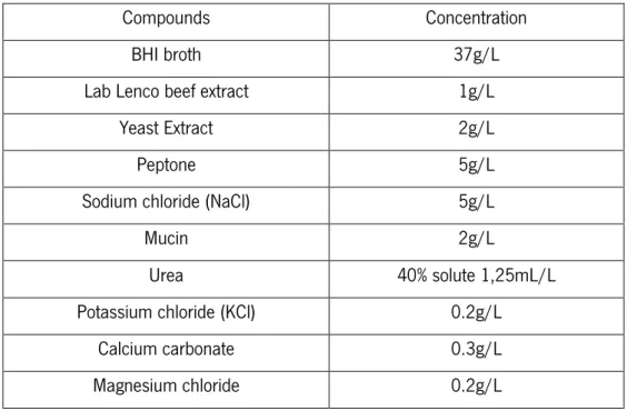
An artificial saliva solution was used in this work, as culture medium for the biofilm formation (Table 1).
Table 1. Composition of artificial saliva
Compounds Concentration
BHI broth 37g/L
Lab Lenco beef extract 1g/L
Yeast Extract 2g/L
Peptone 5g/L
Sodium chloride (NaCl) 5g/L
Mucin 2g/L Urea 40% solute 1,25mL/L Potassium chloride (KCl) 0.2g/L Calcium carbonate 0.3g/L Magnesium chloride 0.2g/L
The culture medium was prepared by dissolving all the components, mentioned in the table 1, in distilled water.
To evaluate the effect of fluoride (F-) in biofilm formation, in the specific assays, a solution of
sodium fluoride (NaF) with a concentration of 0.5g/L was added to the artificial saliva medium, simulating fluoride concentrations in the oral cavity[].
In order to distinguish the different bacteria, several selective media were prepared:
(i) phenylethyl alcohol agar (PAA) enriched with 5% defribinated horse blood - S. salivarius;
(ii) Crystal Violet Erythromycin agar plates (CVE) (Difco, Detroit) - F. nucleatum;
(iii) blood agar plates (Difco, Detroit) enriched with 5% defribinated horse blood, 10 µL/mL haemin, 1 µL/mL menadione and 30 mg/L gentamycine, L-Cystine 0.4g/L, kanamycin 0.1g/L and Vancomycin 7.5mg/L (BAKV) - P. intermedia;
(iv) blood agar plate (Difco, Detroit) enriched with 5% defribinated horse blood, 10 µL/mL haemin, 1 µL/mL menadione and 30 mg/L gentamycine (BAP) - P. gingivalis.