Printed version ISSN 0001-3765 / Online version ISSN 1678-2690 http://dx.doi.org/10.1590/0001-3765201620150874
www.scielo.br/aabc
Capsular Contracture In Silicone Breast Implants: Insights From Rat Models
VIlBeRto J. VIeIRa1
, aRMando d’aCaMpoRa1
, FeRnanda S. neVeS2
, paulo R. MendeS3 , ZulMaR a. de VaSConCelloS3, RodRIgo d’eça neVeS3 and ClaudIa p. FIgueIRedo2
1
Programa de Pós-Graduação em Ciências Médicas, Centro de Ciências da Saúde, Universidade Federal de Santa Catarina, Rua Prof. Maria Flora Pausewang, s/n, Campus Universitário, Trindade, 88040-970 Florianópolis, SC, Brasil
2
Faculdade de Farmácia, Universidade Federal do Rio de Janeiro, Av. Carlos Chagas Filho, 373, 21941-902 Rio de Janeiro, RJ, Brasil
3
Serviço de Cirurgia Plástica e Queimados do Hospital Universitário, Universidade Federal de Santa Catarina, Rua Prof. Maria Flora Pausewang, s/n, Campus Universitário, Trindade, 88040-970 Florianópolis, SC, Brasil
Manuscript received on December 10, 2015; accepted for publication on February 4, 2016
aBStRaCt
Breast augmentation with silicone implants is one of the most common procedures performed by plastic
surgeons around the world. Capsular contracture is a frequent complication in breast augmentation and
reconstructive surgery, that requires invasive intervention. The inflammatory response to implanted
mammary prostheses appears to be directly associated to capsular contracture. This review discusses the
evidences from rat models studies, on the role of inflammation and fibrosis in capsular contraction and its
relation to silicone breast implants surface.
Key words: capsular contracture, silicone breast implant, breast augmentation, smooth silicone implants,
textured silicone implants, polyurethane-coated silicone implants.
Correspondence to: Claudia Pinto Figueiredo E-mail: [email protected]
IntRoduCtIon
Plastic surgery had robust advances in the last
decades, with the development of new surgical
techniques and new materials used as substitutes
of organs and tissues. Breast augmentation with
silicone implants is one of the most common
procedures performed by plastic surgeons in
the world (Sukhova et al. 2012). Few medical
materials were studied on their safety as rigorously
as the silicone gel implants (Barnsley et al. 2006,
Gampper et al. 2007, Spear et al. 2006). Silicone
has been widely used in many areas of medicine
demonstrating its biosafety and biocompatibility
(Sukhova et al. 2012, Barnsley et al. 2006).
Five different generations of silicone implants have
been developed (Maxwell and Gabriel 2009,
Pitan-guy 1991), as demonstrated in Table I.
The first generation of implants (1962-1970)
was characterized by a dense and viscous silicone
gel, surrounded by a thick, smooth implant shell.
Second generation (1970-1982) was rounder,
with less cross-linked gels (less viscous) covered
by a smooth, thinner and slightly permeable shell
(Calobrace and Capizzi 2014). In order to reduce
capsular contracture, third generation implants
(1982-1992) came with a more viscous gel and
thicker either smooth or textured shell, and a
less permeable low-bleeding elastomer barrier
(Maxwell and Gabriel 2009). When the textured
surface came up, the fourth-generation devices
arised (1993 to present) (Adams 2009a). Texturing
of implant surface was due to the experience with
polyurethane (PU)-coated foam implants, which
indicated that rough implants resulted in lower
capsular contracture rates. Finally, cohesive
silicone gel-filled implants can be considered
fifth-generation devices (Adams 2009a, Maxwell and
Gabriel 2009) (Table I). The updates in progressive
generations have correlated with a decreased
incidence in capsular contracture, although it is not
clear if this is entirely because of implant design
(Bengtson et al. 2007, Cunningham 2007, Danino
et al. 2001).
Besides the evolution of silicone implants and
surgical procedures, breast augmentation is still
associated with complications such as hematoma,
seroma, infection, rupture, silicone leakage,
changes in mammary sensitivity, chronic pain,
poor positioning, wrinkling skin, and capsular
contracture (Barnsley et al. 2006, Spear et al.
2006, Thorek 1942., Harris 1961, Lalardrie and
Mouly 1978, Garcia et al. 2002, Edgerton and Mc
1958, Demergian 1963, Calnan 1970). Capsular
contracture is the formation of a fibrous scar
tissue that surrounds a foreign body or surgically
implanted device (Adams et al. 2006). Artificial
taBle I
Evolution of silicone gel-filled breast implants.
Implant generation period Characteristics
First Generation 1962-1970
Thick, two-piece shell Smooth surface Anatomically-shaped (teardrop)
Viscous silicone gel
Second Generation 1970-1982
Thin, slightly permeable shell Smooth surface
Round shape Less viscous silicone gel
Third Generation 1982-1992
Thick, strong, low-bleed shell Smooth surface
Round shape More viscous silicone gel
Fourth Generation 1993-present
Thick, strong, low-bleed shell Smooth and textured surfaces Round and anatomically-shaped More viscous (cohesive) silicone gel
Fifth Generation 1993-present
Thick, strong, low-bleed shell Smooth and textured surfaces Round and diverse anatomical shapes
joints, heart valves, central venous catheter ports,
breast implants, and many additional medical
devices have been involved in capsule formation
and its adverse consequences (Adams et al. 2006).
Capsule formation plays an important role in the
host response against a foreign body. Therefore, the
outcome of this process may pose a serious health
risk and/or aesthetic sequelae (Adams et al. 2006).
Mammary prostheses can induce inflammatory
responses, periprosthetic fibrous capsule formation
and implant encapsulation. Eventually, if capsular
contracture occurs another surgical intervention is
required for implant removal (Balderrama et al.
2009). Capsular contracture remains one of the
most common complication and a leading cause
for patient dissatisfaction of both aesthetic and
reconstructive breast implant surgery (Lee et al.
2014, Wong et al. 2006).
Predisposition to hypertrophic scar and
declining age have been associated to capsular
contracture, hematoma and silicone gel bleed
(Adams 2009b, Vieira et al. 2010). Several
studies also evidence the link between subclinical
infection and capsular contracture occurrence
(Bergmann et al. 2014, Del Pozo et al. 2009, Dobke
et al. 1995). In the same line, many studies support
that bacterial biofilms on breast implants, most
commonly formed by Staphylococcus epidermidis,
can promote chronic inflammation, and stimulate
fibrosis and capsular contracture (Rieger et al.
2013, van Heerden et al. 2009, Dobke et al.
1995, Netscher 2004). Del Pozo et al. (2009), in a
prospective observational assay verified bacterial
infection in breast prostheses from 45 patients who
underwent implant removal for reasons other than
overt infection. The results reveal a significant
association between capsular contracture and the
presence of skin flora bacterias on the implant
(Del Pozo et al. 2009). Recently, Bergmann et al.
(2014) used a rat model to evaluate the influence of
controlled infection in capsular formation around
PU-coated silicone implants, and demonstrated that
both implant surface and bacterial contamination
impact the architecture of capsule formation
(Bergmann et al. 2014). This approach showed
that bacterial infection leads to thicker capsules
with increased inflammatory response evidenced
by the higher amount of inflammatory cells within
the tissue (Tables II and III) (Bergmann et al.
2014). However, Mendes et al. (2008) also using
a rat model, observed no relationship between
Staphylococcus epidermidis infection and capsular
architecture (Table II and III) (Mendes et al. 2008).
Therefore, the possible relationship of bacterial
infection and capsular contracture occurrence
needs further study.
Many studies are committed in unveiling not
only the causes but also the possible preventive
strategies to capsular contracture (Moreira et al.
2009, Poeppl et al. 2007, Ibrahim Canter et al.
2007, Bern et al. 1992, Vieira et al. 2010, Adams et
al. 2006). As showed in Table I, silicone implants
underwent several structural adjustments in the last
decades in order to diminish foreign body reaction
and consequently reduce capsular contraction
incidence (Balderrama et al. 2009). Among other
modifications, smooth surface implants were
replaced by textured linings or PU-coating (Lyras
1993). However, despite these changes in implant
surface being reported by many authors as crucial
in reducing capsular contracture, this approach
is controversial (Pollock 1997, Spear et al. 2000,
Ersek 1991). The real cause of capsular contracture
remains elusive (Rohrich et al. 1999). This review
discusses the evidences obtained from rat models of
silicone breast implants, on the role of inflammation
and fibrosis in capsular contraction pathogenesis
and their relation to the silicone prosthesis surface.
ExPERIMENTAL RAT MODELSFOR CAPSULAR CONTRACTURE STUDY
taBle II
Summary of experimental approaches used to study capsular contracture after implantation of silicone mini-prostheses in rats.
Reference title Study design
Bastos et al. 2007
“Histologic analysis of zafirlukast’s effect on capsule formation around silicone implants”.
animals: 32 adult female Wistar rats.
groups: 3 groups that received both smooth and textured surface silicone mini-implant.
Intervention: Intraperitoneal treatment with either vehicle or zafirlukast
(1.25 or 5 mg/kg/day; for 90 days).
Follow up: 90 days.
Capsular evaluation: Histological (HE, trichrome and picrosirius red stain), and immunohistochemistry (anti-α-SMA antibody) analyses.
Bastos et al. 2012
“Effect of zafirlukast on capsular contracture around silicone implants in rats”.
animals: 40 adult female Wistar rats.
groups: 4 groups that received both smooth and textured surface silicone mini-implant.
Intervention: Intraperitoneal treatment with either zafirlukast (1.25 mg/kg/
day; during 90 days) or vehicle. Follow up: 90 days.
Capsular evaluation: Intra-implant pressure measurement.
Bergmann et al. 2014
“The effect of a bacterial contamination on the formation of capsular contracture with
polyurethane breast implants in comparison with textured silicone implants: an animal study”.
animals: 80 adult female Wistar rats.
groups: 4 groups that received either textured surface or pu-coated
silicone mini-implant.
Intervention: Implant inoculation with vehicle or a standard volume of Staphylococcus epidermidis.
Follow up: 60 days.
Capsular evaluation: Histological (HE, trichrome, naphthol-ASD-acetatesterase and picrosirius red stain), immunohistochemistry (anti-CD3, anti-CD138, anti-Lysozyme, anti-Pax5 and anti-α-SMA antibodies), and microbiological analyses.
Cardenas-Camarena et al. 2005
“Electrostimulation: uses and applications for periprosthetic capsular contracture: experimental model”.
animals: 40 adult female Wistar rats;
groups: 10 groups that received both smooth and textured surface silicone min-implant.
Intervention: Different protocols of local electrostimulation from the 3rd to the 15th postoperative day.
Follow up: 16 days;
Capsular evaluation: Histological (HE) analyses.
Gancedo et al. 2008
“Pirfenidone prevents capsular contracture after mammary implantation”.
animals: 20 adult female Wistar rats.
groups: 2 groups that received both smooth and textured surface silicone mini-implant.
Intervention: Intraperitoneal treatment with vehicle or Pirfenidone (PFD) (200 mg/Kg/day, for 60 days).
Follow up: 60 days.
Capsular evaluation: Histological (HE, trichrome, picrosirius red stain), immunohistochemistry (anti-TGF-β1, anti-α-SMA antibodies), and real-time PCR (TGF-β and collagen αI) analyses.
Mendes et al. 2008
“Histological study on acute inflammatory reaction to polyurethane-coated silicone implants in rats”.
animals: 35 adult female Wistar rats.
groups: 7 groups that received pu-coated silicone mini-implant.
Intervention: Contamination of implant cavity with 101
, 103
, or 105
bacteria/ mL with implant immersions in anti-microbial solution or saline.
Follow up: 30 days.
Reference title Study design
Vieira et al. 2010
“Vascular endothelial growth factor overexpression positively modulates the characteristics of periprosthetic tissue of polyurethane-coated silicone breast implant in rats”.
animals: 34 adult female Wistar rats.
groups: 4 groups that received textured surface or pu-coated silicone mini-implant.
Intervention: no intervention.
Follow up: 30 and 60 days.
Capsular evaluation: Histological (HE, trichrome, picrosirius red stain), immunohistochemistry (VEGF, TGF-β1, α-SMA, and anti-MPx antibodies) analysis.
Zimman et al. 2007
“The effects of angiotensin-converting-enzyme inhibitors on the fibrous envelope around mammary implants”.
animals: 24 adult female Wistar rats.
groups: 4 groups that received either smooth or textured surface silicone mini-implants.
Intervention: angiotensin-converting enzyme inhibitor enalapril administered in drinking water, ad libitum.
Follow up: 90 days.
Capsular evaluation: Histological (HE, trichrome, picrosirius red stain), and immunohistochemistry (anti-TGF-β1 and anti-collagen type III antibodies) analysis.
HE (Hematoxylin-eosine); MPX (myeloperoxidase); Pax5 (paired box protein 5); PU (polyurethane); TGF-β1 (transforming growth factor beta 1); VEGF (vascular endothelial grown factor); α-SMA (alpha smooth muscle actin).
taBle II (continuation)
taBle III
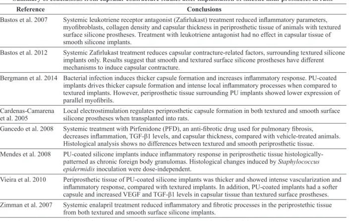
Summary of conclusions from capsular contracture studies after implantation of silicone mini-prostheses in rats.
Reference Conclusions
Bastos et al. 2007 Systemic leukotriene receptor antagonist (Zafirlukast) treatment reduced inflammatory parameters, myofibroblasts, collagen density and capsular thickness in periprosthetic tissue of animals with textured surface silicone prostheses. Treatment with leukotriene antagonist had no effect in capsular tissue of smooth silicone implants.
Bastos et al. 2012 Systemic Zafirlukast treatment reduces capsular contracture-related factors, surrounding textured silicone implants only. Results suggest that smooth and textured surface silicone prostheses have different mechanisms to induce capsular contracture.
Bergmann et al. 2014 Bacterial infection induces thicker capsule formation and increases inflammatory response. PU-coated implants drives thicker capsule formation and intense local inflammatory processes when compared to textured implants. However, periprosthetic tissue surrounding PU implants showed lower expression of parallel myofibrils.
Cardenas-Camarena et al. 2005
Local electrostimulation regulates periprosthetic capsule formation in both textured and smooth surface silicone prostheses when transplanted into rats.
Gancedo et al. 2008 Systemic treatment with Pirfenidone (PFD), an anti-fibrotic drug used for pulmonary fibrosis,
decreases inflammation, TGF-β1 levels, and capsular thickness, compared with vehicle-treated animals. Histological analysis shows no differences between textured and smooth periprosthetic tissue.
Mendes et al. 2008 PU-coated silicone implants induce inflammatory response in periprosthetic tissue histologically-patterned as chronic foreign body granulomas. Histological changes induced by Staphylococcus epidermidis inoculation were dose-independent.
Vieira et al. 2010 Periprosthetic tissue of PU-coated silicone implants was thicker and showed intense vascularization and inflammatory response, compared with textured implants. In addition, PU-coated implants had a softer capsule and increased VEGF and TGF-β1 levels in capsular tissue than textured surface prostheses. Zimman et al. 2007 Systemic enalapril treatment reduced inflammatory and fibrotic processes in the periprostethic tissue
from both textured and smooth surface silicone implants.
studies have used animal models with smooth or
textured prostheses implanted both subcutaneously
and submuscularly, with subsequently histological
evaluation of the neo-formed periprosthetic tissue
(Barone et al. 1992, Bucky et al. 1994, Bern et al.
1992, Clugston et al. 1994, Brohim et al. 1993b).
Importantly, some studies found tighter and thicker
capsules surrounding textured implants compared
to smooth implants (Barone et al. 1992, Bucky et al.
1994, Bern et al. 1992), while others demonstrated
that capsular contracture is less often in textured
surface implants (Clugston et al. 1994, Brohim et
al. 1993b).
Despite these ambiguous results,
miscella-neous of clinical data on capsular contracture
inci-dence in patients (Barnsley et al. 2006), highlights
the relevance of controlled experimental models
to study capsular contracture etiology and many
assays rely on animal models to mimic human
periprosthetic capsular contracture (Barnsley et al.
2006, Wong et al. 2006, Vieira et al. 2010).
Pre-clinical benchwork advantages include the
con-trol of the experimental environment, minimizing
unwanted variables, besides being a faster, less
expensive approach (Bastos et al. 2003, Bucky et
al. 1994, Clugston et al. 1994, Imber et al. 1974,
Ksander et al. 1981, Peters et al. 1980, Vieira et
al. 2010). Animals such as pigs, rabbits, dogs,
rats, and mice have been used with variable results
(Clugston et al. 1994, Fagrell et al. 2001, Ajmal et
al. 2003, Shah et al. 1981, Kossovsky et al. 1984,
Chen et al. 1996, Darouiche et al. 2002, Ksander
et al. 1981, Tang et al. 1998, Adams et al. 2006,
Marques et al. 2012, Brohim et al. 1993a, Minami
et al. 2006, Park et al. 2013, Moyer et al. 2012,
Bastos et al. 2003, 2012), but it is consensus that
the rat is the most appropriate animal model that
provides relevant scientific conclusions with accu
-rate histological extrapolation to the human tissue
(Czerny 1895b, Harris 1961, Lalardrie and Mouly
1978, Garcia et al. 2002). In addition, most studies
agree that a 90-days follow up seems to be suitable
to evaluate capsular contracture in rats (Tables II
and III). The main approaches tested in the studies
reviewed in Tables II and III were the effects of
smooth and/or textured silicone implants on
differ-ent histological and biochemical parameters
(Zim-man et al. 2007, Vieira et al. 2010, Mendes et al.
2008, Gancedo et al. 2008, Cardenas-Camarena et
al. 2005, Bergmann et al. 2014, Bastos et al. 2007b,
2012). The results indicate different degrees of fi
-brosis, fibroblast activation, inflammation, and cap
-sule thickness in periprosthetic tissue (Tables II and
III) (Frangou and Kanellaki 2001, Eltze et al. 2003,
Minami et al. 2006, Eltze et al. 2006, Adams et al.
2006, Cardenas-Camarena et al. 2005, Gancedo et
al. 2008, Chelko et al. 2012, Aparecida da Silva et
al. 2014, Vieira et al. 2010). Some authors did not
find histological differences between smooth and
textured implants surrounding tissue (Gancedo et
al. 2008), but most found thicker capsules with
in-creased cellularity and less frequent contractures in
the textured surface (Bastos et al. 2007a, 2012,
Zim-man et al. 2007, Cardenas-Camarena et al. 2005,
Bergmann et al. 2014) (Tables II and III). There
are some studies with rabbits trying to understand
the capsular contracture process, but interestingly,
most of them use smooth surface mini-implants
only (Shin et al. 2013, Park et al. 2013, Moyer et
al. 2012, Adams et al. 2006). To our knowledge, a
single rabbit study compared textured and smooth
surface silicone mini-implant, and found less
cel-lularity and reduced capsule thickness
surround-ing textured surface prostheses (Uzunismail et al.
2008). Minami et al. (2006) used pigs to show that
capsular contracture in smooth implants have a
significantly higher intra-implant pressure, and the
smooth implant capsule was significantly thicker
than the textured (Minami et al. 2006).
different implant materials (i.e. textured, smooth
or PU-covered) (Chang et al. 1992, Ersek 1991,
Hester et al. 1988, Asplund et al. 1996, Thuesen
et al. 1995), surgical procedures (i.e. subglandular
or submuscular pockets) (Puckett et al. 1987,
Hakelius and Ohlsen 1997, Handel et al. 1995), and
drug delivery systems (Ajmal et al. 2003, Lemperle
and Exner 1993). Tables II and III show some
studies that evaluated the effect of
angiotensin-converting enzyme inhibitors (Zimman et al.
2007), leukotriene receptor antagonist (zafirlukast)
(Bastos et al. 2007b, Bastos et al. 2012), and
anti-fibrotic compound (pirfenidone) (Gancedo et al.
2008) on biochemical and cellular features of
periprosthetic tissue from rats that received smooth
or textured silicone implants (Bastos et al. 2007b,
2012, Gancedo et al. 2008, Zimman 2007). Bastos
et al. (2012) also tested the effects of Zafirlukast
on intra-implant pressure after implantation of
both textured and smooth silicone implant in rats,
and concluded that treatment increases internal
pressure only in textured silicone implants (Tables
II and III). Finally, these distinct pharmacological
outcomes on capsular prevention strengthen the
concept that capsular contracture pathophysiology
is prosthesis type-dependent (Bastos et al. 2007b,
2012, Gancedo et al. 2008, Zimman 2007,
Bergmann et al. 2012, 2014).
Our previous study on the characteristics of
periprosthetic tissue surrounding textured surface
silicone implants in rats demonstrated that
PU-coated implants induce thicker capsules, associated
to intense “foreign body” immune reaction and
up-regulation of vascular endothelial growth factor
(VEGF) (Vieira et al. 2010). We propose that high
vascularization induced by VEGF results in this
thicker capsule, but capsular enlargement would
be due to an increase of the non-collagenoustissue
layer. Furthermore, we concluded that stimulation
of an angiogenic response in periprosthetic
tissue leads to a softer capsule surrounding the
silicone implant, which should decrease capsular
contracture occurrence in breast reconstruction
and augmentation (Vieira et al. 2010). Consistent
with our findings, other studies also hypothesized
that the positive effects of PU implants on capsular
prevention are mainly related to the biochemical
effects of the biomaterial in the surrounding tissue
rather than the prosthesis surface texture itself
(Adams 2009a, Hester et al. 1988, 2001, Lilla and
Vistes 1976, Brand 1984, Dunn et al. 1992, Brohim
et al. 1992, Batra et al. 1995, Picha and Goldstein
1991, Picha et al. 1990, Sank et al. 1993, Santere et
al. 2005, Bucky et al. 1994, Rebello 1996).
ConCluSIonS
(Minami et al. 2006, Clugston et al. 1994). Herein,
we showed that the mean follow up used in most
assays is 90 days. Therefore, long-term studies
using rat models might be necessary to translational
conclusions, especially if one considers that 15
years of follow up in a female human being is
comparable to approximately 9 months in rat
models (Vieira et al. 2010).
In addition, since experimental models are
rarely able to replicate the development of capsular
contracture, most studies evaluate the outcome
of different interventions on normal capsule
formation in their animal models (Vieira et al.
2010, Dobke et al. 1995, Rieger et al. 2013, Bastos
et al. 2003, 2007a, Imber et al. 1974, Ksander et
al. 1981, Peters et al. 1980, Chelko et al. 2012,
Aparecida da Silva et al. 2014, Shin et al. 2013,
Uzunismail et al. 2008, Chang et al. 1992, Hester et
al. 1988, Asplund et al. 1996, Thuesen et al. 1995,
Puckett et al. 1987, Hakelius and Ohlsen 1997,
Handel et al. 1995, Lemperle and Exner 1993,
Zimman 2007, Zimman et al. 2007, Bergmann et
al. 2012, Hester et al. 2001, Lilla and Vistes 1976,
Brand 1984). This fact understates the impact of
conclusions from previous studies, since therapy
should be focused in pathologic capsule formation.
Some authors evaluated parameters such as liquid
infusion and pressure-volume curve or applanation
tonometry, beyond the histological analysis to
assess capsular contracture in animal models
(Bastos et al. 2012, Bucky et al. 1994, Clugston
et al. 1994, Peters et al. 1980, Adams et al. 2006,
Marques et al. 2012, Minami et al. 2006, Moyer et
al. 2012). These studies contribute to the correlation
of capsule histological analysis with dynamic
pressure assays advancing our comprehension of
capsular contracture. To our knowledge, only eight
experimental studies showed indirect presence of
the capsular contracture until now (Bastos et al.
2012, Bucky et al. 1994, Clugston et al. 1994,
Peters et al. 1980, Adams et al. 2006, Marques et
al. 2012, Minami et al. 2006, Moyer et al. 2012).
The translational potential of capsular
contrac-ture animal models still needs to be clarified. How
-ever, pre-clinical studies enable a more controlled
and thorough assessment of the periprosthetic
tis-sue characteristics, which is critical to our
under-standing on how silicone implant surface interacts
with the surrounding tissue. Finally, these advances
can open new avenues on implant type selection,
and in prevention and/or treatment of symptomatic
cases of capsular contracture.
ReFeRenCeS
ADAMS WP. 2009a. Capsular contracture: what is it? What causes it? How can it be prevented and managed? Clinics in plastic surgery 36: 119-126.
ADAMS WP. 2009b. Capsular contracture: What is it? What causes it? How can it be prevented and maneged? Clin Plast Surg 36: 119-126.
ADAMS WP, HAYDON MS JR, RANIERE J JR, TROTT S, MARQUES M, FELICIANO M, ROBINSON JB JR, TANG L AND BROWN SA. 2006. A rabbit model for capsular contracture: development and clinical implications. Plast Reconstr Surg 117: 1214-1219. AJMAL N, RIORDAN CL, CARDWELL N, NANNEY
LB AND SHACK RB. 2003. Chemically assisted capsulectomy in the rabbit model: a new approach. Plast Reconstr Surg 112: 1449-1454.
APARECIDA DA SILVA C, MOLINAR MAUAD CINTRA M, CASTRO COBO E, VINICIUS DA SILVA M, BICHUETTE CUSTODIO F, ROSA MIRANDA CORREA R, ROBERTO CASTELLANO L, ANTONIA DOS REIS M AND REIS MACHADO J. 2014. Renal biopsy: use of biomarkers as a tool for the diagnosis of focal segmental glomerulosclerosis. Dis Markers 2014: 192836.
ASPLUND O, GYLBERT L, JURELL G AND WARD C. 1996. Textured or smooth implants for submuscular breast augmentation: a controlled study. Plast Reconstr Surg 97: 1200-1206.
BALDERRAMA CM, RIBAS-FILHO JM, MALAFAIA O, CZECZKO NG, DIETZ UA, SAKAMOTO DG AND BITTENCOURT LP. 2009. Healing reaction to mammary prostheses covered by textured silicone and silicone foam in rats. Acta Cir Bras 24: 367-376.
BARONE FE, PERRY L, KELLER T AND MAxWELL GP. 1992. The biomechanical and histopathologic effects of surface texturing with silicone and polyurethane in tissue implantation and expansion. Plast Reconstr Surg 90: 77-86. BASTOS EM, NETO MS, ALVES MT, GARCIA EB,
SANTOS RA, HEINK T, PEREIRA JB AND FERREIRA LM. 2007a. Histologic analysis of zafirlukast’s effect on capsule formation around silicone implants. Aesth Plast Surg 31: 559-565.
BASTOS EM, NETO MS, ALVES MTS, GARCIA EB, SANTOS RA, HEINK T, PEREIRA JB AND FERREIRA JM. 2007b. Histologic Analysis of Zafirlukast’s Effect on Capsule Formation Around Silicone Implants. Aesth Plast Surg 31: 559-565.
BASTOS ÉM, NETO MS, FERREIRA LM, GARCIA ÉB, LIEBANO RE AND HAN YA. 2003. Experimental Model of capsular contracture in silicone implants. Acta Cir Bras 18: 22-28.
BASTOS EM, SABINO NETO M, GARCIA EB, VEIGA DF, HAN YA, DENADAI R, SANTOS RDE A AND FERREIRA LM. 2012. Effect of zafirlukast on capsular contracture around silicone implants in rats. Acta Cir Bras 27: 1-6.
BATRA M, BERNARD S AND PICHA G. 1995. Histologic comparison of breast implant shells with smooth, foam, and pillar microstructuring in a rat model from 1 day to 6 months. Plast Reconstr Surg 95: 354-363.
BENGTSON BP, VAN NATTA BW AND MURPHY DK. 2007. Style 410 highly cohesive silicone breast implant core study results at 3 years. Plast Reconstr Surg 120: 40S-48S.
BERGMANN PA, LIODAKI ME, MAUSS KL, LANGE T, GEBHARD M, MAILANDER P AND SIEMERS F. 2012. Histological and immunohistochemical study of capsular contracture in an animal model--a comparison of two implants according to a modification of Wilflingseder’s classification. Handchir Mikrochir Plast Chir 44: 220-226. BERGMANN PA, TAMOURIDIS G, LOHMEYER JA,
MAUSS KL, BECKER B, KNOBLOCH J, MAILANDER P AND SIEMERS F. 2014. The effect of a bacterial con-tamination on the formation of capsular contracture with polyurethane breast implants in comparison with textured silicone implants: an animal study. JPRAS 67: 1364-1370. BERN S, BURD A AND MAY JWJ. 1992. The biophysical
and histologic properties of capsules formed by smooth and textured silicone implants in the rabbit. Plast Reconstr Surg 89: 1037-1042.
BRAND G. 1984. Foam-covered mamary implants. Clin Plast Surg 73: 498.
BROHIM RM, FORESMAN PA, GRANT GM, MERICKEL MB AND RODEHEAVER GT. 1993a. Quantitative monitoring of capsular contraction around smooth and textured implants. Ann Plast Surg 30: 424-434.
BROHIM RM, FORESMAN PA, GRANT GM, MERICKEL MB AND RODEHEAVER GT. 1993b. Quantitative monitoring of capsular contraction around smooth and textured implants. Ann Plast Surg 30: 424-434.
BROHIM RM, FORESMAN PA, HILDEBRANDT PK AND RODEHEAVER GT. 1992. Early tissue reaction to tex-tured breast implant surfaces. Ann Plast Surg 28: 354-362. BUCKY LP, EHRLICH HP, SOHONI S AND MAY JW JR.
1994. The capsule quality of saline-filled smooth silicone, textured silicone, and polyurethane implants in rabbits: a long-term study. Plast Reconstr Surg 93: 1123-1131. CALNAN JS. 1970. Assessment of biological properties of
implants before their clinical use. Proc R Soc Med 63: 1115-1118.
CALOBRACE MB AND CAPIZZI PJ. 2014. The biology and evolution of cohesive gel and shaped implants. Plast Reconstr Surg 134: 6s-11s.
CARDENAS-CAMARENA L, PAILLET JC AND BRISENO R. 2005. Electrostimulation: uses and applications for periprosthetic capsular contracture: experimental model. Aesth Plast Surg 29: 410-414.
CHANG L, CALDWELL E, READING G AND WRAY RC JR. 1992. A comparison of conventional and low-bleed implants in augmentation mammaplasty. Plast Reconstr Surg 89: 79-82.
CHELKO SP, SCHMIEDT CW, LEWIS TH, LEWIS SJ AND ROBERTSON TP. 2012. A novel vascular clip design for the reliable induction of 2-kidney, 1-clip hypertension in the rat. J Appl Physiol 112: 362-366.
CHEN NT, BUTLER PE, HOOPER DC AND MAY JW, JR. 1996. Bacterial growth in saline implants: in vitro and in vivo studies. Ann Plast Surg 36: 337-341.
CLUGSTON PA, PERRY LC, HAMMOND DC AND MAxWELL GP. 1994. A rat model for capsular contracture: the effects of surface texturing. Ann Plast Surg 33: 595-599.
CRONIN TD AND GEROW FJ. 1963. Augmentation mammoplasty:a new natural feel prosthesis. In: Transactions of the Third International Congress of Plastic Surgery, Octuber 13-18, 1963, Amsterdan, p. 41-49. CUNNINGHAM B. 2007. The Mentor Study on contour
profile gel silicone MemoryGel breast implants. Plastic and reconstructive surgery 120.
CZERNY V. 1895. Plastischer ersatz der brustdru se durch ein lipom. Zentralbl Chir 22:72.
DANINO AM, BASMACIOGLU P, SAITO S, ROCHER F, BLANCHET-BARDON C, REVOL M AND SERVANT JM. 2001. Comparison of the capsular response to the Biocell RTV and Mentor 1600 Siltex breast implant surface texturing: a scanning electron microscopic study. Plast Reconstr Surg 108: 2047-2052.
antimicrobe-impregnated saline-filled silicone implants. Plast Reconstr Surg 109: 1352-1357.
DEL POZO JL ET AL. 2009. Pilot study of association of bacteria on breast implants with capsular contracture. J Clin Microbiol 47: 1333-1337.
DEMERGIAN V. 1963. ExPERIENCES WITH THE NEWER SUBCUTANEOUS IMPLANT MATERIALS. Surg Clin North Am 43: 1313-1321.
DOBKE MK, SVAHN JK, VASTINE VL, LANDON BN, STEIN PC AND PARSONS CL. 1995. Characterization of microbial presence at the surface of silicone mammary implants. Ann Plast Surg 34: 563-569.
DUNN KW, HALL PN AND KHOO CT. 1992. Breast implant materials: sense and safety. Brit J Plast Surg 45: 315-321. EDGERTON MT AND MC CA. 1958. Augmentation
mam-maplasty; psychiatric implications and surgical indica-tions; (with special reference to use of the polyvinyl al-cohol sponge ivalon). Plast Reconstr Surg Transplant Bull 21: 279-305.
ELTZE E, BETTENDORF O, RODY A, JACKISCH C, HERCHENRODER F, BOCKER W AND PFLEIDERER B. 2003. Influence of local complications on capsule formation around model implants in a rat model. J Biomed Mater Res A 64: 12-19.
ELTZE E, SCHAFER U, BETTENDORF O, RODY A, HERCHENRODER F, CHIWRITSCH T, JACKISCH C AND PFLEIDERER B. 2006. Radiation-induced capsule tissue reactions around textured breast implants in a rat model. Breast 15: 331-338.
ERSEK RA. 1991. Rate and incidence of capsular contracture: a comparison of smooth and textured silicone double-lumen breast prostheses. Plast Reconstr Surg 87: 879-884. FAGRELL D, BERGGREN A AND TARPILA E. 2001.
Capsular contracture around saline-filled fine textured and smooth mammary implants: a prospective 7.5-year follow-up. Plast Reconstr Surg 108: 2108-2112.
FRANGOU J AND KANELLAKI M. 2001. The effect of local application of mitomycin-C on the development of capsule around silicone implants in the breast: an experimental study in mice. Aesth Plast Surg 25: 118-128.
GAMPPER TJ, KHOURY H, GOTTLIEB W AND MORGAN RF. 2007. Silicone gel implants in breast augmentation and reconstruction. Ann Plast Surg 59: 581-590.
GANCEDO M, RUIZ-CORRO L, SALAZAR-MONTES A, RINCON AR AND ARMENDARIZ-BORUNDA J. 2008. Pirfenidone prevents capsular contracture after mammary implantation. Aesth Plast Surg 32: 32-40.
GERSUNY R. 1980. The classic reprint. Concerning a subcutaneous prosthesis: Robert Gersuny. (Uber eine subcutane Prothese. Zeitschrift f. Heilkunde Wien u Leipzig 21:199, 1900.). Translated from the German by Miss Rita Euerle. Plast Reconstr Surg 65: 525-527. HAKELIUS L AND OHLSEN L. 1997. Tendency to capsular
contracture around smooth and textured gel-filled silicone
mammary implants: A five-year follow-up. Plast Reconstr Surg 100: 1566.
HANDEL N, JENSEN JA, BLACK Q, WAISMAN JR AND SILVERSTEIN MJ. 1995. The fate of breast implants: A critical analysis of complications and outcomes. Plast Reconstr Surg 96: 1521.
HARRIS HI. 1961. Research in plastic implants. Their use in augmentation for amastia or hypomastia. J Int Coll Surg 35: 630-643.
H E N R I K S E N T F, F RY Z E K J P, H O L M I C H L R , MCLAUGHLIN JK, KJOLLER K, HOYER AP, OLSEN JH AND FRIIS S. 2005. Surgical intervention and capsular contracture after breast augmentation: a prospective study of risk factors. Ann Plast Surg 54: 343-351.
HESTER TR JR, NAHAI F, BOSTWICK J AND CUKIC J. 1988. A 5-year experience with polyurethane-covered mammary prostheses for treatment of capsular contracture, primary augmentation mammoplasty, and breast recon-struction. Clin Plast Surg 15: 569-585.
HESTER TR JR, TEBBETTS JB AND MAxWELL GP. 2001. The polyurethane-covered mammary prosthesis: facts and fiction (II): a look back and a “peek” ahead. Clin Plast Surg 28: 579-586.
IBRAHIM CANTER H, KONAS E, BOZDOGAN O, VARGEL I, OZBATIR B, ONER F AND ERK Y. 2007. Effect of slow-release 5-Fluorouracil on capsule formation around silicone breast implants: an experimental study with mice. Aesth Plast Surg 31: 674-679.
IMBER G, SCHAWAGER RG, GUTHTIE RH AND GRAY GF. 1974. Fibrous Capsula formation after subcutaneous implantation of synthetic materials in experimental animals. Plast Reconstr Surg 54: 183.
KJOLLER K ET AL. 2001. Capsular contracture after cosmetic breast implant surgery in Denmark. Ann Plast Surg 47: 359-366.
KOSSOVSKY N, HEGGERS JP, PARSONS RW AND ROBSON MC. 1984. Acceleration of capsule formation around silicone implants by infection in a guinea pig model. Plast Reconstr Surg 73: 91-98.
KSANDER GA, VISTNES LM AND KOSEK J. 1981. Effect of implant location on compressibility and capsule formation around miniprostheses in rats, and experimental capsule contracture. Ann Plast Surg 6: 182-193.
LALARDRIE JP AND MOULY R. 1978. History of mammaplasty. Aesth Plast Surg 2: 167-176.
LEE SD, YI MH, KIM DW, LEE Y, CHOI Y AND OH SH. 2014. The effect of botulinum neurotoxin type A on capsule formation around silicone implants: the in vivo and in vitro study. Int Wound J 13(1): 65-71.
LEMPERLE G AND ExNER K. 1993. Effect of cortisone on capsular contracture in double-lumen breast implants: ten years’ experience. Aesth Plast Surg 17: 317-323.
LYRAS I. 1993. Tissue reaction to rough and smooth silicone implants (A comparative and analytical experimental study in rats. Rev Soc Bras Cir Plast 8: 31-41.
MARQUES M, BROWN S, CORREIA-SA I, MN DSC, RODRIGUES-PEREIRA P, GONCALVES-RODRIGUES A AND AMARANTE J. 2012. The impact of triamcinolone acetonide in early breast capsule formation in a rabbit model. Aesth Plast Surg 36: 986-994.
MAxWELL GP AND GABRIEL A. 2009. The evolution of breast implants. Clinics in plastic surgery 36: 1-13. MENDES PR, BINS-ELY J, LIMA EA, VASCONCELLOS
ZA, D’ACAMPORA AJ AND NEVES RD. 2008. Histological study on acute inflammatory reaction to polyurethane-coated silicone implants in rats. Acta Cir Bras 23: 93-101.
MINAMI E, KOH IH, FERREIRA JC, WAITZBERG AF, CHIFFERI V, ROSEWICK TF, PEREIRA MD, SALDIVA PH AND DE FIGUEIREDO LF. 2006. The composition and behavior of capsules around smooth and textured breast implants in pigs. Plast Reconstr Surg 118: 874-884. MOREIRA M, FAGUNDES DJ, DE JESUS SIMOES M, DE
OLIVEIRA MC, DOS SANTOS PREVIDELLI IT AND MOREIRA AC. 2009. Zafirlukast pocket delivery impairs the capsule healing around textured implants in rats. Aesth Plast Surg 33: 90-97.
MOYER HR, GHAZI BH AND LOSKEN A. 2012. The effect of silicone gel bleed on capsular contracture: a generational study. Plast Reconstr Surg 130: 793-800.
NETSCHER DT. 2004. Subclinical infection in breast capsules. Plast Reconstr Surg 114: 818-820.
PARK SO, HAN J, MINN KW AND JIN US. 2013. Prevention of capsular contracture with Guardix-SG((R)) after silicone implant insertion. Aesth Plast Surg 37: 543-548.
PETERS CR, SHAW TE AND RAJU DR. 1980. The influence of vitamin E on capsule formation and contracture aroud silicone implants. Ann Plast Surg 5: 347-352.
PICHA GJ AND GOLDSTEIN JA. 1991. Analysis of the soft-tissue response to components used in the manufacture of breast implants: rat animal model. Plast Reconstr Surg 87: 490-500.
PICHA GJ, GOLDSTEIN JA AND STOHR E. 1990. Natural-Y Meme polyurethane versus smooth silicone: analysis of the soft-tissue interaction from 3 days to 1 year in the rat animal model. Plast Reconstr Surg 85: 903-916.
PITANGUY I. 1991. Estudo retrospectivo das aplicações clínicas da prótese de silicone gel revestida com poliuretano. Bol Acad Nac Med 151: 67-72.
POEPPL N, SCHREML S, LICHTENEGER F, LENICH A, EISENMANN-KLEIN M AND PRANTL L. 2007. Does the surface structure of implants have an impact on the formation of a capsular contracture? Aesth Plast Surg 31: 133-139.
POLLOCK H. 1997. Breast capsular contracture. Plast Reconstr Surg 100: 1619-1620.
PUCKETT CL, CROLL GH, REICHEL CA AND CONCANNON MJ. 1987. A critical look at capsule contracture in subglandular versus subpectoral mammary augmentation. Aesth Plast Surg 11: 23-28.
REBELLO C. 1996. Mamaplastia de aumento: algumas considerações científicas sobre a validade do emprego dos implantes de gel de silicone. Rev Bras Cir 86(5): 261-283. RIEGER UM, MESINA J, KALBERMATTEN DF, HAUG M,
FREY HP, PICO R, FREI R, PIERER G, LUSCHER NJ AND TRAMPUZ A. 2013. Bacterial biofilms and capsular contracture in patients with breast implants. Brit J Surg 100: 768-774.
ROHRICH RJ, KENKEL JM AND ADAMS WP. 1999. Pre-venting capsular contracture in breast augmentation: in search of the Holy Grail. Plast Reconstr Surg 103: 1759-1760.
RUBIN LR. 1951. Polyethylene--a three year study. Plast Reconstr Surg 7: 131-142.
SANK A, CHALABIAN-BALIOZIAN J, ERTL D, SHERMAN R, NIMNI M AND TUAN TL. 1993. Cellular responses to silicone and polyurethane prosthetic surfaces. J Surg Res 54: 12-20.
SANTERE JP, WODHOUSE K, LAROCHE G AND LABOW RS. 2005. Understanding the biodegradation of polyurethanes: From classical implants to tissue engineering materials. Biomaterials 26: 7457-7470. SHAH Z, LEHMAN JA AND TAN J. 1981. Does infection
play a role in breast capsular contracture? Plast Reconstr Surg 68: 34-38.
SHIN KC, CHUNG KI, PARK BY, KIM HK, KIM WS, BAE TH AND KIM MK. 2013. The effect of antiadhesion agent on peri-implant capsular formation in rabbits. Ann Plast Surg 71: 600-604.
SMAHEL J, SCHNEIDER K AND DONSKI P. 1977. Bizarre implants for augmentation mammaplasty: long term human reaction to polyethylene strips. Brit J Plast Surg 30: 287-290.
SPEAR SL, BULAN EJ AND VENTURI ML. 2006. Breast augmentation. Plast Reconstr Surg 118: 188S-196S. SPEAR SL, ELMARAGHY M AND HESS C. 2000.
Textured-surface saline-filled silicone breast implants for augmentation mammaplasty. Plast Reconstr Surg 105: 1542-1552.
SPEAR SL, PARIKH PM AND GOLDSTEIN JA. 2009. History of breast implants and the food and drug administration. Clin Plast Surg 36: 15-21.
STEIERT AE, BOYCE M AND SORG H. 2013. Capsular contracture by silicone breast implants: possible causes, biocompatibility, and prophylactic strategies. Medical devices (Auckland, NZ) 6: 211-218.
Breast implants--current trends and new concepts. Handchir Mikrochir Plast Chir 44: 240-253.
TANG L, JENNINGS TA AND EATON JW. 1998. Mast cells mediate acute inflammatory responses to implanted biomaterials. Proceedings of the PNAS 95: 8841-8846. THOREK M. 1942. Amastia, hypomastia, and inequality of
the breasts. In: Plastic surgery of the breast and abdominal wall. Springfield (IL): Thomas. In: Plastic surgery of the breast and abdominal wall, p. 369-386.
THUESEN B, SIIM E, CHRISTENSEN L AND SCHRODER M. 1995. Capsular contracture after breast reconstruction with the tissue expansion technique. A comparison of smooth and textured silicone breast prostheses. Scand J Plast Reconstr Surg Hand Surg 29: 9-13.
UZUNISMAIL A, PERK C, FINDIK H, BEYHAN G AND DINCLER M. 2008. Effect of a fascial tissue interface on silicone implant capsule formation--a preliminary experimental study. JPRAS 61: 1199-1204.
VAN HEERDEN J, TURNER M, HOFFMANN D AND MOOLMAN J. 2009. Antimicrobial coating agents: can biofilm formation on a breast implant be prevented? JPRAS 62: 610-617.
VIEIRA VJ, D’ACAMPORA AJ, MARCOS AB, DI GIUNTA G, DE VASCONCELLOS ZA, BINS-ELY J, D’ECA NEVES R AND FIGUEIREDO CP. 2010. Vascular endothelial growth factor overexpression positively modulates the characteristics of periprosthetic tissue of polyurethane-coated silicone breast implant in rats. Plast Reconstr Surg 126: 1899-1910.
WONG CH, SAMUEL M, TAN BK AND SONG C. 2006. Capsular contracture in subglandular breast augmentation with textured versus smooth breast implants: a systematic review. Plast Reconstr Surg 118: 1224-1236.
YOUNG VL AND WATSON ME. 2001. Breast implant research: where we have been, where we are, where we need to go. Clin Plast Surg 28: 451-483.
ZIMMAN OA. 2007. A rabbit model for capsular contracture: development and clinical implications. Plast Reconstr Surg 119: 1955-1956.