Universidade Nova de Lisboa
Instituto de Higiene e Medicina Tropical
Activity of human macrophages when exposed to visceral and
cutaneous species of Leishmania spp.
Author: Geraldina Cândida Manjate
Thesis to obtain the master’s degree in Medical Parasitology
NOVEMBER, 2016
Universidade Nova de Lisboa
Instituto de Higiene e Medicina Tropical
Activity of human macrophages when exposed to visceral and
cutaneous species of Leishmania spp.
Author: Geraldina Cândida Manjate
Supervisor: Prof. Dr.Gabriela Santos-Gomes Co-supervisor: Dr. Ana Maria Armada
Thesis submitted for compliance with the requirements to obtain the
master’s degree in Medical Parasitology.
ii
“Não que eu já tenha obtido tudo isso ou tenha sido aperfeiçoado, mas prossigo para alcançá-lo, pois para isso também fui alcançado por Cristo Jesus.
Irmãos, não penso que eu mesmo já o tenha alcançado, mas uma coisa faço: esquecendo-me das coisas que ficaram para trás e avançando para as que estão adiante,
prossigo para o alvo, a fim de ganhar o prémio do chamado celestial de Deus em Cristo Jesus.”
1 I.
CONTENTS
I.CONTENTS ... 1 II. ACKNOWLEDGEMENTS ... 3 IV. RESUMO ... 7 V. ABSTRACT ... 8 Introduction ... 9 1.1.Epidemiology ... 9 1.1.1.Visceral Leishmaniasis ... 10 1.1.2.Cutaneous Leishmaniasis ... 10 1.1.3.Mucocutaneous Leishmaniasis ... 10 1.2. The Parasite ... 11 1.2.1. Morphology ... 11 I.2.2.Life Cycle ... 12I.2.3.Vertebrate Host-parasite Interaction ... 13
1.3. Immune Response ... 14
1.3.1. Innate Immune System ... 14
1.3.2. Acquired Immune Response ... 15
1.4. Diagnostic Methods... 19 1.4.1. Parasitological Methods ... 19 1.4.2. Seroimmunological Methods ... 20 1.4.3. Molecular Diagnostic ... 20 1.5. Treatment ... 20 1.6. New approaches ... 22 2.OBJECTIVES ... 24
3.Material and methods ... 25
3.1.Experimental design (flowchart) ... 25
3.2.Biological samples ... 26
3.2.1.Mononuclear cells ... 26
3.2.2.Direct agglutination test ... 27
3.2.3.Flow cytometry ... 28
3.2.4.Peripheral blood monocyte differentiate macrophage ... 29
3.2.5.Isolation of lymphocytes ... 30
3.3. Parasites ... 30
3.3.1.Macrophage Infection ... 31
2
3.3.2.1.Scanning electron microscopy ... 32
3.3.2.2.Histone detection by immunolabeling ... 33
3.3.3.Production of nitric oxide by macrophages ... 33
3.3.4.Evaluation of MHC in infected-MØ when in contact with lymphocytes ... 34
3.3.5.Statistical analysis ... 35
4.Results and Discussion ... 36
4.1.Participants did not evidence previous contact with L. infantum parasites... 36
4.2.Macrophages ... 36
4.2.1.Complete macrophage differentiation was achieved after 72h of incubation ... 36
4.2.2.Cutaneous species of Leishmania show high infectivity to human macrophages ... 37
4.2.3.Leishmania spp. do not promote macropahe oxidative burst ... 40
4.2.4.When in contact with lymphocytes Leishmania spp. stimulates MHC expression ... 41
4.2.5.L. infantum parasites downregulate MET emission ... 43
4.2.7 L. infantum parasites restrain the release of histone to the extracellular space ... 45
4.Final remarks ... 47
3
II. ACKNOWLEDGEMENTS
First of all, I would like to express my sincere gratitude to my supervisor Dr. Gabriela Santos-Gomes. For her guidance, knowledge, support, patience and constructive suggestions, without her kind advice throughout my research this dissertation would not have been possible. I am grateful for her support on the good and bad moments.
I also would like to thank to my co-supervisor Dr. Ana Maria Armada, for her guidance, availability and support throughout my research studies.
My sincere appreciation to Maria de Aires Pereira, for her constructive suggestions and knowledge shared indispensable for the accomplishment of this thesis. I thank Maria Armanda Rodrigues and Dr. Graça Alexandra-Pires for their patient and support.
To Dr. Luiz Felipe Passero, for making the same of the materials used available.
To my fellow colleagues Pedro Ruas, Áurea Gabriel, Madalena Duarte, Ana Rita Pedrosa, João Tavanez and Dr. Celso Cunha, for all the laughs that we have shared. To my closest friends Lurdes, thanks for understanding, encouragement and being always present in all moments. And to all my kind friends that supported and accompanied me in this journey.
At last, but by no means the least, I would like to express my appreciation to my loved ones and thank you for being always there for me, for giving me motivation and, most of all, for supporting me spiritually throughout this entire process.
4
Index of Figures
Figure 1.1. Life stages of Leishmania spp.……….12
Figure 1.2. The life cycle of Leishmania pp………...13
Figure 3.1. Isolation of blood peripheral mononuclear cells by density gradient...…27
Figure 3.2. Schematic representation of flow cytometry process………...28
Figure 3.3. The principal components of a flow cytometer………..…..29
Figure 4.1. Macrophage differentiation………...37
Figure 4.2. Macrophages infected with Leishmania spp……….…..….38
Figure 4.3. Intensity of macrophage infection caused by Leishmania spp……….39
Figure 4.4. Nitric oxide production by macrophage exposed to Leishmania spp……..40
Figure 4.5. Frequency of Leishmania-infected MØ expressing MHCI when in presence of lymphocytes………..41
Figure 4.6. Frequency of Leishmania-infected MØ expressing MHCII when in presence of lymphocytes………..42
Figure 4.7. MHCII and MHCI surface expression of Leishmania-infected MØ when in the presence of autologous lymphocytes.….………....43
Figure 4.8. L. infantum regulates the emission of MET by human MØ……….…...44
5 III. LIST OF ABBREVIATIONS
ºC - degree Celsius
APCs - Antigen presenting cells CC - cytotoxic concentration
CDC - Centers for Diseases Control and Prevention‖ CL - cutaneous leishmaniasis
CO2 -Carbon dioxide
CPD-1 - citrate-phosphate-dextrose CSFs - Colony stimulating factors CTLs - cytotoxic T lymphocytes DAPI - 4',6-diamidino-2-phenylindole DCs - dendritic cells
DMSO - 1% dimethyl sulfoxide DNA - deoxyribonucleic acid IFN-γ - Interferon gamma
iNOS - inducible nitric oxide synthase FBS - fetal bovine serum
Gp63 - Glycoprotein of 63kDa HCL - Hydrochloric acid LCan - canine leishmaniasis LPG – Lypophosphoglycan LPS - lipopolysaccharide
MAC - membrane attack complex MCL - mucocutaneous leishmaniasis
6
MILT - Miltefosine
METs - macrophage extracellular traps MHC - Major histocompatibility complex MØ - macrophages
NaCl - Sodium chloride NO - nitric oxide
OM - optical microscope OsO4 - osmium tetroxide
PBS - phosphate buffered saline
PBMC: Peripheral blood mononuclear cells. PMA - phorbol myristate acetate
PMN - polymorphonuclear cells
RPMI - Roswell Park Memorial Institute medium SEM - scanning electron microscope
SCHN - Schneider's Insect Medium TNF-α - tumoral necrosis factor U.S.A – United States of America VL - visceral leishmaniasis VWR- We Enable Science
7
IV. RESUMO
As leishmanioses são doenças parasitárias que têm como agente etiológico um protozoário do género Leishmania. São transmitidas através da picada de insetos vetores, pertencentes à família Phlebotominae. É considerada um problema de saúde pública em vários países, e dependendo da espécie infetante origina diversas manifestações clínicas. O sistema imunitário é um importante componente do nosso organismo que tem a capacidade de identificar, inativar e eliminar os agentes patogénicos. Contudo, após ser fagocitado por macrófagos (MØ) Leishmania sobrevive, multiplica-se e estabelece uma infeção crónica no hospedeiro mamífero, subvertendo a atividade desta célula da imunidade inata que é também responsável pela apresentação antigénica e pela ligação à imunidade adquirida. Deste modo, é importante explorar a atividade macrofágica na presença de diferentes estirpes de Leishmania spp., tendo em vista a identificação de novos alvos profiláticos e terapêuticos, sobretudo que possibilitem novas soluções para colmatar a resistência aos fármacos antileishmania. No presente estudo foram avaliadas as taxas de infeção em macrófagos humanos expostos a promastigotas de L. infantum e de especies cutâneas de Leishmania. Adicionalmente, outros parâmetros importantes para a caracterização destas infeções, nomeadamente a produção do óxido nítrico (NO), análiseda libertação de armadilhas extracelulares por macrófagos (MET) e a capacidade de apresentarem antigénios através de moléculas do complexo maior de histocompatibilidade (MHC) foram investigados. Foi verificado que as espécies cutâneas originaram maiores taxas de infeção face à espécie visceral, o que conduziu a uma maior intensidade de infeção dos MØ. Foi também possível descrever pela primeira vez a libertação de MET na presença de L. infantum, ainda que aparentemente de uma forma controlada, e seguir o percurso da histona H1 desde o núcleo até ao espaço extracelular. As espécies de Leishmania analisadas induziram a expansão das subpopulações de MØMHCI+ e MHCII+ e o aumento destas moléculas na superfície dos MØ, indicando a existência de apresentação antigénica e a possibilidade de ocorrer a estimulação quer dos linfócitos T helper quer dos linfócitos T citotóxicos. Apesar de o MØ dispor de um conjunto de mecanismos de controlo directo da infeção e de poder activar a resposta imune mediada por células, o parasita parece modelar estes mecanismos, provavelmente conduzindo a seleção dos parasitas mais virulentos, capazes de estabelecer a infeção e causar doença do hospedeiro
Palavras-chaves: Macrófagos, Leishmania spp., Armadilhas extracelulares por macrófagos, Moléculas do complexo maior de histocompatibilidade
8
V. ABSTRACT
Leishmaniasis is a parasitic disease caused by an intracellular protozoan of genus
Leishmania and is transmitted by the bite of an insect vector, the phlebotomine sand fly.
It is considered a Public Health problem in numerous countries and depending on the infecting species, the disease can present multiple clinical manifestations; cutaneous, visceral and mucocutaneous leishmaniasis. Cutaneous leishmaniasis can be produced by
L. amazonensis, L. shawi and L. guyanensis among many other species. Leishmania infantum is the agent responsible for zoonotic visceral leishmaniasis, considered the
most severe form that can be fatal if not treated. The mammal immune response is an important barrier that can prevent the establishment of a long-lasting chronic infection. Macrophages (MØ) play an important role being phagocytic and antigenic presenting cells, and moreover the definitive host cells of Leishmania parasite. It is important to analyze the activity of human MØ in presence of visceral and cutaneous species of
Leishmania spp. in order to better understand the strategies used by the parasite to
subvert MØ mechanisms of defense. This information is relevant to find new targets for the development of prophylactic and therapeutic tools, mainly the establishment of new anti-Leishmania therapeutics, since drug resistance has been reported. Having that in mind, the behavior of human MØ exposed to Leishmania promastigotes was investigated. Infection levels, oxidative burst, release of extracellular traps (MET) and antigenic presentation by major histocompatibility complex (MHC) were in vitro evaluated. It was found that the cutaneous species have a higher rate of infection, which drive a higher infection intensity. It was also possible to describe for the first time that
L. infantum parasites induce the release of MET, although apparently in a controlled
way, and to follow the histone H1 from the nucleus to the extracellular environment. The four species of Leishmania induced the expansion of MHCI+MØ and of MHCII+MØ subsets as well as the increase of these molecules at MØ surface, pointing towards the existence of antigenic presentation and the possibility of stimulation of both helper and cytotoxic T lymphocytes. Although MØ has a set of mechanisms that can control the infection and be able to induce the activation of cell-mediated immune response, the parasite seems to negatively modulate these mechanisms, probably directing the selection of the most virulent parasites able to establish infection in the host and promote disease development.
Keywords: Macrophages, Leishmania spp., Macrophage extracellular traps; Molecules of major histocompatibility complex
9
Introduction
Leishmaniasis is a parasitic disease caused by the intracellular protozoan of genus
Leishmania Ross, 1903 (Basano et al. 2004; Croft et al. 2006), order Kinetoplastida and
family Trypanosomatidae. This parasite is transmitted by the bite of an insect vector, the phlebotomine sand fly (Andrade et al. 2007). At least 30 species of Leishmania are identified and 20 of them are pathogenic for humans (Banuls et al., 2007).
Most species of Leishmania that cause human disease are zoonotic, meaning that animals are reservoirs of the parasite, such as the domestic dog and sylvatic animals, including rodents, monkeys, sloths and foxes. However, a few species give origin to anthroponotic diseases as is the case of L. donovani that is characteristically transmitted man-to-man through the sand fly bite.
Leishmaniasis is spreading over the Mediterranean to tropic and subtropic regions worldwide. The presence or absence of parasite species is determined by the geographic distribution of the respective vector species.
1.1.Epidemiology
The estimated number of new cases of leishmaniasis per year is more than 2 million, according with WHO. A total of 98 countries of Latin America, Africa, Southern Europe and Asia reported human cases of leishmaniasis. However, the worldwide incidence and prevalence still are difficult to establish since this parasitic disease is not an obligatory reported in every endemic country. In fact, only 33 of 98 endemic countries present regular reports specifying the annual number of leishmaniasis cases diagnosed (Desjeux, 2004; Pigott et al. 2014).
Depending on the infecting species, the disease can present multiple clinical manifestations (Desjeux and Alvar, 2003) that can be differentiated into three major clinical syndromes: visceral leishmaniasis (VL), which is also known as kala-azar (Desjeux, 1996; Murray, 2005; Chappuis, 2007), cutaneous leishmaniasis (CL) and mucocutaneous leishmaniasis (MCL) (Banuls et al. 2011).
10
1.1.1. Visceral Leishmaniasis
Visceral leishmaniasis (VL) is considered the most severe form that can be fatal if not treated. According to WHO (2015), more than 90% of cases occur mainly in Bangladesh (600 new cases), Brazil, Ethiopia, India, South Sudan and Sudan. In the Old World (Europe, Africa and Asia), L. donovani and L. infantum are VL etiologic agents (Berman, 1997). The main sand fly vector responsible for the transmission of
Leishmania spp. belongs to the genus Phlebotomus (Killick-Kendrick and Rioux, 2002).
The typical symptoms are prolonged intermittent fever, abdominal distension, with splenomegaly and hepatomegaly, lymphadenopathy, decreased appetite, weight loss and in many cases, signs of chronic kidney disease. Some cases that remain asymptomatic or subclinical may resolve spontaneously (Dawit et al., 2013; Farrar et al., 2014).
1.1.2.
Cutaneous Leishmaniasis
Cutaneous leishmaniasis (CL) is the most common clinical form characterized by the presence of localized cutaneous lesions that, depending on the infecting species, can originate lifelong scars. The self-healing scenario is also possible but in some cases it may take several months and some forms leave permanent lesions(Reithinger et al., 2007). Countries like Afghanistan, Algeria, Brazil, Colombia, the Islamic Republic of Iran, Pakistan, Peru, Saudi Arabia and the Syrian Arab Republic are the most affected (WHO 2015).
L. braziliensis, L. panamensis, L. guyanensis, L. shawi, L. peruviana, L. mexicana, L. amazonensis, L. venezuelensis,L. lainsoni, L. naiffi and L. lindenbergiof New World
and, L. tropica, L. major and L. aethiopica of Old World are the species involved in CL(Ashford, 2000; Dedet and Pratlong, 2003; Reithinger et al., 2007).
1.1.3.
Mucocutaneous Leishmaniasis
Mucocutaneous Leishmaniasis (MCL, also called espundia) usually refers to a metastatic cutaneous infection caused by the parasite spread to the naso-oropharyngeal mucosa. The majority of cases occurs in Bolivia, Brazil and Peru.
11
Although L. braziliensis is the species usually associated with MCL L. panamensis, L.
colombiensis, L. peruviana and, less often L. guyanensis can also cause MCL (Dedet
and Pratlong, 2003). The initial manifestations are erythema and ulceration at the nares that if left untreated can spread, involving the nasal septum, and in some cases, the pharynx or larynx (Dawit et al., 2013.), which can lead to the partial or total destruction of mucous membranes, causing serious disability.
1.2. The Parasite
1.2.1. Morphology
Leishmania is an eukaryote unicellular protozoan parasite that replicates by binary
fission. This digenetic microorganism differentiates in two main morphological forms during its life cycle, the amastigote and the promastigote.
Amastigotes are small, round to oval bodies not exhibiting an external flagellum. This intracellular obligatory parasitic form is found inside the mononuclear phagocytic cells of infected vertebrate hosts.
Promastigote forms can be found in the midgut of the insect vector. These slender microorganisms are extracellular and present single anterior flagellum that confers motility (Figure 1.1). Furthermore, it is possible to differentiate two stages in promastigote development (Bates and Rogers, 2004). Procyclic promastigote which has an elongated shape, are bigger than amastigote and, usually have a shorter flagellum. This parasite form is not infectious, but is in constant replication. Metacyclic promastigotes present a short form, but have a long flagellum that can be two to three times longer than the cell body. This long flagellum confers to this highly infectious parasite form great mobility (Ashford, 2000).
12
Figure 1.1. Life stages of Leishmania spp. Schematic representation of the amastigote (a) and
promastigote (b) morphological forms. F - flagellum; B - flagelar pocket; K- kinetoplast; RE- endoplasmatic reticulum; M- mitochondria; G- golgic complex; mt- multivesicular tubule; N- nucleus; L- lysosome.
I.2.2. Life Cycle
Leishmania spp. alternates between two morphological forms highly adapted to distinct
environments. Promastigote, that is in sintony with the midgut of the insect vector and amastigote, that is adjusted to live inside the phagolysosome of mammalian macrophage (MØ) (Rivas et al., 2004).
The infection occurs when a female sand fly takes a blood meal in the vertebrate host and flagellate promastigotes gain access to the vertebrate skin. Then parasites are rapidly internalized by macrophages MØ. Once inside the cell they differentiate into amastigote, which replicates actively. The cycle is completed when another sand fly ingests parasitized MØ in a new meal. When inside the vector gut, amastigotes rapidly convert back into promastigotes (Carlsen et al., 2015).
13
Figure 1.2. The life cycle of Leishmania spp. Schematic representation of parasite life cycle in the
mammalian (right) host and inside the vector (left). Adapted from CDC, 2016 and
http://www.parasitologie.univ-montp1.fr/english_vers/en_leish2.htm#
I.2.3. Vertebrate Host-parasite Interaction
The invasion of the vertebrate host by Leishmania spp. involves several factors of the humoral and cellular immune system. Macrophages are the preferred target, but parasites can be found in other cells like neutrophils and dendritic cells (DCs) (Bogdan
et al., 2000a; Feijó et al., 2016).
It is important to have an effective immune response considering that Leishmania parasites are preferentially intracellular and it is expected that properly activated MØ kill the parasite. Amastigotes stay in the parasitophorous vacuoles that fuse with lysosomes rich in proteolytic enzymes, generating the phagolysosomes (Bogdan and Röllinghoff, 1998) that should promote parasite lysis. Furthermore, respiratory burst activity that includes the production of reactive nitrogen and oxygen species is one of the mechanisms used by phagocytes to destroy intruders (Murray and Nathan, 1999).
14
However, Leishmania has developed a set of strategies able to subvert MØ activity and resist to the host defense system. The parasite has a mechanism that allows protein kinase to phosphorylate components important to the lytic activity of complement, such as C3, C5 and C9 (Zambrano-Villa et al.,2002). This enzyme modifies other proteins by chemically adding phosphate groups, resulting in functional changes of the target protein. Inhibiting phagosome-lysosome fusion by the action of lipophosphoglycan (Desjardins and Descoteaux, 1997), parasites protect themselves from oxidative stress
(Bogdan and Röllinghoff, 1999). Furthermore, amastigote forms are resistant to
enzymes and acidic pH of the phagolysosome (Bogdan et al., 1990; Kima, 2007).
Another way used by the parasite is to interfere with the immune mechanisms. This parasite difficult the crosstalk between MØ and T cell by avoiding the antigen presentation trough class I (MHCI) and class II (MHCII) molecules of major histocompatibility complex. Consequently, interfering with antigen presentation the parasite prevent T cell activation, avoid the release of pro-inflammatory cytokines (Podinovskaia and Descoteaux, 2015) and, in some cases enhance the production of anti-inflammatory interleukin (IL)-4, the regulatory tumor growth factor (TGF)- and IL-10 (Bhattacharya and Ali, 2013).
All these mechanisms keep infected MØ inactivated, resulting in increased host susceptibility to the parasite (Podinovskaia and Descoteaux, 2015).
1.3. Immune Response
The invasion of vertebrate host by Leishmania parasites causes a set of reactions involving all defense components of innate and acquired immunity.
1.3.1 Innate Immune System
After inoculation in the skin, the parasite has to face the cytotoxic action triggered by the innate immune response. It promotes an immediate robust and nonspecific immune response, which plays an important role in controlling or eliminating pathogenic invaders (Dunkelberger and Song, 2010).
15
The complement system consists of a wide range of proteins organized hierarchically in proteolytic cascades that can promote pathogen destruction or target the pathogen, directing its annihilation by effector cells (Sacks and Sher, 2002; Dunkelberger and Song, 2010). However, metacyclic promastigotes have mechanisms that allow them to block the lytic activity of the complement system. Furthermore, the parasite also can use the complement factors to facilitate MØ recognition and enhance promastigote phagocytosis (Brittingham and Mosser, 1996; Cunningham, 2002), subverting immune system function. Lypophosphoglycan (LPG), an abundant constituent of parasite surface, covering the entire organism, including the flagellum has an important role in the immune escape, preventing the insertion in the parasite membrane of the lytic complex of complement factors (membrane attack complex, MAC) (Sher, 2002; Dunkelberger and Song, 2010) avoiding its own destruction.
Glycoprotein of 63kDa (gp63), a metalloprotease present in the surface of Leishmania parasites promotes the conversion of the complement factor C3b to its inactive form (iC3b). The presence of iC3b in parasite membrane enhances promastigote uptake by phagocytes (Brittingham and Mosser, 1996).
Leishmania protein kinases also can phosphorylate several components of the
complement system, such as C3, C5 and C9, causing the subsequent inhibition of classic and alternative complement pathways (Sher, 2002
).
1.3.2 Acquired Immune Response
The mammal immune response is an important barrier that can prevent the establishment of a long-lasting chronic infection. Macrophages play an important role in the initiation, modulation and regulation of the acquired immune response since they participate in antigenic presentation, leading to T cell activation.
16
Antigen Presentation
There are three cells recognized as professional antigen presenting cells (APCs): MØ, dendritic cells (DCs) and B lymphocytes. However, only MØ are simultaneously the
Leishmania definitive host cell and APC.
Differentiated from bone marrow precursor cells, bloodstream monocytes can migrate through the blood vessel and settle in the tissues as mature MØ, constituting the mononuclear phagocyte system (Roitt et al., 2006; Duque and Descoteaux, 2014). Macrophages recognize and phagocyte pathogens, process the antigens and present the complex antigen - MHCII to T helper cells, resulting in T lymphocyte activation, and the consequent release of cytokines than can activate other cells like B cells. Activated B lymphocytes initiate the production of antibodies specific to the antigens presented by MØ (Duque and Descoteaux, 2014).
MHC molecules synthesized in the APCs endoplasmic reticulum are conducted along the Golgi complex, subsequently transported to endosomes / lysosomes toward the plasma membrane.
According with Germain and Margulies (1994) there are two principal ways to complex with the antigen. MHCI bound to peptides, which are broken down within the cell and transported to the endoplasmic reticulum. While peptides derived from the extracellular proteins are related to MHCII molecules. Peptides resulting from proteins that have been internalized by the cell and then degraded in phagolysosomes are transferred to a vesicle called MIIC which binds to MHCII. The MHCII-peptide complex moves to the cell surface, indicating the existence of intracellular infection. The T helper cell (TCD4+) interact with antigen presenting molecules, triggering an appropriate immune response (Roitt et al. 2006).
For the interaction between MØ and T cells have the right conformational stability required to drive lymphocyte activation, the additional intervention of several types of molecules, such as co-stimulatory molecules B7-1 (CD80) and B7-2 (CD86), present on the surface of APCs and recognized by receptors CD28 (positive co-stimulatory signal) and CD152 (CTLA-4) (negative co-stimulatory signal) on the surface of T cells are crucial (Santos-Gomes et al., 2008).
17
The modulation of T cell functional activity is another mechanism that allows the parasite to evade the host immune response. Several in vitro studies with infected MØ showed that amastigote forms of Leishmania lead to a MHCII decrease in the MØ surface (Reiner et al., 1987; Kwan et al., 1992). Furthermore, these parasites prevent the access of MHCII to parasite antigens, promoting the internalization and degradation (L.
amazonensis) of MHCII molecules inside the parasitophorous vacuole (Leao et al.,
1995). Several assays with different parasites, L. major, L. donovani and L.
amazonensis, also showed inhibition of antigen processing and binding to MHC
molecules, as well as the retention of parasite antigens in the endocytic compartments (Kima et al., 1996; Bogdan and Röllinghoff, 1999; Zambrano-Villa et al., 2002).
Using these mechanisms, the parasite subverts the activity of host acquired immune response, increasing their own ability to survive.
Mature T lymphocytes, also express CD8 molecules, which are important to monitor all the cells of the body and control viral infections. Once active,CD8+ cytotoxic T lymphocytes (CTLs) recognize antigen presented by MHC class I and can rapidly kill the infected cell considered to be a threat to the integrity of the host. Moreover, CTLs have the ability to identified quantitative and qualitative antigenic differences in transformed cells, eliminating spontaneous malignant tumors (Andersen et al., 2006, Roitt et al., 2006).
Induction of a lymphocyte activation
TThe resistance or susceptibility to Leishmania infection depends on the ability of T lymphocytes to specifically proliferate. After stimulation, CD4+ T cells can differ functionally in two sub-populations, T helper 1 (Th1) and T helper 2 (Th2), which produce a particular blend of cytokines Th1-type response, a protective immune response, is associated with the production of proinflammatory cytokines, such as interferon (IFN)- that induces the production of nitric oxide (NO) by MØ, promoting parasite killing (Green et al., 1991; Liu and Uzonna, 2012). However, other cells are involved in cytokine production, DCs produce IL-12 and natural killer cells (NK) also produce IFNSacks and Noben-Trauth, 2002; Teixeira et al., 2006). While the
18
cytokines IL-4, IL-5 and IL-10 are typical of a Th2 type immune response, stimulating the antibody production by B cells (Mosmann and Coffman, 1989; Sacks and Noben-Trauth, 2002; Duque and Descoteaux, 2014). The prevalence of Th2 non-protective immune response in a Leishmania infection is associated with disease severity (Heinzel
et al., 1989; Reiner and Locksley, 1995) and supports parasite persistence.
Macrophage extracellular traps
In addition to the production of oxidative radicals, MØ also have other mechanisms to combat Leishmania parasites. As a multifunctional cell, MØ are able to emit extracellular chromatin webs, known as macrophage extracellular traps (METs) (Chow
et al., 2010; Boe et al., 2015).These traps are considered to be an antimicrobial strategy
used to entrap and kill a wide spectrum of microbial organisms, protozoan included (Goldmann and Medina, 2012; Mohanan et al., 2013). Beside chromatin, METs are also constituted by nuclear DNA, histones, antimicrobial peptides and proteases (Boe et al., 2015).
MØ stimulated by lipopolysaccharide (LPS), cytokines, such as IL-8 and tumoral necrosis factor (TNF-α) and chemicals, as is the case of phorbol myristate acetate (PMA) present chromatin decondensation followed by the mix up of euchromatin and heterochromatin and disruption of the nuclear membrane, losing the cell the normal organized configuration/morphology. Generation of vesicles in the nuclear and cytoplasmic membrane ends with release of DNA fibers into the extracellular environment (Goldmann and Medina, 2012; Mohanan et al., 2013; Boe et al., 2015). Although the production of radical oxidative specimens (ROS) and the activity of NADPH oxidase are essential for the release of extracellular traps (ET) it has been reported that microorganisms like Staphylococcus aureus and L. donovani induce ET release through a molecular process that is independent of ROS (Goldmann and Medina, 2012).
Extracellular traps are a different process of cell death, morphologically and functionally, from programmed cell death and necrosis.
19
1.4. Diagnostic Methods
Ultimately, the diagnosis of leishmaniasis is considered positive when amastigotes are detected in relevant tissue aspirates, as bone marrow and lymph nodes or biopsies of skin lesions in the case of LV or LC, respectively.
There are basically three types of diagnostic methods: parasitological methods (parasite detection), seroimmunological methods (detection of anti-Leishmania antibodies) and molecular methods for detecting parasite DNA.
1.4.1. Parasitological Methods
Parasitological methods allow parasite direct examination or parasite isolation in culture media.
Direct examination under the optical microscope of Giemsa stain smears of proper biological samples allows the amastigote visualization (Kilic et al. 2008). Although this method presents a high specificity its sensitivity is mostly depending on infection intensity (Harith et al. 1987).
Cultural examination is the conventional parasitological method. Classically, the biological sample is inoculated into the biphasic blood/agar “Novy-MacNeal-Nicolle” (NNN) medium. However, other commercial growth liquid media supplemented with bovine sera can also be used. However, this is a time-consuming method that exhibit reduced sensitivity in the initial stage of the disease, when parasite levels are low. As the media used to grow Leishmania parasites are rich in nutrients, the lack of care during the entire process can lead to contamination by other microorganisms, such as fungi and bacteria that prevent parasite growth, compromising the diagnosis (Ashford et al., 1995).
20
1.4.2. Seroimmunological Methods
Among the seroimmunological methods used in VL stands out the indirect immunofluorescence (IFI), the enzyme-linked immunosorbent assay (ELISA), the direct agglutination test (DAT) and the immunochromatographic test rK39 (Alvar et al. 1997; Desjeux, 2004; Maia and Campino, 2008). However, most of the serological methods used in leishmaniasis diagnosis do not allow the distinction between the past and the recent infections. Moreover, these tests are not reliable in immunocompromised patients (Piarrouxet al., 1995).
1.4.3. Molecular Diagnostic
Despite the aforementioned diagnostic techniques are often used in the laboratory, none of them is completely satisfactory. Looking for simplest procedure, high sensitivity and specificity, faster and non-invasive diagnostic method makes PCR one of the alternatives to the traditional diagnostic techniques. PCR based methodologies are increasingly used and accepted as a diagnostic method, since it detects parasite DNA in a wide variety of samples and in all clinical forms of the disease (Minodieret et al. 1997; Schallig and Oskam, 2002).
1.5. Treatment
Leishmaniasis treatment is complex and depends upon the clinical syndrome and the causative organism. However, there are only few specific anti-leishmanial drugs and the treatment exhibits several disadvantages such as high costs, same drugs used must be given parenterally, high toxicity and eventual development of drug resistance (Croft et
al., 2006; Singh et al., 2012).
Antimonials
The first-line drugs for the treatment against all forms of leishmaniasis, which stands for more than 70 years are the pentavalent antimonial compounds (Sbv) sodium stibogluconate (Pentostam®) and meglumine antimoniate (Glucantime®) (Singh et al.,
21
2012; Farrar et al., 2014). Currently, these compounds are still being considered as the first-line antileishmanial drugs in countries of South America, North Africa, Middle Eastland also in some European countries (Gradoni et al., 2008). These drugs inhibit parasite metabolic enzymes by blocking the generation of guanosine triphosphate (GTP) and adenosine triphosphate (ATP), the energy required for the processes that allow the survival of the parasite, as well as the pyruvate dehydrogenase and also trypanothion reductases, whose importance for the parasite is crucial in protection against host reactive oxygen and nitrogen species (Singh et al., 2012).
Significant drug resistance can be explained by inappropriate use, as in the case of sub-therapeutic doses that allow the parasite to develop tolerance to the drug or the use of drugs of poor quality manufacture (Sundar, 2001; Croft et al., 2006).
Amphotericin B (AmB)
Amphotericin B is a natural antibiotic with antifungal and anti-parasitic activity first isolated from Streptomyces nodosus. This aromatic diamine binds irreversibly to ergosterols in the membrane of Leishmania parasites, causing pores that leak ions, promoting the subsequent cell death (No, 2016). AmB is mostly used in resistant cases of leishmaniasis as a second-line drug, but severe adverse effects may also occur, since a prolonged period of treatment is required. The liposomal formulation of AmB (AmBisome ®) presents a high efficacy in short-course regimens and has fewer secondary effects, but the high cost and the parenteral administration are the main disadvantages (Sundar et al., 2001; Murray et al. 2005; Farrar et al., 2014).
Miltefosine(MILT)
Miltefosine initially developed in the 1980s as an anticancer drug has been used as orally effective anti-leishmanial agent. Although the mechanism of action is unclear, it seems to be related to alterations in the lipid metabolism, inducing the change of phospholipid metabolism and causing pathogen apoptosis. Although there are serious inconveniences, this drug requires a long period of administration with adverse effects, including vomiting and diarrhea with occasional hepatic and renal toxicity, which can lead to the abandonment of treatment and therefore low patient compliance. In addition to the fact that it is a highly teratogenic drug, MILT it is not suitable for pregnant
22
women (Sundar et al., 2012; Singh et al., 2012; No, 2016). Another disadvantage of this drug is the long half-life, so its use in monotherapy should be avoided in order to prevent the emergence of resistance (Croft et al., 2006; Farrar et al., 2014).
Pentamidine
This aromatic diamidina was the first drug to be used in refractory patients following treatment with the antimonial. However, this drug has been abandoned in the treatment of human LV due to the progressive declined rate of treatment failure, their high toxicity and cost (Murray, 2005; Croft et al.,2006; No, 2016).
1.6. New approaches
Several other classes of drugs have been tested in order to improve antileishmanial therapy, including lower toxicity, greater effectiveness and low cost (Sundar et al., 2001; Jesus et al., 2015). Several plant compounds have shown a therapeutic effect against Leishmania such as alkaloids; particularly indoles, naphtylisoquinolines, bisbenzylisoquinolines, benzoquinolizidines; the terpenoids, including the triterpenes, steroids, saponins, sesquiterpenes and diterpenes; and the flavonoids, including several chalconas and isoflavones (Passero et al., 2013).
Notably, two of the most important triterpenoid compounds, oleanolic acid and ursolic acid exhibit a broad spectrum of antifungal activity (Liu, 1995). As well as leishmanicidal activity against L. amazonensis and L. braziliensis (Passero et al., 2011),
L. donovani, L. major (Begum et al., 2014) and L. infantum (Musayeib et al., 2013)
promastigotes. Triterpenoid compounds present similar therapeutic effect as AmB, associated with moderated cytotoxic effects. These drugs are being strongly considered as an alternative treatment for the disease (Passero et al., 2011; Yamamoto et al., 2014). Others compounds to be taken into account are the flavonoids quercetin and chalcone 8. Both these drugs have revealed important advantages in the treatment of leishmaniasis when compared with the current drugs. For example, quercetin was able to control lesion size in L. amazonensis-infected mice, having similar effects as glucantime (Passero et al., 2013). Also, synthetic chalcone analogs showed high activity against
23
intracellular L. infantum and L. braziliensis (Mello et al., 2014), and in some cases proved to be more effective than Pentostam, the reference drug used (Boeck et al., 2006).
Take into account the above considerations, the experimental compounds are strongly suggested as good potential candidates for the development of new antileishmanial drugs.
24
2. OBJECTIVES
The immune system is crucial as a host defense against a variety of pathogens. However, Leishmania parasites preferentially infect MØ. These cells have a key role in the immune system and are involved in both innate and cell-mediated immunity. Therefore, this study aims to characterize the activity of human MØ when exposed to visceral and cutaneous species of Leishmania spp.
Using L. infantum, L. amazonensis, L. shawi and L. guyanesis parasites, the following specific goals were taken into account:
1. Differentiate MØ from monocytes and isolate lymphocytes from peripheral blood of healthy adults;
2. Characterize in vitro MØ infection by optical microscopy; 3. Assess MØ activation by:
3.1. Examine MØ morphological changes, including METs emission through scanning electron microscopy and fluorescence microscopy; 3.2. Analyze the production of nitric oxide by Griess reaction;
25
3. Material and methods
3.1. Experimental design (flowchart)
Induction of MET release and Histone
detection Infected-MØ + Lymphocytes Analysis of the supernatant Infection: Percentage of cells infected and intensity Differentiation Isolation mononuclear cells Blood colection fom healthy volunteers
Peripheral blood
Monocytes
Macrophages: Analyzed by flow cytometry Macrophages + Leishmania spp. : L. infantum L. amazonensis L. shawi L. guyanensis Production of nitric oxide MHC I+ and MHC II+ subsets and level of surface
expression
Lymphocytes
Analized by SEM and Immunolabeling
26
3.2. Biological samples
For this study, peripheral blood samples were obtained from healthy volunteers of both genera, over 18 years of age and actually living in Portugal. The participants were informed of the objectives of the study and approved consents were obtained. Assays were performed in, at least 10 volunteers and the direct agglutination test (DAT) was applied to identify a possible previews contact with L. infantum.
3.2.1. Mononuclear cells
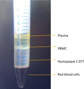
Mononuclear cells (lymphocytes and monocytes) were obtained following a simple method according with Bøyum (1976). This method takes advantage of density differences between mononuclear cells and other cellular constituents (red cells and PMN). Hystopaque® (density 1.077 g.mL-1, Sigma-Aldrich), a solution of polysucrose and sodium diatrizoate, was used to create a density gradient.
The blood sample was collected into 50 mL tubes with the anticoagulant citrate-phosphate dextrose adenine (CPD-1) (Polymed). In a 15 mL tube were placed 3 mL of Hystopaque and carefully layered on the same quantity of blood, avoiding any mixture. The tubes were centrifuged at 425 ×g, 20 min at room temperature with no brake. Differential migration during centrifugation results in the formation of layers of different constitution. Erythrocytes are aggregated by polysucrose and rapidly sediment, granulocytes become slightly hypertonic, which increases their sedimentation rate, resulting in pelleting at the bottom of the centrifuge tube. Mononuclear cells and lymphocytes (PMBC - peripheral blood mononuclear cells), which have a lower density than the other cells are found at the interface between the plasma and the Hystopaque constituting a white layer (Figure3.1). The layer was gently removed using a Pasteur pipette and washed three times (300 ×g for 10 min at room temperature) in phosphate buffered saline (PBS) to remove the excess of Hystopaque. Cells were then resuspended in RPMI 1640 (Lonza) at pH 7.2 supplemented with 2 mM L-glutamine (Merck), 100 μg.mL-1
of streptomycin (Sigma-Aldrich) (complete RPMI 1640 medium), 10% (v/v) of heat-inactivated fetal bovine serum (FBS) (BioWhittaker) and 20% (v/v) of colony stimulating factor (CSF) to induce monocyte differentiation into MØ. Concentration of viable cells was assessed by trypan blue staining method. This dye enters passively into
27
non-viable cells that stain blue while viable cells can actively exclude the dye. Concentration of viable cells was estimated in a Neubauer-counting chamber under optical microscope (OM). The plasma was also collected into a dry tube and stored at -20ºC until be used.
Figure 3.1. Isolation of blood peripheral mononuclear cells by density gradient. After centrifugation
on the hystopaque ® gradient the ring of monocuclear cells, enriched in lymphocytes can be visualized.
3.2.2. Direct agglutination test
Harith et al. (1986) developed an agglutination test (DAT) that can be used in large scale, easy to perform, read and interpret. When compared with other serologic tests, presents high levels of sensitivity and specificity and does not require any major equipment (Haliu, 2002) or expensive reagents/compounds
DAT procedure
The agglutination reaction detects L. infantum specific antibodies. In a positive control, the antigen forms a pale blue film over the well, but if the result is negative, antigen accumulates at the bottom of the plate, forming a dark blue spot. After an incubation period, the result is expressed as the highest dilution in which agglutination occurs. Using a 96 well V-bottom, 50 L of diluent solution previous prepared [0.15 M HCL, with 0.2% gelatin (Sigma-Aldrich) in distilled water and 0.2 M 2-mercaptoetanol
28
(Sigma-Aldrich)] were plated in the wells. Plasma samples (50 L) were added to the first column and 1:10 serial dilutions were made. For negative control were use wells without plasma. Serum samples from individuals with visceral leishmaniasis kindly provided by Prof. Carlos Henrique Nery Costa from the Federal University of Piauí, Teresina, Brazil, were used as positive controls. The antigen used was gently provided by Prof. Saul Semião Santos, Tiradentes University, Aracaju, Brazil. Then, the plates were incubated for 1 h at 37ºC and 50 L of antigen (5 μg.mL-1) were added to each well. Plates were incubated overnight at room temperature. The results were assessed visually, and are expressed as the maximum dilution at which agglutination is observed.
3.2.3. Flow cytometry
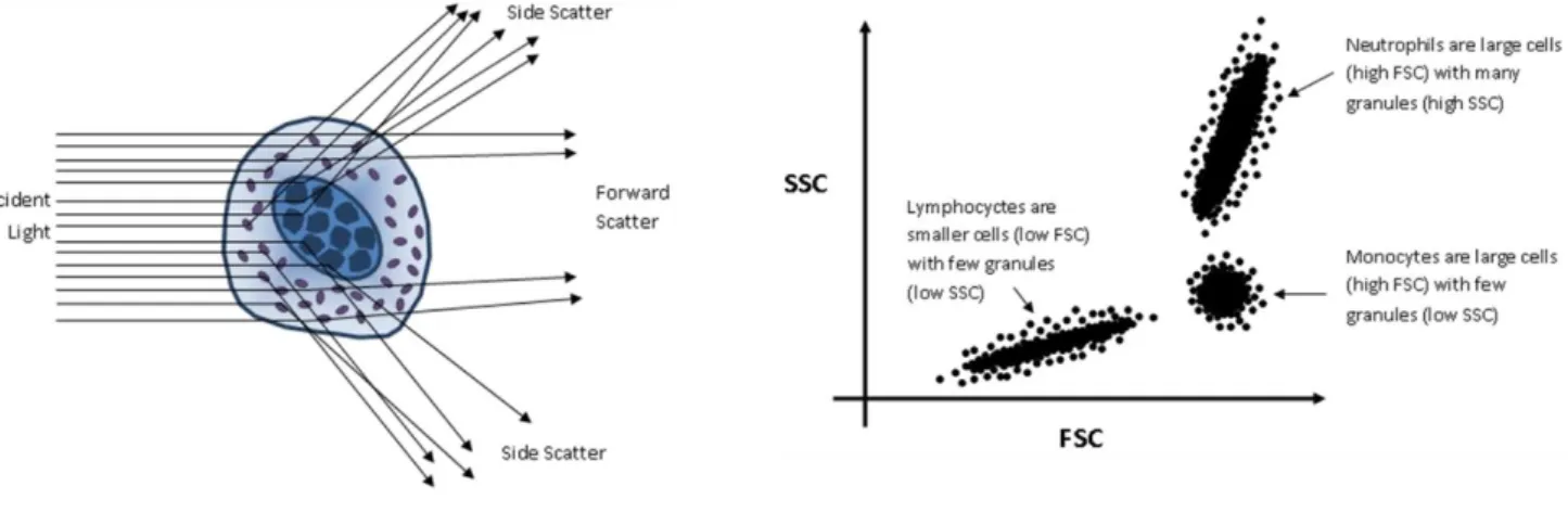
Flow cytometry is a technique widely used to analyze multiple characteristics of single particles in a fluid. This method allows the individual analysis of cellular multiple characteristics, such as size (represented by forward-angle light scatter), internal complexity (represented by right-angle scatter), and the immunophenotyping by evaluating the expression of intracellular and cell surface markers, using specific monoclonal antibodies complexed with fluorescent reagents (fluorochroms) (Figure 3.2).
Figure 3.2. Schematic representation of flow cytometry process. Cell can be evaluated by forward
scatter (FSC), which is proportional to the cell surface area or size (A) and by side scatter (SSC) that is proportional to the degree of internal complexity of the cell. Plotted results show different leukocyte populations based on size and complexity.
29
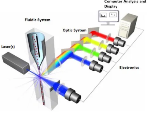
In the flow cytometer (Figure 3.3), each cell flowing in a liquid stream is intercepted by a laser light and fluorochromes release fluoresce in all directions. Optics direct the light to series of beam splitters and filters to appropriate detectors. The electronic signals produced are digitized for computer analysis (Brown and Wittwer, 2000). Since fluorochromes can have extended emission curves, different light wavelengths can be simultaneously scattered from one cell. When the emission curves overlap the proper data analysis become affected. Thus, spectral compensations obtained by subtracting a percentage of signal from one detector to the other and aligning populations affected by spillover are a very important way to solve this issue.
Figure 3.3. The principal components of a flow cytometer: The flow system includes the fluidics, optical system, light sensing, electronic system and the signal processing. https://www.labome.com/method/Flow-Cytometry-A-Survey-and-the-Basics.html
3.2.4. Peripheral blood monocyte differentiate macrophage
Monocytes circulate in the bloodstream until migrated to other tissues, differentiating into MØ able to develop and execute specialized functions. In order to maximize the number of MØin culture, it was necessary to determine the best incubation time.
In a six wells plate, 3 mL of monocyte suspension (described in 3.2.1) was seeded per well and the plates were incubated in an incubator at 37 ºC ± 1 ºC, with 5% CO2 in a
30
humidified atmosphere. At different time points (24 h, 48 h and 72 h) obtained samples were acquired by flow cytometry.
Macrophages are cells with ability to adhere to a substrate as is the case of polyethylene terephthalate, depositing on the bottom of the plate. At each time point, cells were washed with 0.9% NaCl and removed by thermal shock (30 min on the ice) using a sterilized cell scraper (VWR) in a gentle way to ensure the integrity of plasmatic membrane.
The cell suspension was centrifuged (300 ×g for 10 min at room temperature) and the pellet was resuspended in 500 µl of 2% paraformaldehyde, and then set on ice for 20 min protected from light. Cells were then washed (400 ×g, for 10 min at + 4 ºC), the supernatant were rejected and the pellet resuspend in 500 µl of PBS 1×. Cells were kept in the dark at + 4 ºC until be analyzed by flow cytometry.
3.2.5. Isolation of lymphocytes
As previously mentioned (3.2.4) MØ adhere to polyethylene terephthalate, so it is possible to isolate lymphocytes, non-adherent cells (supernatant), after 24 h of incubation time.
The cellular suspention was centrifuged at 300 ×g for 10 min at room temperature. Then re-suspended in complete RPMI 1640 medium supplemented with 10% (v/v) FBS and 1% of DMSO and preserved at -20 ºC until be used.
3.3. Parasites
Four species of Leishmania were used in the present study. The visceral L. infantum (MCAN/2012/IHMT0003SG) isolated from a case of canine leishmaniasis (LCan) from Seixal municipality (Setúbal, Portugal) and maintained in BALB/c mice by successive passages. To assure the use of virulent parasites, only L. infantum promastigotes with less than five passages in culture (Santos-Gomes and Abranches, 1996) was used throughout the study.
Cutaneous strains of Leishmania were kindly provided by Dr. Luiz Felipe Passero. L.
31
diffuse leishmaniasis, living in Pará state (Brazil). L. shawi (MHOM/BR/96/M15789) was isolated from a patient with LC from Buriticupu (Maranhão state, Brazil) and L.
guyanensis (MHOM/BR/2001/M19663) was isolated from a patient with LCL, living in
Santarém (Pará state, Brazil). The parasites were maintained in BALB/c mice, being subsequently isolated from lesions on the footpads. Parasite identification was based on monoclonal antibodies and multilocus enzyme electrophoresis at the Evandro Chagas Institute (Passero et al., 2012).
Parasites were cultured in Schneider's Insect Medium (SCHN, Sigma-Aldrich) supplemented with 10% FBS and penicillin-streptomycin (Biochrom, Germany) at 100 U.mL-1 and 100 µg.mL-1 respectively (complete SCHN medium), and incubated at 24ºC ± 1 °C in a refrigerated incubator (Lovibond, Germany). Concentration of promastigotes in culture (promastigotes.mL-1) was determined under OM using a Neubauer-counting chamber and calculated according to the following equation:
N= promastigotes.mL-1
n= total number of promastigotes counted
50=factor associated with the number of squares counted FD =Dilution factor
=Volume correction factor
This equation was adapted for counting on the central square of Neubauer-counting chamber.
3.3.1. Macrophage Infection
In a chamber slide (LabteK Chamber SlideTM, Nunc) was added 300 L of cell suspension in complete RPMI medium supplemented with 20% (v/v) CSF and 10% (v/v) FBS. Every day the culture medium was replaced with fresh medium until MØ achieved complete differentiation on the 3th day of culture. Cells )
32
were then exposed to L. amazonensis, L. guyanensis, L. shawi or L. infantum promastigotes at MØ/parasite ratio of 1:5. After 5 h at 37°C in a 5% CO2 humidified
atmosphere, cells were washed with PBS 1× to remove non internalized free promastigotes. Plastic chamber was detached from slide and left to dry. Slides were fixed with methanol and stained with Giemsa. The amount of infected cells (i.e. number of infected cells per 100 MØ) and the intensity of infection (i.e. number of amastigotes per infected cells) were determined by light microscopy at ×1000 magnification by counting at least 100 cells per well.
3.3.2. MET
3.3.2.1.
Scanning electron microscopy
As scanning electron microscope (SEM) uses a focused beam of high-energy electrons to interact with the solid surface of the sample, generating high resolution 2D-images. In the present study, SEM was used to evaluate the ability of Leishmania spp. to induce MET release.
Samples for SEM were prepared according to Brinkmann et al. (2010).
In a 24-well cell plate, sterile glass cover slip was placed and plated. Then L. amazonensis, L. guyanensis, L. shawi or L. infantum promastigotes were added at macrophage/parasite ratio of 1:5. In parallel, LPS-stimulated MØ PMA-stimulated MØ and PMA-stimulated MØ exposed to Leishmania spp. were used as positive controls. After 5 h at 37°C in a 5% CO2 humidified atmosphere, the coverslips
were fixed with 2.5 % glutaraldehyde (Sigma-Aldrich).
Later on, coverslips were washed three times with water and transferred to new 24 plate with 0.5% osmium tetroxide (OsO4, Sigma-Aldrich) and incubated for 30 min at room
temperature. Water washing steps were realized before 30 min of incubation at room temperature with 1% tannic acid (Sigma-Aldrich). Slides were then submitted to new washing, incubated again with 0.5% OsO4 solution and washed again. Cells were then
dehydrated by successive passages in increasing concentrations of ethanol (30%, 50%, 70%, 80%, 90% and 100%) and conserved at +4ºC in 100% ethanol. At last, samples were dried by using the critical point drying method and metallized with gold
33
palladium. Cells were observed under a SEM (JEOL 5200-LV, Japão) and images were acquired.
3.3.2.2.
Histone detection by immunolabeling
Immunolabeling is a biochemical process that enables the detection and localization of an antigen in a particular site within a cell. In the present study immunolabeling was used to localize histone during macrophage infection and treatment.
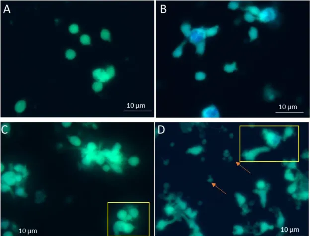
For this procedure, after the coverslips being fixed in the well with 2.5% glutaraldehyde (as described in 3.3.2.1.), cells were washed three times with PBS 1× and permeabilized using 0.5% Triton X-100 (Sigma-Aldrich). Then, coverslips were washed again (PBS 1×) and transferred to new 24 well plate with blocking buffer. The plate was incubated in a humid chamber for 30 min at 37ºC. After incubation, cells were stained with the primary antibody Histone H1 (AE-4, Santa Cruz Biotechnology, Germany) diluted in blocking buffer and incubated (1 h, 37ºC). The coverslips were submitted to a new washing and the secondary antibody goat anti-mouse IgG (Santa Cruz Biotechnology, Germany) was added. Cells were incubated for 1 h at 37ºC, washed and stained with DAPI (4',6-diamidino-2-phenylindole) (Sigma-Aldrich). DAPI is a fluorescent dye that strongly binds to the A-T regions of DNA and can be used to stain both live and fixed cells. The slides were maintained in the dark at + 4 ºC until be analyzed by fluorescence microscopy.
3.3.3. Production of nitric oxide by macrophages
In general, infected Ø can produce nitric oxide (NO). NO is generated by inducible NO synthase (iNOS), an enzyme that catalyzes the oxidation of L-arginine in NO and L-citrulline.
Supernatants of resting-MØ, PMA-stimulated MØ and MØ exposed to Leishmania promastigotes for 5 h were used to quantify total nitrate plus nitrite by Nitrate/Nitrite Colorimetric Assay Kit (Abnova, Taiwan).
34
This method converts the ion NO3- present in the supernatants to ion NO2- by the
enzyme nitrate reductase. Subsequent addition of Griess reagent converts NO2- in to azo
chromophore pink/purple compound, allowing the determination of total concentration of both metabolites in the sample.
The assay was made according to the manufacturer’s instructions without sample dilution. A standard curve was built to estimate the concentration of the metabolites in the sample, by reading the absorbance of known NO3 amounts (final concentrations
between 35 M and 5 M). Absorbance was measured at 550 nm using a microplate reader (Anthos 2010, Austria).
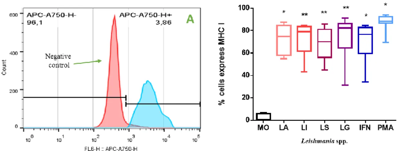
3.3.4. Evaluation of MHC in infected-MØ when in contact with
lymphocytes
Lymphocytes, previously isolated were added to homologue infected-MØ and incubated for 20 h at 37 ºC in a 5% CO2 humidified atmosphere. The cells were removed
according with 3.2.4. Then, the cell suspension was centrifuged (300 ×g for 10 min at room temperature) and the pellet was resuspended in 500 µl of PBS 1×. Cell suspensions were equally divided into two 2 mL Eppendorf tubes (DeltaLab),the first one, stained with APC human HLA-A,B,C (BioLegend) and the other, FITC anti-human HLA-DR, DP, DQ (BioLegend). Then, cells were set on ice for 20 min protected from light. Cells were washed (400 ×g, for 10 min at + 4 ºC), the supernatant was rejected and the pellet was resuspend in 500 µl of 2% paraformaldehyde. Afterwards, cells were set on ice for 20 min and protected from light. At the end, one more wash was made, and the pellet was resuspend in 500 µl of PBS 1× and cells were kept in the dark at + 4 ºC until be analyzed by flow cytometry (Cytoflex, Beckman coulter life science). The data analyses were made in FlowJo vX 0.7.
35
3.3.5. Statistical analysis
Statistical analysis was performed with the IBM SPSS Statistics version 23.0 (IBM, USA).The non-parametric Wilcoxon test was used to compare variables of two dependent samples from 10 samples of Leishmania-infected macrophages evaluated in triplicate. Differences were considered significant with a 5 % significance level (p<0.1).
36
4. Results and Discussion
4.1. Participants did not evidence previous contact with L. infantum
parasites
DAT was negative for all participants, evidencing no previous contact with L. infantum. These results indicate that the individuals that participated in this study, did not develop antibodies anti-L. infantum, suggesting that they were never infected.
4.2. Macrophages
4.2.1. Complete macrophage differentiation was achieved after 72h of
incubation
Macrophages derived from peripheral blood monocytes and are strategically located throughout lymphoid and non-lymphoid body tissues However, not only tissue MØare difficult to obtain as it is also hard to get in a high number to be used as a research tool. Thus, isolate monocytes from peripheral blood and differentiate into MØ is a way to overcome this barrier.
After peripheral blood monocyte isolation (PBM) the period of time needed for MØ differentiation was established by flow cytometry analysis. Taken into account that MØ present higher forward (size) and side scatter (complexity) levels than monocytes, it was estimated a period of 72h for MØ achieve complete differentiation. Furthermore, morphologically these cells showed an extensive ruffled plasma membrane and a large purple-stained nucleus, presenting an irregular shape (Figure 4.1) characteristic of MØ and, also exhibited the phagocytic ability. Consequently, further assays were performed with 72 h monocyte derived MØ.
37
Figure 4.1. Macrophages differentiation. Peripheral blood monocytes were left to differentiate into MØ
in the presence of CSF. Forward Scatter (x axis) VS Side Scatter (y axis) dot plots generated at 24 h, 48 h and 72 h of culture are showing. The selected area R1 correspond to monocytes, R2 to other smaller and less complex cells and R3 correspond to fully differentiate MØ. After 72 h of differentiation, cells were stained with Giemsa and observed by optical microscopy. Acquired images (× 1000 magnification, scale bar corresponds to 10 µm) exhibit cells with morphological characteristics of macrophages (A).
4.2.2. Cutaneous species of Leishmania show high infectivity to human
macrophages
The percentage of MØ infected with Leishmania parasites and the number of intracellular parasites per macrophage using the macrophage : parasite proportions of 1:5 for 5 h of incubation time was determined by morphological evaluation under optical microscope (Figure 4.2).
38 L e i s h m a n i a s p p . P e r c e n ta g e o f in fe c te d c e ll s L A L I L S L G 0 2 0 4 0 6 0 8 0 1 0 0
Figure 4.2. Macrophages infected with Leishmania spp. Macrophages were exposed to promastigotes
of L. amazonensis (LA), L. infantum (LI), L. shawi (LS) and L. guyanensis (LG) at a MØ/parasite ratio of 1:5 for 5h. The percentage of MØ infected was determined by light microscopy (× 1000 magnification) by counting at least 100 cells per well in slides stained with Giemsa. The results are expressed as standard deviation of ten samples. *(p<0.05) indicates statistical differences between LI and the cutaneous species.
The percentage of cells infected with cutaneous species have a higher rate in comparison to the visceral species(p=0.0286). L. infantum had no more than 43%. This result was similar to the percentage of human PBM infected with L. infantum promastigotes obtained by Ogunkolade et al. (1990) and to the human PBM infected with Leishmania strains (IMT-151/ IMT-373) obtained by Maia et al. (2007).
And clearly, L. amazonensis is the species with the highest rate of infection (89%), followed by L. shawi (78%) and L. guyanesis (74%). Similar results were obtained for Campos et al. (2008) in both strains (L. shawi and L. guyanesis), although significant differences were not found.
39 L e i s h m a n i a s p p . N o . a m a s ti g o te s / 1 0 0 m a c r o p h a g e s L A L I L S L G 1 0 1 0 0 1 0 0 0 *
Figure 4.3. Intensity of macrophage infection caused by Leishmania spp. Macrophages were exposed
to promastigotes of L. amazonensis (LA), L. infantum (LI), L. shawi (LS) and L. guyanensis (LG) at a MØ/parasite ratio of 1:5. The number of intracellular amastigotes per 100 cells (A) was determined at 5 h post infection by light microscopy (× 1000 magnification) in slides stained with Giemsa. The results are expressed as the mean and standard deviation of ten samples. * (p<0.05) indicates statistical differences between LI and the cutaneous species. Optical microscopy images of macrophages showing intracellular amastigotes of L .amazonensis (A), L. shawi (B) and of L. guyanensis (C) were also acquired.
The number of amastigotes present in the cells for each strain intensifies the infection rates. Once again, L. infantum have the lowest value (median = 58 amastigotes), showing less permissiveness(p=0.0286), in contrast with L. amazonensis (median = 428 amastigotes), intermediate values for L. shawi (median = 264 amastigotes) and L.
40
4.2.3. Leishmania spp. do not promote macrophage oxidative burst
The production of nitric oxide (NO) by the enzyme NO synthetize (iNOS) is one of mechanism used by MØ to destroy parasites that can be induced by cytokines and other immune stimuli. However, it has been reported that Leishmania spp. inhibit macrophage NOS2expression (Bogdan et al., 2000). NO production by MØ exposed to visceral and cutaneous species of Leishmania were evaluated (Figure 4.4).
Figure 4.4. Nitric oxide production by macrophage exposed to Leishmania spp. Supernatants of
blood monocyte derived macrophages incubated with promastigotes of L. amazonensis (LA), L.
infantum (LI), L. shawi (LS) and L. guyanensis (LG) at a MØ/parasite ratio of 1:5 for 5h were used to
evaluate NO release. In parallel, supernatants of resting MØ and of PMA-stimulated MØ were also assessed. *(p<0.05) indicate statistical differences between LI and other conditions.
It was possible to detect the presence of NO in the infected MØ, and as expected these results were low because Leishmania spp. inhibits the NO production in the infected cells, facilitate the initial survival of the parasites in the host, significant differences were not found. Leishmania infatum was the species that caused higher inhibition of MØ oxidative burst (p=0.0148). L e i s h m a n i a s p p . [N it r a te + N it r it e ] m M M O L A L I L S L G M O + P M A 0 .0 0 .2 0 .4 0 .6 *