Susana Maria Ribeiro e Sousa Mendes de Freitas
Albumin and fibronectin adsorption
and osteoblast adhesion on titanium oxides
Tese submetida à Faculdade de Engenharia da Universidade do Porto
para candidatura à obtenção do grau de Doutor em Engenharia Metalúrgica e de Materiais
Faculdade de Engenharia Universidade do Porto
Professor Mário Adolfo Barbosa (supervisor) Faculdade de Engenharia, Universidade do Porto
INEB – Instituto de Engenharia Biomédica, Laboratório de Biomateriais, Universidade do Porto
Professor Pedro Moradas Ferreira (co-supervisor)
Instituto de Ciências Biomédicas Abel Salazar, Universidade do Porto IBMC – Instituto de Biologia Molecular e Celular, Universidade do Porto
The research described in this thesis was conducted at:
INEB – Instituto de Engenharia Biomédica, Laboratório de Biomateriais
IBMC – Instituto de Biologia Molecular e Celular, Cellular and Applied Microbiology laboratory and Advanced Light Microscopy facility
IST, Instituto Superior Técnico, Centro de Química Estrutural IST, Instituto Superior Técnico, Departamento de Física
The research in this thesis was financially supported by:
I would like to express my sincere gratitude to the persons who helped me during this long-term project, and made this thesis possible.
I will start acknowledging my supervisors, Professor Mário Barbosa and Professor Pedro Moradas Ferreira for the opportunities they gave me, and for all of their help, support, patience, and friendship during the development of this work. It was for me a privilege to have the opportunity of working in renowned institutions, INEB and IBMC that have hosted me in a very friendly way, and always made available every necessary mean to carry out my research work. At these institutions I found exceptional researchers with whom I shared experiences and learned with, but I also made friends for life.
I would like to acknowledge Professor Fernando Jorge Monteiro for his friendship along these years.
I would also like to acknowledge the collaboration of several persons from the following institutions:
At INEB: I will probably face the most difficult of the tasks, which is to express my sincere gratitude to all my remarkable colleagues and friends at INEB, with whom I had the privilege of working with. As much as I could write here would never be enough to show how grateful I am to them all.
I would like to thank to a long time and dear friend Cristina Ribeiro, whose friendship goes far beyond laboratory relationship. Thank you for being there whenever I needed!
I also like to thank Isabel Amaral for being a special friend that also provided me with a considerably enrichment of knowledge of my work do to her excellent competence.
I am also very grateful to Manuela Brás and Meriem Lamghari for their friendship and support and for work collaboration in AFM and cell culture experiments.
I am thankful to Ana Paula Pêgo, Cristina Barrias, and Cristina Martins, for their encouragement, prompt advices, patience, and above all for their friendship. Ana Paula Filipe, Ana Queiroz, Judite Barbosa, Maria Ascenção Lopes, Maria Pia Ferraz, and Pedro Granja thank you for being always my friends and supported me in this work.
contributed with their help and friendship and made my life at the lab much easier. I am very grateful to them all: Anabela Dias, Andreia Cabral, Alis Mateus, Ana Rosa Carvalho, Carla Monteiro, Carlos Fonseca, Cristina Matos, Daniel Duarte, Diamantino Falcão, Dulce Carqueijó, Eliana Vale, Fátima Pina, Hugo Oliveira, Inês Gonçalves, Lino Ferreira, Januário Lima, José Paulo Pereira, Marta Lima, Patrícia Cardoso, Pedro Sá, Rui Azevedo, Sandra Teixeira, Serafim Oliveira, Sofia Teixeira, Susana Carrilho, and Virgínia Fonseca.
At ISEP: I would like to express my sincere gratitude to my colleagues and friends, especially to Prof. Cristina Delerue Alvim Matos for all the support, and encouragement, but above all for being my friend.
At IBMC: To Dra. Amélia Amorim, to Victor Costa, Frederico Silva, for their availability whenever needed; to Paula Sampaio for her help in Confocal Laser Scanning Microscopy analyses; to Isabel Cardoso and M Rosário Almeida who guide me at the beginning of my radiolabelling work.
At CEMUP: To Doctor Carlos Sá for his assistance in XPS analyses.
At IST: To Professor Benilde Saramago, from Departamento de Engenharia Química, for scientific support in the characterization of surface energetics. To Professor Luís Viseu Melo, from Departamento de Física, for his assistance in AFM measurements.
At INESC: To Professor Paulo Freitas and Doctor Susana Freitas for the titanium oxide sputtered sample preparation.
Many thanks to all my other friends for their support, particularly in the most difficult times. No one is forgotten even if it is not mentioned! I would like to especially express my gratitude to Nina Maio who was always present and for being a true friend.
Eugénia Leitão and Neal for being exceptional friends all along these years, and for english assistance in the final phase of this thesis.
want to acknowledge all the support and friendship.
At last, but above all, special thanks to Miguel for your support and patience, for being always unconditionally present when I needed you most, and for supporting my choices! Thank you dear Leonor and Francisca for your patience and comprehension for every weekends and holidays I had to work in my thesis instead of being available to be with you both. Your prayers were finally heard!
Several promising multifunctional biomaterials are currently being investigated and applied to new types of artificial organs and advanced medical devices. However, metals are still the more suitable biomaterials to replace hard tissue due to good bone matching mechanical properties. The physiological stability of titanium associated with its corrosion resistance as well as exceptional mechanical properties and favorable osteointegration, makes it one of the most used biomaterials for bone and cartilage replacement.
When biomaterials are implanted in the body, proteins immediately adsorb onto the implant surfaces. The tissue response to implanted materials arises mainly from differences in the adsorption of proteins that depend on surface parameters, such as chemical composition, surface roughness, and surface charge, among others.
The main objective of this thesis was to identify the main features that govern the protein adsorption on titanium oxide surfaces using different topographies, at the nanometric scale. The experimental approach involved two key proteins, albumin (HSA) and fibronectin (FN), adsorbed both from single protein, as well as from complex protein solutions. Albumin is the predominant plasma protein, and is generally considered to “passivate” surfaces preventing cell adhesion and greatly reducing the acute inflammatory response to the material. On the contrary, FN is an adhesive protein of the extracellular matrix (ECM) required for integrin-receptor-based cell adhesion, cytoskeleton reorganization, proliferation, and differentiation. This work was based on the preparation and characterization of titanium oxide surfaces chemically similar to those used in clinical practice, with different roughnesses. Two surfaces, TiO2 sputtered on Si (TiO2 sp), and TiO2 formed on commercially pure titanium after H2O2 immersion (TiO2 cp), were developed. The two surfaces presented identical chemical composition but different topography, as shown by XPS, wettability studies, and AFM. Surface roughness was always significantly higher on the TiO2 cp surfaces.
Subsequently, the above TiO2 surfaces were evaluated regarding their ability to adsorb human serum albumin, HSA, or human plasma fibronectin, FN, both from single protein solutions, plasma, and diluted plasma. FN showed a contrasting affinity to the TiO2 surfaces, as compared to HSA. A higher affinity of the FN solution to the TiO2 cp surface was found, as compared to the TiO2 sp. The radiolabelling data also shown more FN adsorbed onto TiO2 cp than onto TiO2
surface, as suggested by the work of adhesion and by the exchangeability studies, following the trend observed for albumin. AFM allowed direct visualization of the adsorbed FN in air and in liquid, on both surfaces tested. Upon adsorption, FN appeared to form globular aggregates or ellipsoids. The FN aggregates coalesced forming clusters as time of adsorption and concentration increased. AFM results provided novel evidence that FN adsorbs spontaneously as self-assembly at TiO2 surfaces, as a function of time. The aggregate structure is an intermediate feature shared by some protein fibrillar assemblies at interfaces, which is believed to promote cell adhesion, and cytoskeleton organization.
Finally, the effect of competitive preadsorption of HSA and FN on both TiO2 substrates from binary protein mixtures and from 10% plasma on osteoblast adhesion and morphology was investigated, using MC3T3-E1 osteoblasts. Our results showed a direct correlation between osteoblast adhesion and the amounts of FN and HSA adsorbed on TiO2 surfaces. In addition, fewer osteoblasts adhered after 4 and 24h to both substrates after prepreadsorbed with HSA, HSA/FN=200 and 10% plasma, as compared to the surfaces preadsorbed with FN and HSA/FN=1. For the latter, FN was able to compensate the inhibitory effect of HSA on osteoblast adhesion. Therefore, the presence of lower amounts of co-adsorbed albumin may improve presentation of FN in a more integrin-recognized conformation. This fact suggests that some degree of molecular packing prevents loss of integrin-binding activity.
FN reversibility does not seem to be dependent on the HSA/FN adsorption mass ratio in solution, suggesting that FN competitively adsorbs to TiO2 in a favorable conformation allowing exchange with other FN molecules in solution.
In conclusion, the HSA and FN adsorption pattern on TiO2 and the subsequent response of osteoblasts (cell adhesion and morphology), depends on the type of TiO2 surface. The presence of lower amounts of co-adsorbed albumin may improve the exposure of FN in a more integrin-recognized conformation, suggesting that some degree of molecular packing is likely to prevent loss of integrin-binding activity.
Plusieurs biomatériaux multifonctionnels sont actuellement étudiés et aussi utilisés dans différents types d’organes artificiels ainsi que dans des outils médicaux. Cependant, les métaux sont toujours considérés plus adéquats pour substituer le tissue osseux due à la proximité de leur propriétés mechaniques. La stabilité physiologique du titanium associée à sa résistance à la corrosion, ses propriétés mechaniques expetionnelles et son potentiel d’ostéointégration ont permis qu’il soit le biomatériau le plus utilisé pour la substituition de l’os et du cartilage.
Quand les biomatériaux sont implantés dans l’organisme, les protéines adsorbent immédiatement à la surface de l’implant. La réponse du tissu hôte aux matériaux implantés résulte éssentiellement des différences dans l’adsorption des protéines. Cette réponse dépend des paramétres de la surface de l’implant dont la composition chimique, la rugosité et la charge. Le principal objectif de ce travail a visé d’identifier les caractéristiques principales qui régulent l’adsorption des protéines aux surfaces du titanium oxidé en utilisant différentes topographies à l’échelle nanométrique. L’approche expérimentale inclu deux protéines clefs, l’albumine (HSA) et la fibronectine (FN). L’adsorption de ces deux protéines a été réalisée à partir d’une solution de HSA ou de FN ou d’une solution protéique plus complexe. La HSA est la protéine prédominante du plasma et généralement considérée de type passivant des surfaces, évitant l’adhésion céllulaire et réduisant la réponse inflammatoire au matériau implanté. Contrairement à la HSA, la FN est une protéine d’adhésion de la matrice extracéllulaire (MEC) nécessaire à l’adhésion céllulaire via les récepteurs d’integrines, à la réorganisation du cytosquelette, à la prolifération et à la differenciation des céllules.
Ce travail a ciblé la préparation et la charactérisation des surfaces du titanium oxidé, chimiquement similaires à celles utilisées en clinique, avec différentes rugosités. Deux surfaces, TiO2 ‘sputtered’ en Si (TiO2 sp) et TiO2 obtenu à partir du titanium pure, commercialement disponible, après immersion dans H2O2 (TiO2 cp), ont été developpées. Les deux surfaces présentent une composition chimique identique mais une topographie différente comme a été démontré par le XPS, les études de mouillage et l’AFM. La surface du TiO2 cp a montré une rugosité plus élevée.
Ultérieurement, l’étude de la capacite des deux surfaces de TiO2 décrites ci-dessus d’adsorber l’albumin du serum humain, la HSA, la fibronectine du plasma humain ou la FN à partir d’une
différente aux deux surfaces de TiO2 en comparaison avec celle de l’HSA. La solution de FN a exhibé une plus grande affinité aux surface de TiO2 cp en comparaison à celle observée en présence du TiO2 sp. De plus, les études de radioactivité ont montré que la FN adsorbe préfèrentiellement aux surfaces TiO2 cp qu’à celles du TiO2 sp. Cependant, les molécules de FN semblent être plus fortement attachées à la surface du TiO2 cp, comme a été suggéré par les études d’adhésion et de permutation, avec une tendance analogue à celle observé en présence d’albumin. La technique d’AFM a permis une visualisation directe de la FN adsorbée dans les conditions air ou liquide sur les deux surfaces testées. Après l’adsorption, la FN semble former des aggrégats globulaires ou éllipsoides. Ces aggrégats s’unissent et forment des "clusters" au fur et à mesure que le temps d’adsorption et la concentration augmentent. Les résultats d’AFM ont apporté une nouvelle évidence montrant que la FN adsorbe spontanément avec une autoorganisation aux surfaces de TiO2 en fonction du temps. La structure des aggrégats est une caractéristique intermédiaire partagée par certains rassemblement de protéines fibrillaires au niveau des interfaces considérées comme promotrices d’adhésion céllulaire et organisation du cytosquelette.
L’effet de la compétitivité de préadsorption de la HSA et de la FN sur les deux substrats de TiO2 à partir de solution binaire de protéines ou de 10% de plasma sur l’adhésion et la morphologie des ostéoblastes a été étudié. Les ostéoblastes MC3T3-E1 ont été utilisés. Les résultats montrent une corrélation directe entre l’adhésion des ostéoblastes et les concentrations de la FN et de la HSA adsorbées au niveau des surfaces de TiO2. De plus, un faible nombre des ostéoblastes adhérent après 4 et 24h aux deux surfaces préalablement préadsobées avec la HSA, HSA/FN=200 et 10% de plasma en comaparison avec les surfaces préadsobées avec la FN et HSA/FN=1. Cependant, la FN a pu compenser l’effet inhibiteur de l’HSA sur l’adhésion des ostéoblastes. Par conséquent, la présence de faible concentrations de l’HSA adsorbée peut améliorer la présentation de la FN sous forme d’une conformation permettant une reconnaisance de la protéine via les integrines. Ceci suggére qu’un certain degré de compactage moléculaire évite la perte de l’activité résultante de la liaison des integrines.
L’effet réversible de la FN ne semble pas être dépendant de l’adsorption de la masse du ratio HSA/FN en solution, suggérant que la FN adsorbe compétitivement au TiO2 avec une
de TiO2. La présence de faibles concentrations d’albumin co-adsorbée peut améliorer l’éxposition de la FN sous une conformation permettant une reconnaisance de la protéine via les integrines. Ceci suggére qu’un certain degré de compactage moléculaire paraît esquiver la perte de l’activité résultante de la liaison des integrines.
Presentemente existe uma vasta gama de biomateriais promissores que estão a ser investigados ou aplicados no desenvolvimento de novos tipos de órgãos artificiais e dispositivos médicos. No entanto, os metais, devido às suas propriedades mecânicas, são ainda considerados os biomateriais mais apropriados para substituição de tecido duro. A estabilidade fisiológica do titânio associada à sua resistência à corrosão, assim como às propriedades mecânicas e osteointegração favorável, contribuem para que este seja um dos biomateriais mais utilizados para a substituição do osso e da cartilagem.
Quando um biomaterial é implantado, as proteínas adsorvem imediatamente à sua superfície. A resposta do tecido aos materiais implantados surge principalmente das diferenças na adsorção das proteínas que, por sua vez, dependem de parâmetros de superfície, composição química, rugosidade e carga de superfície, entre outras.
O objectivo principal desta tese foi o de identificar as principais características que medeiam a adsorção de proteínas em superfícies do óxido titânio com diferentes topografias à escala nanométrica. O trabalho envolveu a adsorção de duas proteínas chave na adesão celular, a albumina (HSA) e a fibronectina (FN), provenientes de soluções simples com uma só proteína, de soluções com duas proteínas, assim como de plasma. A albumina é a proteína predominante do plasma, e é conhecida como “passivante” das superfícies prevenindo a adesão celular e reduzindo a resposta inflamatória ao material implantado. Por outro lado, a FN é uma proteína de adesão da matriz extracelular (ECM) conhecida como fundamental na adesão, na reorganização do citoesqueleto, na proliferação, e na diferenciação celular.
A primeira parte desta tese visou a preparação e caracterização de superfícies de óxido de titânio quimicamente idênticas às usadas na prática clínica, com duas rugosidades distintas. Para isso preparam-se duas superfícies de óxido de titânio, TiO2 “sputtered” em Si (TiO2 sp) e TiO2 formado por imersão de titânio comercialmente puro em H2O2 (TiO2 cp). Através das técnicas de espectroscopia de fotoelectrões de raios-X (XPS), dos estudos de molhabilidade, e por microscopia de força atómica (AFM), foi possível confirmar uma composição química idêntica e topografia diferente. A rugosidade de superfície foi sempre significativamente mais elevada nas superfícies de TiO2 cp.
2 partindo de soluções simples de proteína, de plasma e de plasma diluído. A FN mostrou diferente afinidade para as superfícies de TiO2, quando comparada com a HSA. Através da técnica de marcação radioactiva observou-se que a FN adsorve mais à superfície de TiO2 cp do que à superfície de TiO2 sp. No entanto, as moléculas de FN pareceram estar mais fortemente aderentes à primeira superfície, como sugerido pelo trabalho de adesão e pelos estudos de desorção, seguindo a mesma tendência observada para a HSA. Os estudos de AFM permitiram a visualização directa ao ar e em água da FN adsorvida, para as duas superfícies testadas. Após adsorção, foi possível observar agregados globulares de FN, que coalesceram com o tempo de adsorção assim como com o aumento de concentração. Os agregados são estruturas intermediárias partilhadas por algumas proteínas fibrilares em interfaces, que se crê promoverem a adesão celular e a organização do citoesqueleto.
Por último, avaliou-se o efeito da competição na preadsorção de HSA e FN aos substratos de TiO2, a partir de soluções binárias das duas proteínas e do plasma a 10%, na adesão e morfologia de osteoblastos MC3T3-E1. Os resultados mostraram que existe uma correlação entre a adesão dos osteoblastos e as quantidades de FN e HSA adsorvidas às superfícies de TiO2. Existem menos osteoblastos aderentes aos dois substratos às 4 e 24h após preadsorção com HSA, HSA/FN=200 e 10% plasma, quando comparados com a preadsorção com FN e HSA/FN=1. Para esta última condição, a FN conseguiu compensar o efeito inibidor da HSA na adesão de osteoblastos. A presença de pequenas quantidades de albumina a adsorverem simultaneamente com a FN, parece contribuir para que esta última assuma uma conformação adequada ao reconhecimento pelas integrinas, sugerindo que um certo grau de empacotamento molecular parece prevenir a perda de actividade por ligação às integrinas. A reversibilidade da FN não parece ser dependente da razão de HSA/FN em solução, facto que sugere que a FN adsorve competitivamente ao TiO2 numa conformação favorável à permuta com outras moléculas de FN em solução.
Em resumo, a adsorção de HSA e FN ao TiO2 e subsequente resposta dos osteoblastos (adesão celular e morfologia), dependem do tipo de superfície de TiO2. A presença de pequenas quantidades de HSA a coadsorverem com a FN parecem contribuir para que esta proteína adsorva numa conformação favorável ao reconhecimento por integrinas, sugerindo
Acknowledgements i
Abstract v Résumé vii Resumo xi Contents xv
List of Tables and Figures xvii
List of abbreviations xxiii
Chapter I Introduction 1
1. Titanium as a biomaterial for bone replacement 1
2. Titanium oxide 3
2.1 Physico-chemical properties of titanium oxide 3 2.2 Composition, thickness and structure of oxide films on pure titanium 5 2.3 Modification and physico-chemical properties of titanium oxide films in
contact with electrolytes and body fluids 10
2.4 Surface treatments of titanium 14
3. Protein adsorption at solid/liquid interfaces 15
3.1 Protein structure 15
3.2 Protein adsorption 16
3.2.1 Type of interactions 18
3.2.1.1 Electrostatic interactions 18
3.2.1.2 Hydrophobic interactions 19
3.2.2 Conformational stability of proteins 19 3.2.3 Structure of the adsorbed protein layer 20
3.2.4 Competitive protein adsorption 21
3.2.5 Reversibility of protein adsorption 21
4. Cell/material interactions 22
4.1. The role of adsorbed proteins in tissue response to biomaterials 22
5. Techniques for protein adsorption study at biomaterials surfaces 26
6. Aim and structure of the thesis 29
7. References 34
Chapter II Human Serum Albumin Adsorption on TiO2 from Single Protein
Solutions and from Plasma 47
Chapter III TiO2 type influences fibronectin adsorption 83
Chapter IV Dynamics of fibronectin adsorption on TiO2 surfaces 101
Chapter V Osteoblast adhesion and morphology on TiO2 depends on the
competitive preadsorption of HSA and FN 131
Chapter I
Table 1 - Purity grades of CP Titanium according to ISO 5832-2:1993. The C and H wt % are, for all grades, 0.1 and 0.015, respectively. [adapted from ref3]………....2 Figure 1 – Crystallographic unit cell of the two most common forms of titanium oxide, TiO2, a)
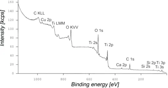
rutile and b) anatase. Grey small balls represent titanium cations, Ti4+, black balls represent oxygen anions, O2-.[adapted from ref.8]…………...……….…4 Table 2 - Selected physico-chemical properties of the most stable titanium oxide, TiO2.1,4,8,9…..4 Figure 2 – Typical XPS survey spectrum of Ti CP mechanically polished with a native, room
temperature grown oxide film.12.. …………...…………...…………...6 Table 3 – Typical XPS oxide film composition (at %) and oxide layer thickness of TiCP after
different surface treatments. [adapted from ref.12]…………..………...6 Figure 3 – XPS Ti2p emission peak recorded on an evaporated high-purity titanium film. The
oxide film composition shows contributions from Ti in oxidation states +IV (TiO2), +III (Ti2O3), +II (TiO) and 0 (Ti metal).8………...………7 Table 4 – XPS Binding energies for the titanium/titanium oxide film5-7,10,13-17…………...…..…..8 Figure 4 – Schematic representation of the early interfacial processes taking place at the
interface between the titanium oxide and the biological fluid in contact with the implant. The dimensions of features are not to scale. The oxide films are shown as ordered structures for convenience, but are often amorphous or only partially crystalline.8………...13, 14 Figure 5 – The primary, secondary, tertiary, and quaternary structure of the hemoglobin
molecule.31………16
Table 5 - Properties that determine protein adsorption at titanium surface [adapted from
references37,38]……… 17
Figure 6 - Interactions between cells and the extracellular matrix. The association between cells and the proteoglycan of the ECM is mediated by a membrane protein (integrin) and by FN an ECM protein with binding sites for both integrin and the proteoglycan.
functionality of various domains. Two subunits are shown, with amino termini to the left and carboxyl termini to the right. Regions implicated in adhesion and assembly are shown. [adapted from ref.101]...24 Figure 8 – Schematic diagram showing the synergy site and the RGD-loop that are located on
the 9th and 10th type III modules, FN-III9 and FN-III10, respectively.112………25 Table 6 - Selected techniques for protein adsorption study at biomaterials surfaces
35-37,84,119,120...27, 28
Chapter II
Figure 1 - 3D AFM images of a (a) TiO2 sputtered surface, (b) Ti cp surface (before H2O2 treatment), and (c) TiO2 cp surface (after H2O2 treatment)……….56 Table 1 - Ra, Rq, and Rmax Values for all the Ti substrates studied………..57 Table 2 – Contact angles and their standard deviations of water and diiodomethane (n=10) on
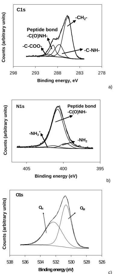
TiO2 surfaces, at t=0 s……….………....59 Figure 2 - Representative deconvolution of the XPS envelope for (a) C1s, (b), N1s and (c) O1s
peaks (after 60 min of 0.20 mg/mL HSA adsorption at a TiO2 cp surface) at 0º…….61 Figure 3 - Evolution of the XPS element envelope with increased time of albumin immersion
([HSA]=0.20 mg/mL)……….……….62 Figure 4 - Changes in atomic % with |HSA| in solution after 60 min of adsorption at a TiO2 cp surface: (a) C%; (b) N%; (c) Ti% and O%...63 Figure 5 - Time dependence contact angle of several solutions at a TiO2 cp surface…………..65 Table 3 - Contact angles and their standard deviation of PBS and different protein solutions on
TiO2 surfaces at t=600 s………66
Figure 6 - Work of adhesion between TiO2 substrates and (a) water, (b) |HSA|=4.0 mg/mL, and (c) plasma………..…….67 Figure 7 - Work of adhesion between TiO2 cp and HSA at different concentrations and between
TiO2 cp and plasma solutions………...68 Figure 8 - Work of adhesion between HSA molecules and the TiO2 substrates as a function of
2 2
HSA concentration in solution. The elution time was 24 h or 72 h for the TiO2 cp and
24 h for the TiO2 sp……….70
Figure 11 - Comparison of the kinetics of HSA adsorption (0.20 and 4.0 mg/mL) and desorption (with HSA) from a TiO2 cp surface……….…….71 Figure 12 - Comparison of the kinetics of HSA adsorption (0.20 and 4.0 mg/mL) on TiO2 sp and desorption (with HSA)………72 Figure 13 - Comparison between adsorption of albumin onto a TiO2 cp surface by 125I-HSA radiolabeling and XPS measurements of N%...75
Chapter III
Figure 1 - Representative deconvolution of XPS envelope for (a) C1s, (b) N1s and (c) O1s peak (after 60 min of FN 200 μg/mL adsorption at TiO2 sp surface) at 0º……….…...…….90 Figure 2 - Atomic % as a function of |FN| in solution after 60 min of adsorption on a TiO2 cp
surface……….91 Table 1 - Contact angle values of different solutions at TiO2 surface (n = 10)………...92 Figure 3 - Work of adhesion between TiO2 substrates and FN (|FN| = 200µg/mL)………..93 Figure 4 - Work of adhesion between TiO2 cp and protein solutions………..………93 Figure 5 - Adsorption isotherms of FN on TiO2………...94 Figure 6 - Elution of FN molecules by other FN molecules from TiO2 sp and from TiO2 cp as a
function of FN concentration in solution. The elution time was 24 h……….95
Chapter IV
Figure 1 – AFM images and cross sections of a clean TiO2 cp substrate surface, a) topography and b) phase image. The image was performed using MAC mode in water……….109 Figure 2 – AFM height and phase images of TiO2 cp surfaces, before and after protein
contrast from the surface is high enough to observe protein…………..……….110 Figure 3 - Surface roughness of TiO2 cp and TiO2 sp determined in air and water, before and
after protein adsorption (scan area: 1000x1000 nm2), a) R
q and b) Rmax…………..111 Figure 4 - AFM height (a) and phase (b) images of FN adsorbed on a TiO2 sp surface after 10
min of incubation with 50 µg/mL of FN and correspondent cross sectional profiles [1000x1000 nm2]. The images were performed using MAC mode in water………..112 Figure 5 – Topographic AFM images recorded in air (a and c) and in water (b and d) of TiO2 sp
surfaces, before and after 30 min. with 75 µg/mL of FN adsorption (lateral scan size: 2000x2000 nm2 and 1000x1000 nm2 for the higher magnification images)...113 Table 1 – Particle dimensions of FN adsorbed on TiO2 sp, under different adsorption
conditions………..114 Figure 6 – Evolution of XPS element envelope with increased time of FN immersion (|FN|=20
µg/mL): a) Ti% and O%; b) N% and C%...115 Figure 7 – Kinetics of FN adsorption (20 µg/mL) on TiO2 surfaces………116 Figure 8 – Elution of FN molecules after adsorption of FN at 20 µg/mL (60 min.) from TiO2 sp
and from TiO2 cp. a) Elution of FN by other FN molecules or by HSA, and b) Elution of FN by other FN in solution, as a function of time of adsorption. The elution time was 24 h………117 Figure 9 – Comparison between adsorption of FN as a function of time onto a TiO2 cp surface
by 125I-FN radiolabelling and XPS measurements of N%...120
Chapter V
Figure 1 – Effect of protein preadsorption on MC3T3-E1 osteoblast cell adhesion on TiO2 after 4 and 24 h of incubation, expressed as OD/cm2. TCPS was used as a control (data not shown). Values reported are the average ± SD of four to six cultures. Asterisks (*) indicate a significant difference from a substrate to those pre-adsorbed with FN (p≤0.05). The hash symbol (#) indicates a significant difference between a substrate
HSA/FN=200. F-actin is indicated in green while cell nuclei were counterstained in blue with DAPI dye. Scale bar = 20 µm...141 Figure 3 - MC3T3-E1 cell morphology and cytoskeletal organization on TiO2 cp imaged with
confocal microscopy after 24 h, with the preadsorption conditions: a) HSA, b) FN, c) 10 % plasma. F-actin is indicated in green while cell nuclei were counterstained in blue with DAPI dye. Scale bar = 20 μm………142 Figure 4 - Competitive adsorption of HSA and FN to TiO2 sp substrates as a function of HSA/FN mass ratio in solution (1, 10, 50 and 200). Calculations were performed considering as 100 % adsorption the concentration of HSA or FN adsorbed from a pure albumin solution or from a pure fibronectin solution, respectively………..144 Figure 5 - Relative a) HSA and b) FN amount competitively adsorbed from 10 % plasma
solutions to TiO2 sp substrates. Calculations were performed considering as 100 % adsorption the concentration of HSA or FN adsorbed from a pure albumin solution or from a pure fibronectin solution, respectively……….145 Figure 6 - Exchangeability of HSA and FN competitively adsorbed to TiO2 sp surfaces, following
125I Iodine-125
125I-FN Radiolabelled fibronectin with iodine-125 125I-HSA Radiolabelled albumin with iodine-125 AES Auger electron spectroscopy
AFM Atomic force microscopy
ATR Attenuated total reflectance technique
ATR-FTIR Attenuated total reflectance - Fourier transform infrared technique
BCA Bicinchoninic acid
BE Binding energy
BSA Bovine serum albumin
CCD Charge-coupled device
cp Chemical polishing
CP Commercially pure
ECM Extracellular matrix
FN Human plasma fibronectin
HSA Human serum albumin
IR Infrared
IRAS Infrared reflection-absorption spectroscopy
MAC Magnetic alternated current
NR Neutral red
OD Optical density
OWRK Owens-Wendt-Rabel and Kaelbe method
PBS Phosphate buffered saline
RGD Arg-Gly-Asp sequence
SA Surface area
SEM Scanning electron microscopy sp Sputtering
SPR Surface plasmon resonance technique STM Scanning tunneling microscopy
TCA Trichloroacetic acid
TEM Transmission electron microscopy
Tof-SIMS Time-of-flight secondary ion mass spectrometry XPS X-ray photoelectron spectroscopy
Introduction
1. Titanium as a biomaterial for bone replacement
At present the metallic implants have a significant contribution in treatment of health problems. Therefore this utilization generates a high economical impact. The worldwide market for all types of biomaterials was estimated at over 4 billion euros in the late 1980’s and is likely to exceed 18 billion euros by 2005. Approximately 3.6 million orthopedic operations per year are made in the United States, and four of the ten most frequent involve metallic implants.1 The class of implantable metallic biomaterials can be divided into four subgroups: stainless steels, the cobalt-based alloys, titanium metals, and miscellaneous others, including tantalum, gold, dental amalgams as well as other specific metals. The main considerations in selecting metallic implants for biomedical applications are biocompatibility, appropriate mechanical properties, corrosion resistance and reasonable cost.2
In the last 30 years titanium has become the best material for implants in bone. This is in part because Ti is among the most biocompatible biomaterials and partly because of its high strength and low weight. Commercially pure (CP) titanium is mainly used as dental implant material and in osteosynthesis, because the strength of CP titanium is too low for load-bearing orthopedic applications. For orthopedic load-bearing implants alloying elements are added to titanium to increase the strength of the material. CP titanium is available in four different purity grades (according to ISO 5832-2:1993), which are listed in Table 1.3 The four basic grades of pure titanium differ primarily in oxygen and iron content and represent about 24% of the titanium production. Grade 1 has the highest purity and the lowest strength.4
_______________________________________________________________________________________________
Table 1 – Purity grades of CP Titanium according to ISO 5832-2:1993. The C and H wt % are, for all grades, 0.1 and 0.015, respectively. [adapted from ref 3]
Grade Ti (wt %) O (wt %) N (wt %) Fe (wt %) 1 >99.5 max. 0.18 max. 0.03 max. 0.15 2 >99.4 max. 0.25 max. 0.03 max. 0.20 3 >99.2 max. 0.35 max. 0.05 max. 0.25 4 >99.0 max. 0.45 max. 0.05 max. 0.30
The elastic moduli of Ti and Ti-6Al-4V are much lower than that of stainless steels or cobalt-chromium molybdenum alloys, but still at least an order of magnitude higher than that of natural bone. As a consequence bone resorption (stress-shielding effect) in the vicinity of metallic implants may occur. This clinical complication arises because preferential distribution of mechanical loading through the metallic prosthesis withdraws bone of the mechanical stimulation needed to maintain homeostasis.2
The earliest applications of titanium as a material for medical, surgical and dental devices started after the progress in titanium manufacturing processes for aviation industry and military requirements on the post-World War II. The four CP titanium grades (ASTM F 67), the extra low interstitial Ti6Al4V ELI (ASTM F 136) and standard Ti6Al4V (ASTM F 1472) were the first titanium biomaterials used in implantable components and devices. The corrosion resistance and biocompatibility for those “non-aerospace” titanium grades was studied and documented and increasing interest was generated since they were more suitable for medical, surgical and dental devices than other metals used until then. Titanium manufacturers, taking advantage of titanium properties, created new devices and improved versions of old ones. This stimulated the need for a better understanding the titanium formulations available and initiated research into improved titanium biomaterials more suited for the specific needs of dental applications, total joint replacement and fracture fixation devices. The widespread and successful clinical use of titanium as an implant material, indicated that titanium showed improvements in materials
used.3 The observation of corrosion, wear problems, bone resorption and inflammation in stainless steels, especially in long term implantation, accelerated the progressive use of titanium.
2. Titanium oxide
Titanium biomaterials (pure titanium as well as several alloys), rely upon the formation of an extremely thin (3-7 nm thick),5-7 hard, adherent, protective oxide film.
This oxide forms naturally and spontaneously and is responsible for the passive properties of titanium surfaces. The excellent chemical inertness, corrosion resistance and repassivation ability of titanium and its alloys are a direct consequence of the chemical stability of the titanium oxide film.8
2.1 Physico-chemical properties of titanium oxide
A variety of stoichiometries of titanium oxide are known, being the most common TiO, Ti2O3, and TiO2. The wide range of oxygen to titanium ratios is a consequence of two issues: the existence of various and relatively stable titanium oxidation states and the fact that oxygen presents a reasonable solubility in titanium. The titanium oxides, especially TiO2, are thermodynamically very stable. The Gibbs free energy for the formation of the oxide is highly negative (ΔG0298=-888.8 KJ/mol of oxide formed) for oxidation media such as oxygen, water or oxygen containing organic molecules. However, the most stable oxide of titanium is TiO2, where titanium is in the preferred oxidation state, +IV.8
The TiO2 film formed at room temperature on pure titanium is amorphous or nanocrystalline. TiO2 exists in three different crystallographic forms: rutile and anatase with tetragonal and brookite with orthorhombic structures. In the two more common forms of TiO2, anatase and rutile, the titanium ion is coordinated to six oxygen anions (Figure 1).8
_______________________________________________________________________________________________
Figure 1 – Crystallographic unit cell of the two most common forms of titanium oxide, TiO2, a) rutile and b) anatase. Grey small balls represent titanium cations, Ti4+, black balls represent oxygen anions, O2-.[adapted from ref.8]
There are some physico-chemical properties of the oxide that are believed to confer the outstanding stability and biological performance to TiO2 (Table 2).1,4,8,9 The titanium oxide presents a low overall solubility in water with a slightly negative charge at physiological pH, and with no significant tissue response. The mean dielectric constant is similar to the one of water (εw=78) with the consequence that the Coulombic interaction of charged species is similar to that of water molecules.1,4,8,9
Table 2 - Selected physico-chemical properties of the most stable titanium oxide, TiO2.1,4,8,9
TiO2
Isoelectric point (ISP) ca. 6.2
Charge at pH=7.4 negative
Dielectric constant of oxide 48 (anatase) 110 (rutile) 78 (brookite) Solubility at pH=7 (mol/L) 3.10-6 Typical tissue response inertness
Hydroxide and chemisorbed water are strongly bound to the titanium cations at the outermost surface with properties depending on the surface fabrication conditions and history of the sample; additionally, a film of weakly bound, physisorbed water is present.8
2.2 Composition, thickness and structure of oxide films on pure titanium
The composition, thickness and structure of native room-temperature grown films are the properties that are believed to be the most important in what concerns the performance of titanium as biomaterial. For that reason there are an extensive number of papers on the subject in the literature and only a fraction of them is being cited here.5,6,8-11
Quantitative surface composition
The determination of surface composition of titanium oxide can be obtained from X-ray photoelectron spectroscopy (XPS) data. This method gives information from depths in the range of 2-10 nm, provides semi-quantitative to quantitative surface concentrations and senses different chemical environments of the atoms present at the surface ('chemical shifts'). Survey spectrum can be created and the overall surface composition determined. Normally, peaks that can be attributed to titanium, oxygen and carbon dominate the spectrum. The carbon peak is mainly adventitious hydrocarbon contamination that cannot be avoided for samples that have been exposed to the ambient atmosphere. An example of a XPS survey spectrum of Ti cp mechanically polished with a native, room-temperature grown oxide film is shown in Figure 2.12
_______________________________________________________________________________________________
Figure 2 – Typical XPS survey spectrum of Ti CP mechanically polished with a native, room-temperature grown oxide film.12
The overall quantification of the referred surface is shown in Table 3. Carbon concentrations of 20-30 at% determined with XPS seem high, but are typical for even well cleaned surfaces that have been exposed to air for a period of time.
Table 3 – Typical XPS oxide film composition (at %) and oxide layer thickness of Ti CP after different surface treatments. [adapted from ref.12]
Ti CP
Element Mechanically polished (at %) Plus HNO3 passivation (at %)
Ti 14.8±1.6 26.1±0.9
O 46.8±1.9 54.4±2.0
C 30.9±2.1 19.0±2.9
N 0.6±0.2 0.5±0.1
Additional detected elements Si, Ca, Zn, Cu, Pb -
An additional cleaning step may be applied immediately before the surface analysis, such as oxygen plasma treatment or UV/ozone treatment, showing carbon concentrations as low as 5-15 at%. However, if surfaces are subsequently exposed to air, the carbon concentration increases again, attaining a steady state value that depends on the environmental conditions.
Oxide film stoichiometry
A typical high-resolution spectrum for Ti2p is shown in Figure 3.8 The Ti2p signal can provide detailed information on the chemical composition of titanium oxide, namely oxidation state and stoichiometry, after peak deconvolution. The Ti2ppeak is a doublet peak, Ti2p3/2 and Ti2p1/2, revealing a Ti4+ oxide state for CP Ti surfaces exposed to air (Binding energies, BE, in Table 4) 5-7,10,13-17
Figure 3 – XPS Ti2p emission peak recorded on an evaporated high-purity titanium film. The oxide film composition shows contributions from Ti in oxidation states +IV (TiO2), +III (Ti2O3), +II (TiO) and 0 (Ti metal).8
_______________________________________________________________________________________________
However, this spectrum gives clear evidence that although at lower concentration, TiO2 can coexist with other sub-oxides, like TiO and Ti2O3 at the Ti surface. If the film is not thick enough, photoelectrons from the bulk metal can reach the surface and Ti (0) can also be detected. The peak positions can be attributed to Ti in the following oxidation states: +IV (TiO2), +III (Ti2O3), +II (TiO) and 0 (Ti metal).8
Table 4 – XPS Binding energies for the titanium/titanium oxide film5-7,10,13-17 Ti 2p3/2 Ti 2p1/2 TiO2 459.0 465.0 459.4 465.2 459.1 - 458.9 464.6 459.1 465.0 Ti2O3 457.5 462.5 457.9 463.6 457.2 - 457.2 462.9 457.6 463.4 TiO 455.5 461.0 460.0 461.7 455.4 - 455.2 461.0 455.7 461.4 Ti 453.9 460.0 454.2 460.3 454.2 - 453.6 460.0
All values in eV relative to C1s=285.0 eV
The high resolution O1s spectra obtained from Ti CP surface often reveals a shoulder on the higher binding energy side, justifying, the separation of O1s spectrum into two or more components. The BE assigned to titanium oxide (OM) (530.4 eV)18,19 appears in the low-energy range followed by other components at higher BE, namely hydroxide/hydroxyl groups plus
Armstrong et al.20 and Leitão et al.21 found that binding energy differences between two major transitions may indicate the chemical composition of a metal oxide. The BE difference between the O1s and the Ti2p3, is ΔBE = O (1s) - Ti (2p/3)= 71.5 ev, which, according to the data obtained by the same authors, corresponds to TiO2.
Thickness
The oxide thickness on machined titanium surfaces can be estimated either by using the relative intensities of the oxide and metallic contributions in the Ti2p spectra, or by XPS or Auger electron spectroscopy, AES, sputter depth profiling.5
As it was previously referred the TiO2 film thickness is between 3 ant 7 nm. However, the exact value for the oxide thickness clearly depends on the history of the sample, as it was mentioned before. Factors known or believed to influence oxide layer growth during exposure to the atmosphere include temperature, time and humidity. At room temperature the native oxide film forms within a very short time on fresh titanium metal surfaces, but afterwards grows only very slowly (only ca 1 nm after 20 days of exposure to ambient room temperature atmosphere).6 More noticeable changes in oxide film thickness occur at elevated temperature including thermal sterilization and in particular during anodic oxidation.
Oxides, hydroxides and water
Despite surface composition and oxide film thickness, the nature of oxygen species present at the outermost oxide surface is believed to be important to the behavior of materials in contact with biological environments. Hydroxide is frequently associated with the outermost oxide layer and the oxide/hydroxide surface accounts with water strongly adsorbed to titanium ions (chemisorbed) and water molecules weakly adsorbed (physisorbed) as mono or multilayer. The combined effect of hydroxide formation and water adsorption is often called as “hydration”. The
_______________________________________________________________________________________________
XPS O1s spectrum can demonstrate the presence of titanium oxide at 530.3 eV, hydroxide at ca. 531.3 eV and water at ca. 532.5 eV, as it was previously referred.7,16,17,19
The oxide layer presents in many cases oxygen-containing organic compounds arising from contamination and as a result from reaction of organic molecules with titanium oxide, forming alcoholates (Ti-O-R) or carboxylates (Ti-OOC-R).5,10,22 These compounds easily adsorb or reactively form at titanium oxide surface, as a consequence of the high oxide adsorptivity and high reactivity of oxide towards organic species.
It is also possible to found oxygen-containing inorganic species such as nitrate, phosphate, silicate or sulfate as traces.8
2.3 Modification and physico-chemical properties of titanium oxide films in contact with electrolytes and body fluids
Titanium oxide layer is very stable in air but, there is clear evidence that in contact with aqueous body fluids more significant changes, such as oxide thickness growth, and inorganic and organic (protein) molecule adsorption, takes place. It is obvious that is the interface titanium oxide/biological fluids the most important to the use of titanium in biomedical field. However, there are much more information on the interface titanium oxide/air than the interface titanium oxide/biological fluids. This mainly reflects the fact that solid/liquid interfaces are much more difficult to study and that there are still lack of suitable and reliable techniques to investigate such interfaces.8 In general, the processes that are expected to occur at the titanium oxide solid/liquid interface are the hydration and the dissolution and growth of oxide films, formation of electric charges at the oxide/fluid interface following acid/base reactions, interaction of inorganic ions, such as calcium and phosphate and organic molecules like proteins, peptides and polysaccharides, with the titanium oxide.
The titanium oxide in contact with water or aqueous solutions is believed to be highly hydroxylated and to carry a slight negative charge near the physiological pH. The hydroxides of
multivalent cations like Ti4+ exhibit both acid and alkaline properties. The reactions showing the formation of such hydroxides may be represented as,
Ti-OH + H2O ↔ [Ti-O]- + H3O+ acidic reaction Ti-OH + H2O ↔ [Ti-OH2]+ + OH- basic reaction
The acidic reaction leads to the formation of negative charges at the surface, and the basic reaction to the positive charges. The overall charge of the surface will depend on the equilibrium constant (Ka) of the reactions and on the pH of the aqueous solution. In water at neutral pH titanium oxide will be moderately negative due to the formation of a fraction of acidic hydroxides being deprotonated, while almost all of the basic and a large part of acidic groups are still present in neutral form.
Regarding the mechanism of the oxide growth in contact with aqueous fluids, both oxygen diffusion through the oxide film to the titanium/metal interface,23 electric-field assisted transport of titanium ions into the oxide lattice7 and chemical reactions of the titanium surface with enzymaticaly-produced peroxides (H2O2) and superoxides24,25 have been proposed for in vitro. The generally observed increase of titanium oxide thickness in vivo has been partly attributed to metabolic activity and to the formation of reactive oxygen metabolites. Oxide film growth and changes in its chemical composition have been reported for in vitro and for in vivo investigations.26,27
In what concerns the microstructure of titanium oxide, changes have been observed upon immersion in electrolytes, such as growth of the oxide crystals. These crystals are believed to be polycrystalline or single crystals of rutile and anatase, assessed by transmission electron microscopy (TEM). The growth is believed to be due to newly-formed oxide during immersion and not due to crystallization of the originally-present native oxide film.8
When the solution contacting titanium oxide presents ions the charge will be modified. For example, in simulated body fluid containing calcium ions the surface charge of titanium oxide will rise as a consequence of calcium ions being adsorbed. An electric double layer will be built up that depends on the oxide surface and the type and concentration of cations and anions in
_______________________________________________________________________________________________
solution.8,9 Ca2+ is believed to electrostatically bind to the negatively charged surface.28 On the contrary PO43-, HPO42- and H2PO4- ions seems to replace hydroxides at the basic hydroxides sites, forming a strong complexation bond.
The surface processes that lead to the adsorption of organic species such as for example proteins, glycoproteins, glycolipids, lipoproteins are also very important in what concerns the interaction of titanium oxide film with biological fluids. The two main arguments why an improved knowledge of the interfacial biomolecule/surface processes may be important are: cells are unlike to sense directly the titanium oxide surface and proteins are believed to be the mediating layer and to the complexity of the interfacial zone between the bone and titanium implants, that has been found to be constituted by titanium oxide with calcium phosphate deposits, followed by a layer of proteoglycans and proteins and then a layer of collagen. The complexity of this interface has motivated investigation towards simpler biomolecules, namely amino acids and proteins.
Textor et al.8 summarise the interfacial processes that might occur at the titanium electrolyte interface in a simplistic diagram (Figure 4) although very illustrative of what might happen.
Hydroxylation and hydration of the titanium oxide film in contact with aqueous fluid.
Adsorption of cations and anions from the electrolyte constituents of the body fluid, in particular calcium and phosphate
Adsorption of biomolecules (proteins, glycoproteins, glycolipids, lipoproteins, proteoglycans, polysaccharides), attachment of platelets, formation of fibrin network in contact with blood.
Figure 4 – Schematic representation of the early interfacial processes taking place at the interface between the titanium oxide and the biological fluid in contact with the implant. The dimensions of features are not to scale. The oxide films are shown as ordered structures for convenience, but are often amorphous or only partially crystalline.8
_______________________________________________________________________________________________
Figure 4, cont.
Further modifications of the interface: growth of the oxide/hydroxide layer (porous, gel-like outer layer) and precipitation of calcium phosphates containing also other species (magnesium, carbonate, fluoride, etc.) present in the fluid. Exchange reactions and reorganization within the proteinaceous film (“biofilm”).
Formation of an interface architecture in the course of cellular activity: matrix deposition, collagen network formation, mineralization.
2.4. Surface treatments of titanium
There is clear evidence that the biological performance of a biomaterial is a function of its surface properties. In fact, it is generally accepted that the outermost atomic/molecular layers of the material can play a key role. This is the primordial reason why surface treatments such as mechanical, thermal, chemical, and electrochemical methods, are performed. The main effects obtained with each type of method can be broadly divided in three categories: cleaning or
modification of composition and structure of the oxide layer or controlled formation of a new surface layer.
Mechanical methods are useful mainly for modifying surface topography and roughness. Standard chemical treatments are used essentially for etching or chemical passivation, and generally lead to thin (<10 nm) oxides composed mainly of TiO2 for pure Ti. Alternative chemical or electrochemical treatments namely heat treatments, immersion in H2O2, alkaline etching, apatite precipitation, electropolishing, anodic oxidation, vacuum techniques, can be used for preparing in a controlled way a wide range of surface films with varying morphology, thickness, microstructure and/or chemical composition.29
3. Protein adsorption at solid/liquid interfaces
3.1 Protein structure
Proteins consist of amino acid copolymer chains that interact with each other to give the molecule a three-dimensional structure.30 They play a key role in nearly all biological processes. Enzymes, the catalysts of biochemical reactions, are mostly proteins. Proteins also facilitate a wide range of other functions, such as transport and storage of vital substances, coordinated motion, mechanical support, and protection against diseases. The human body contains an estimate of 105 different kinds of proteins each of which has a specific physiological function. The chemical composition and structure of these complex natural polymers are the basis of their specificity.31
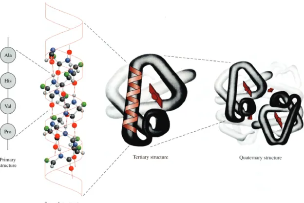
Protein structure has been described at four different scales31,32 (Figure 5).31 The primary structure refers to the order and number of amino acids in a copolymer chain. From the 20 amino acids building blocks (residues), 8 have nonpolar (hydrophobic) side chains, 7 have neutral polar side chains and 5 have charged polar side chains,2 rendering the protein amphoteric and amphiphilic.33 It is therefore not surprising that most proteins are highly surface active.34 Proteins do not exist simply as long, extended chains of amino acids. Instead, interactions between amino acids cause folding, bending, and coiling of the chain to give a
_______________________________________________________________________________________________
specific three dimensional structure (conformation). Secondary structure results from hydrogen bonding associated with the amide linkages in the backbone of the proteins to form structures as α-helix and β-pleated sheet. Tertiary structure refers to the three-dimensional structure of the whole protein subunit resulting from intramolecular associations, including ionic interactions, salt bridges, hydrophobic, hydrophilic interactions, hydrogen bonding and covalent disulphide bonds. Quaternary structure refers to interactions between subunits, or individual polypeptide chains, in multichain proteins.2
Figure 5 – The primary, secondary, tertiary, and quaternary structure of the hemoglobin molecule.31
3.2 Protein adsorption
Most reactions in biology occur, not in solution, but at interfaces. Typical interfaces of biological importance include the cell surface/synthetic biomaterial, extracellular matrix(ECM)/biomolecule,
surfaces offer are high accessibility for reaction, a low energy barrier to mobility in the plane of the surface facilitates complex reactions (clustering and conformational changes), high surface area geometries enhance reaction turnover rates, surface energy minimization can orient specific structures to interfaces and molecular recognition can be readily implemented.35
Biomaterials readily adsorb proteins from biological fluids when inserted in the human body. Indeed, the protein adsorption event occurs well before cells arrive at the surface. Therefore, cells see primarily a protein layer, rather than the actual surface of the biomaterial. Since cells respond specifically to proteins, this interfacial protein film is believed to be the event that controls subsequent reaction to implants. Protein adsorption is also concern for biosensors, immunoassays, array diagnostics, haemodialysis, food processing, marine fouling and a large number of other phenomena.36 It is evident that the scope of the investigations in the different areas above must be very different. A common interest is, however to measure and understand protein adsorption, protein layer structure, and exchange reactions on the solid surfaces. Some important properties for protein-metal surface interactions are presented in Table 5.37,38
Table 5 - Properties that determine protein adsorption at titanium surface [adapted from references37,38]
Titanium surface properties Protein properties
Free energy Overall hydrophobicity
Charge Charge
Ion binding Ion binding
Redox/corrosion potential Prosthetic groups
Hydrogen bonding capacity Hydrogen bonding residues
Surface water structure/ binding strength Water binding
Acid/basic groups (“surface pH”) Isoelectric point (pI)
Surface impurities Specific interacting residues
Surface topography Quaternary, tertiary and secondary
structure
Oxide (thickness, growth, dielectric constant, water and ion sorption) Number of disulphide bridges
_______________________________________________________________________________________________
The properties are lined up although in practice it is not such a simple relation between the adsorption of a given protein molecule on a given metal surface. In fact protein adsorption is very complex, being electrostatic and hydrophobic interactions and the intrinsic structural stability of the protein the most relevant phenomena.33
In addition to the protein and surface properties described above, adsorption also depends on the availability of molecules for interaction with substrate. Molecules can be brought to the surface by one or more of four major transport mechanisms: diffusion, thermal convection, flow, and coupled transport, and as the combination of convection and diffusion. Variables such as concentration, velocity, and molecular size are important in determining the arrival of protein molecules at surface.2
Several papers have been published on protein adsorption on titanium dealing with specific proteins such as albumin,28,39-41 fibronectin,23,42,43 vitronectin,44,45 fibrinogen,39 glycosaminoglycans,46 laminin.47-49 However there are no general accepted conclusions as to the importance of the protein film, or specific proteins, on biological reaction to Ti.50
3.2.1 Type of interactions
3.2.1.1 Electrostatic interactions
Protein adsorption to hydrophilic surfaces is usually determined by electrostatic interactions.51,52 Although interactions may occur at the atomic scale, the global charges on proteins and surfaces appears to dominate electrostatically driven adsorption.33,53-55 This is observed especially with adsorption to hydrophilic surfaces of proteins that are less susceptible to structural changes.56,57
It is intuitive that opposite charges will be mutually attracted and this is indeed observed with respected to protein adsorption. However, the situation becomes complicated in aqueous solutions, in which surface charges are shielded by hydrating water, modulated by pH and
adsorption generally occurs at the isoelectric point of the protein-surface system (pH at which the net charge is minimized).56
Coadsorption of counter-ions may contribute to this process and, indeed, the presence of such ions is implied by adsorption of proteins to like-charged surfaces.57 This may account for the observed involvement of calcium ions in some adsorption processes.28,58,59
3.2.1.2 Hydrophobic interactions
Hydrophobic interactions play an important role in protein adsorption. Protein molecules tend to minimise exposition of their hydrophobic groups to the aqueous environment. However, Haynes and Norde33 estimate that 40-50 % of the accessible surface of proteins is often occupied by non-polar groups. For a protein to adsorb, both adsorbate and surface must (at least partially) dehydrate.54,60 Dehydration of hydrophobic parts of the protein and the sorbent surfaces is driven by entropy gain and, therefore, promotes adsorption to occur spontaneously. 57,61,62 The adsorption to hydrophobic substrates may induce conformation change of the protein by hydrophobic-hydrophobic interactions with the substrate contributing to irreversible adsorption.61,62
Hydrophobic dehydration is relatively unimportant for hydrophilic surfaces and/or rigid hydrophilic proteins.61,63
Apparently due to a greater number of possible adsorption-promoting interactions, hydrophobic surfaces are usually reported to adsorb more protein than hydrophilic surfaces.64-67
3.2.2 Conformational stability of proteins
Protein molecules must not be thought of as rigid structures, as they are flexible chains that have been coiled, folded, and bent to assume a particular conformation. Changes in the microenvironment of the proteins, such as pH, temperature, and ionic strength, can alter the conformation of the molecule.2 Structural stability of proteins is of particular importance in the
_______________________________________________________________________________________________
adsorption process and reversibility of adsorption. This is dependent on intramolecular forces within the protein molecules that may lead to an alteration of protein conformation.63,68
Protein molecules with low conformational stability (soft protein), such as albumin, are found to adsorb on a great variety of surfaces (hydrophilic and hydrophobic) even under adverse electrostatic condition.67 In contrast, proteins with high conformational stability (hard protein), such as lysozyme, adsorb mainly on hydrophobic surfaces, adsorbing on hydrophilic surfaces only under favourable electrostatic interactions.55,63,67,69-73
Upon adsorption on hydrophobic surfaces, soft proteins change their conformation to a greater extent than hard proteins, due to a loss of secondary structure.74-77 Although conformational alterations may result in protein denaturation and/or loss of functional activity in some situations they can be beneficial rather than detrimental.78-80 As an example enzymes are often more stable in immobilized formulations than in solution, and enzymatic activity can be enhanced in the presence of an interface.78,80,81
3.2.3 Structure of the adsorbed protein layer
Maximum plateau adsorption is usually assumed as a more or less well-packed layer of proteins. The surface concentration corresponding to a monolayer falls in the range expected for a close-packed monolayer of protein (ca. 1 and 5 mg/m2). This value is dependent on the protein, its molecular orientation, conformational state, and the type of substrate.55,71,82
In a simple way it is generally considered that non-spherical proteins may adsorb with two different orientations, i.e., one with the long axis (end-on type) and the other with the short axis (side-on type) perpendicular to the surface.57,83,84
Though proteins often adsorb as monolayers, multilayer adsorption can also occur, especially when the solution concentration is high.82
3.2.4 Competitive protein adsorption
Body fluids, including blood, tears, and saliva, contain numerous types of biomolecules. For example, blood contains more than 150 proteins, apart from lipids, carbohydrates, and hormones.2 Adsorption from these types of fluids is very complex since there are many proteins competing for the surface. However adsorption from mixtures of proteins is selective, leading to enrichment of the surface phase in proteins that have higher surface affinity. Studies of plasma interaction with solid surfaces results in a hierarchical adsorption process known as the “Vroman effect”.85-87 The high concentration proteins dominate the surface at short times due to the higher collision rates. As time passes, various exchange processes occur and proteins with higher surface affinities dominate the surface. Finally, at very long times only the highest affinity proteins are present on the surface, even if their bulk solution concentration is very low.55,85,88
3.2.5 Reversibility of protein adsorption
Protein adsorption to hydrophobic surfaces is usually reported as irreversible.57,64,89 The irreversibility of adsorption may occur from a range of causes. One explanation could be that hydrophobic peptide moiety in the protein molecule changes its orientation in order to interact with the hydrophobic region on the surface, resulting in a multipoint attachment.2,71,90 Alsoby increasing the adsorption period, the protein undergoes structural deformations.74,89 The extent of conformation alterations after adsorption increases as protein solution concentration decreases and therefore with the decrease of the surface coverage.90 At low solution concentration, the protein has no neighbours on the surface and thus can optimally adapt to the surface, maximising the number of binding interactions. At high solution concentration, any adsorbed protein is surrounded by neighbours, minimising the probability that it can conformationally adapt to the surface. Consequently more protein may be present on the surface, but with each molecule having fewer surface contacts.2,55
_______________________________________________________________________________________________
4. Cell/material interactions
4.1. The role of adsorbed proteins in tissue response to biomaterials
Cell adhesion to adsorbed proteins is primarily mediated by integrin receptors.91,92 Integrins represent a widely expressed family of heterodimeric (α and ß subunits) transmembrane protein receptors that bind to adhesive motifs present in various extracellular matrix proteins, including fibronectin (FN), vitronectin (VN), laminin, and collagen.32 Following ligand binding, integrins cluster and associate with cytoskeletal elements to form focal adhesions, supramolecular assemblies of structural and signaling proteins that provide anchorage forces and activate signaling cascades regulating cell cycle progression and differentiation in developing tissue (Figure 6).32
Figure 6 - Interactions between cells and the extracellular matrix. The association between cells and the proteoglycan of the ECM is mediated by a membrane protein (integrin) and by FN an ECM protein with binding sites for both integrin and the proteoglycan. [adapted from ref 32]
Serum or plasma contains proteins such as albumin and IgG that decrease or inhibit cell attachment while FN and VN can facilitate the adhesion of cells.2,55,93-96 It is believed that the efficiency of cell adhesion and growth is dependent on the balance between of adhesion-promoting versus adhesion-inhibiting proteins which competitively adsorb to a surface.
The protein conformation upon adsorption plays a key role in controlling cell behavior including cell adhesion, spreading, migration, and differentiation. For example, fibronectin adsorption to different surfaces alters the structure of the adsorbed protein influencing cell adhesion, spreading, migration, and differentiation.97,98
4.1.1 Adhesion proteins
Fibronectin
Fibronectin is one of the most abundant extracellular matrix glycoproteins that adsorbs to biomaterials, mediating cell adhesion. Apart from FN, there are other glycoproteins containing cell-binding domains such as vitronectin, laminin, and collagen. However, FN is an important substratum for cell adhesion, migration, and wound healing, inflammation, and metastasis.99 FN exists in a soluble dimeric form in micromolar concentration in blood plasma and in an insoluble multimeric form in ECM. In soluble form, van der Waals and electrostatic interactions between modules stabilize FN in a compact conformation, unreactive to other extracellular matrix proteins and to self-assembly.99 FN partially opens its arms upon surface adsorption, exposing adhesion sites that are buried in the soluble form.100 Fibronectin is composed of two homologous subunits, each with a molecular weight of ca. 220 kDa, connected near their carboxy termini by a pair of disulfide bonds. Each dimer arm is approximately 70 nm long and consists of a series of globular modules of three types of structural homology (classified as fibronectin repeats FN-I, FN-II, and FN-III). The modules are grouped together into functional domains that bind to cell surfaces (integrins), fibrin, heparin, and collagen (Figure 7).101