PEX5 binds to the components
of the peroxisomal docking and
export machinery
Vera Hagmann
1, Stefanie Sommer
2, Patrick Fabian
1, Jan Bierlmeier
1, Nadine van Treel
2,
Henning D. Mootz
2, Dirk Schwarzer
1, Jorge E. Azevedo
3& Gabriele Dodt
1Peroxisomal matrix proteins contain either a peroxisomal targeting sequence 1 (PTS1) or a PTS2 that are recognized by the import receptors PEX5 and PEX7, respectively. PEX5 transports the PTS1 proteins and the PEX7/PTS2 complex to the docking translocation module (DTM) at the peroxisomal membrane. After cargo release PEX5 is monoubiquitinated and extracted from the peroxisomal membrane by the receptor export machinery (REM) comprising PEX26 and the AAA ATPases PEX1 and PEX6. Here, we investigated the protein interactions of monoubiquitinated PEX5 with the docking proteins PEX13, PEX14 and the REM. “Click” chemistry was used to synthesise monoubiquitinated recombinant PEX5. We found that monoubiquitinated PEX5 binds the PEX7/PTS2 complex and restores PTS2 protein import in vivo in ΔPEX5 fibroblasts. In vitro pull-down assays revealed an interaction of recombinant PEX5 and monoubiquitinated PEX5 with PEX13, PEX14 and with the REM components PEX1, PEX6 and PEX26. The interactions with the docking proteins were independent of the PEX5 ubiquitination status whereas the interactions with the REM components were increased when PEX5 is ubiquitinated.
Mammalian peroxisomes are single membrane-bound organelles that do not contain DNA or RNA. All matrix proteins are nuclear encoded and translated on free ribosomes and hence all these proteins must be posttransla-tionally imported into peroxisomes1. Their correct sorting to the organelle is ensured by peroxisomal targeting signals (PTS), small peptide sequences present in their primary structure that are recognized by shuttling recep-tors. There are two types of PTSs, the so-called PTS1 and PTS2. The PTS12,3 is a short C-terminal signal peptide that is used by most peroxisomal matrix proteins. PTS1 proteins are recognized by the shuttling receptor PEX5 while still in the cytosol. This interaction involves the PTS1 itself, on one side, and the C-terminal tetratricopep-tide repeat (TPR) domain of PEX54,5, on the other side. The PTS2 is a degenerated nonapeptide present at the N-terminus of only a few mammalian enzymes6,7. PTS2 proteins are also transported to the peroxisome by PEX5. However, in this case the cargo protein-PEX5 interaction requires an extra factor, the co-receptor PEX78–10. We note that mammalian PEX5 is expressed in at least two forms, PEX5L and PEX5S, which are generated by alterna-tive splicing of the PEX5 transcript11,12. PEX5L contains an insert of 37 amino acids that is positioned after amino acids 214 of PEX5S. This insert contains part of the binding-site for the PEX7/PTS2 cargo complex and thus only PEX5L (hereafter referred to as simply PEX5) can transport PTS2 proteins to the peroxisome13,14.
After binding their cargos in the cytosol, PEX5 or PEX5/PEX7 interact with the peroxisomal membrane dock-ing translocation module (DTM), which in mammals comprises the peroxins 14 and 1315 and the three RING (really interesting new gene) finger proteins PEX2, PEX10, and PEX1216. The N-terminal disordered region of PEX517 harbours several binding motifs for PEX14 and PEX13 that have been extensively studied by several groups13,18–21. The interaction process may involve partly cooperative and sequential steps22. Although the com-position of the DTM has been analysed in detail, the stoichiometry of each of its components is still unclear and
1Interfakultäres Institut für Biochemie (IFIB), Universität Tübingen, Tübingen, Germany. 2Institut für Biochemie,
Universität Münster, Münster, Germany. 3Instituto de Investigação e Inovação em Saúde (I3S), Instituto de Biologia
Molecular e Celular (IBMC), and Instituto de Ciências Biomédicas Abel Salazar (ICBAS), University of Porto, Porto, Portugal. Correspondence and requests for materials should be addressed to G.D. (email: [email protected])
Received: 28 June 2018 Accepted: 12 October 2018 Published: xx xx xxxx
undergoes deubiquitination in the cytosol. A new protein transport cycle can then start41.
Although our knowledge on how proteins are sorted to the peroxisomal matrix is rather comprehensive, there are still many mechanistic aspects of this pathway that remain poorly defined. An important one regards the extraction step of PEX5 from the DTM. It is known for long that monoubiquitination of PEX5 at the DTM is mandatory for its extraction into the cytosol but exactly how PEX5-linked ubiquitin is released from the DTM by the PEX1-PEX6 heterohexamer is unknown. A major problem in addressing this issue is related to the fact that pure monoubiquitinated PEX5 suitable for protein-protein interaction studies has not been available thus far. Here, we describe the chemical synthesis of such a protein. Using a copper(I)-catalysed alkyne-azide cycload-dition (CuAAC, also known as “click” chemistry), a strategy frequently employed to produce proteins modified with ubiquitin and ubiquitin-like proteins42–44, we have generated large amounts of recombinant human PEX5 possessing a single ubiquitin molecule covalently attached to its residue 11.
We show that this monoubiquitinated PEX5 protein is functional in PTS2 import in vivo and in vitro and interacts with several components of the DTM and the REM in vitro. Importantly, monoubiquitinated PEX5 binds better to the REM components than the non-ubiquitinated PEX5 protein. Finally, our data also suggest that PEX26 interacts with PEX14 and weakly with PEX13, thus raising the possibility that PEX26 might be a part of the DTM, at least transiently, when monoubiquitinated PEX5 is exported back into the cytosol.
Results
Synthesis of monoubiquitinated PEX5 using the copper(I)-catalysed alkyne-azide
cycloaddi-tion (CuAAC).
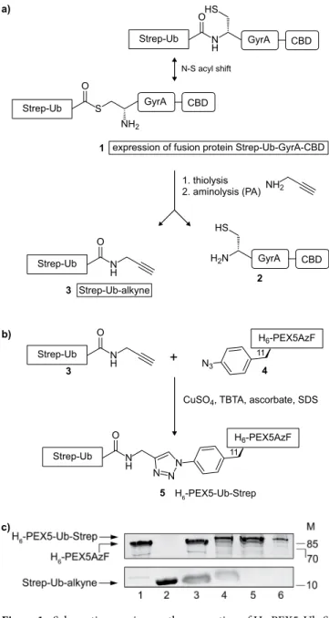
After delivering its cargos to the peroxisome matrix PEX5 gets monoubiquitinated at cysteine 11, a mandatory modification for its subsequent extraction from the DTM by the receptor export machinery REM. To gain more insights in the interaction of monoubiquitinated PEX5 with the components of the export machinery, it was necessary to have suitable amounts of the pure compound. Considering that naturally occur-ring monoubiquitinated PEX5 is a labile and a very low abundance protein we decided to chemically synthesise large amounts of it by linking ubiquitin to recombinant human PEX511 using a CuAAC reaction. A brief descrip-tion of the procedure used is provided below.We first produced a PEX5-azide by incorporating the unnatural amino acid p-azidophenylalanine (AzF) at position 11 of its polypeptide chain. This resulted in H6-PEX5C11AzF, from here on named H6-PEX5AzF (4, see
Fig. 1b), which was then purified (Fig. 1c, lane 1).
To generate a ubiquitin-alkyne we generated a fusion protein (1) comprising (from the N- to the C-terminus) a Strep-tag, yeast ubiquitin, an intein (GyrA from Mycobacterium xenopi) and a chitin binding domain (CBD). Note that the ubiquitin moiety in this fusion protein lacks the last two glycines in order to approximate the native spacing between PEX5 and ubiquitin after the formation of a 1,2,3-triazole linkage by the CuAAC reaction (5)42 (see Supplementary Fig. S1 for the comparison of the native and chemical linkage). The fusion protein Strep-UbΔGG-GyrA-CBD, referred to as Strep-Ub-GyrA-CBD (1), was subjected to thiolysis by treat-ment with 2-mercaptoethanesulfonic acid (MESNA). Subsequent addition of propargylamine (PA) resulted in Strep-Ub-alkyne (3) that was then purified by size exclusion chromatography (SEC) and verified by ESI-MS (Fig. 1c, lane 2 and Supplementary Fig. S2).
Next, a CuAAC reaction was performed using purified Strep-Ub-alkyne (3) and H6-PEX5AzF (4) at
a ratio of 3:1 (Fig. 1b). The reaction was monitored by SDS-PAGE analysis which revealed the appearance of a new species migrating 10–15 kDa above H6-PEX5AzF (4), indicating formation of the desired product
H6-PEX5AzF-Ub-Strep, referred to as H6-PEX5-Ub-Strep (5) (Fig. 1c, compare lanes 3 and 4). Residual starting
materials (i.e., non-clicked Strep-Ub-alkyne and H6-PEX5AzF) were removed by subjecting the reaction mixture
to two purification steps (Fig. 1c, lanes 5 and 6; see materials and methods for details). The chemically ubiquiti-nated structure differs slightly in the linkage between the PEX5 and ubiquitin motif from the natural one but was expected to retain all the key interactions of ubiquitinated PEX5 (see Supplementary Fig. S1 for the comparison of the native and chemical linkage).
H
6-PEX5-Ub-Strep binds a PEX7/PTS2 protein complex in vitro and is functional in importing
PTS2 proteins in vivo.
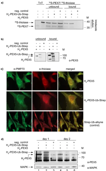
To test the functionality of the chemically synthesised H6-PEX5-Ub-Streppro-tein several experiments were performed. In one of these we asked whether H6-PEX5-Ub-Strep can interact
with the PEX7/PTS2 protein complex in vitro. Figure 2a shows the results of a pull-down assay performed with magnetic Ni-beads coupled with purified H6-PEX5 or H6-PEX5-Ub-Strep or no recombinant protein
(nega-tive control). The prepared Ni-beads were incubated with 35S-pre-thiolase-myc (a PTS2 protein, referred to as 35S-thiolase) and 35S-myc-PEX7 (referred to as 35S-PEX7) which were synthesised in vitro in the presence of
35S-methionine. The results show that both H
6-PEX5 and H6-PEX5-Ub-Strep, can bind the two radiolabelled
pro-teins (Fig. 2a). Note that in contrast to 35S-thiolase (which contains 12 methionines) 35S-PEX7 contains only two
methionines in its primary structure, hence the weak signal in the autoradiograph. We obtained similar results with other PTS2-reporter proteins such as PTS2-CAT (a bacterial chloramphenicol transferase) and PTS2-GFP (see Supplementary Fig. S3). Unexpectedly, similar pull-down assays using radiolabelled PTS1 proteins, such as PTS1-GFP and pre-SCP2 (pre-sterol carrier protein 2), revealed that the clicked H6-PEX5-Ub-Strep is not able to
bind these proteins (see Supplementary Fig. S4a,c) Subsequent experiments suggest that the click chemistry con-ditions affect the function of the TPR domain of PEX5 (see Supplementary Fig. S4c lane 4 and 5 and discussion). Figure 1. Schematic overview on the generation of H6-PEX5-Ub-Strep. (a) The expressed fusion protein
Strep-Ub-GyrA-CBD (1) was bound to a chitin matrix by its CBD and incubated with 2-mercaptoethanesulfonic acid (MESNA) to release the Strep-Ub-MESNA thioester. Treatment of this thioester with propargylamine (PA) resulted in the alkyne (3). (b) A CuAAC reaction was performed with purified Strep-Ub-alkyne (3) and H6-PEX5AzF (4). The proteins were mixed together at a ratio of 3:1 in the presence of CuSO4,
TBTA, ascorbate and SDS resulting in a stable H6-PEX5-Ub-Strep (5). (c) Analysis of the purification and click
reaction steps of H6-PEX5AzF (4), Strep-Ub-alkyne (3) and H6-PEX5-Ub-Strep (5) on a 17% SDS-gel: (4) after
Ni-column (lane 1), (3) after SEC (lane 2), control of CuAAC reaction (lane 3), CuAAC reaction of (3) and (4) resulting in (5) (lane 4). (5) was purified twice using a Ni-column to eliminate (3) (lane 5) and a Strep-Tactin
®
-column to remove (4) (lane 6). The arrows to the left indicate H6-PEX5-Ub-Strep (82 kDa), H6-PEX5AzF(72 kDa) and Strep-Ub-alkyne (11 kDa). The SDS-gels were stained with coomassie brilliant blue. Numbers to the right indicate the molecular mass (M) of proteins in kDa. The cropped images originate from one gel. The full-length gel is presented in Supplementary Fig. S6. GyrA = intein from Mycobacterium xenopi, CBD = chitin binding domain, AzF = p-azidophenylalanine, TBTA = tris(benzyltriazolylmethyl)amine.
Figure 2. Functional studies of recombinant H6-PEX5-Ub-Strep in vivo and in vitro. (a) Pull-down assay with
magnetic Ni-beads that were coupled with purified H6-PEX5 and H6-PEX5-Ub-Strep, respectively or without
protein (control). After incubating the beads with in vitro synthesised 35S-PEX7, 35S-thiolase, buffer A and ATP,
the samples were eluted with imidazole. The bound (50%) and the unbound (10%) fractions as well as the input (TNT) were analysed by SDS-PAGE/autoradiographic detection. (b) The bound (2.9%) and unbound (1.1%) fractions of the pull-down assay in (a) were also analysed by immunoblot detection with α-PEX5. The lower migrating band of PEX5 is a common degradation product in E. coli that also gets ubiquitinated. The two cropped blots originate from one gel. The pull-down is representative for four experiments. (c) Human ΔPEX5 fibroblasts were electroporated with purified H6-PEX5, H6-PEX5-Ub-Strep and Strep-Ub-alkyne (control), respectively. On
the second day, an immunofluorescent staining was performed against thiolase (Alexa 594) and the peroxisomal membrane protein PMP70 (Alexa 488). Both proteins, H6-PEX5 and H6-PEX5-Ub-Strep could import thiolase
into peroxisomes which is co-localised with PMP70, while no import was detected in the control. The experiment was performed twice with H6-PEX5-Ub-Strep and four times with H6-PEX5. Complementation rates were
calculated by counting punctate thiolase positive cells. At least 500 cells were counted for each electroporation. The scale bar represents 20 µm. (d) The stability of purified H6-PEX5 and H6-PEX5-Ub-Strep after electroporation in
ΔPEX5 fibroblasts was analysed after the first and the second day by immunoblot detection using α-PEX5. MAP-Kinase (MAPK) was used as a loading control and detected with α-MAPK. Both cropped blots are from one blot that has been cut horizontally, developed with the different antibodies and exposed on a single film together. The numbers to the right indicate the molecular mass (M) of proteins in kDa (a,b and d). The full-length versions of all gels are presented in Supplementary Fig. S6. (▲): The triangle to the right represents an unspecific protein band.
Finally, we examined the capacity of H6-PEX5-Ub-Strep to complement the peroxisomal protein import
defect of ΔPEX5 fibroblasts. In these cells, the import of PTS1 and PTS2 proteins is impaired and therefore many of these proteins display a cytosolic localization45,46. For this purpose, we used electroporation to introduce puri-fied H6-PEX5-Ub-Strep, H6-PEX5 or Strep-Ub-alkyne (control) into ΔPEX5 fibroblasts. Two days after
electro-poration, immunofluorescent staining was performed using antibodies against endogenous thiolase and against the peroxisomal membrane protein PMP70. As shown in Fig. 2c electroporation of both proteins, H6-PEX5 and
H6-PEX5-Ub-Strep, resulted in the import of thiolase into peroxisomes as determined by its colocalization with
PMP70 (Fig. 2c, “merged” panels). We note that larger complementation rates were obtained with H6-PEX5
(32.4%) than with H6-PEX5-Ub-Strep (11.3%). No complementation could be detected when Strep-Ub-alkyne
was electroporated. Immunoblot analysis of lysates from cells harvested one and two days after electroporation revealed that already on the first day the amount of H6-PEX5-Ub-Strep was reduced compared to H6-PEX5
(Fig. 2d, lanes 2 and 1, respectively). Furthermore, two days after electroporation we could still detect H6-PEX5
in cell lysates whereas H6-PEX5-Ub-Strep was barely detectable (Fig. 2d, lanes 4 and 5, respectively). Apparently,
although H6-PEX5-Ub-Strep can complement the PTS2-import defect in ΔPEX5 fibroblasts it seems to be more
unstable than H6-PEX5 (see discussion). We also asked whether H6-PEX5-Ub-Strep could restore protein import
of EGFP-PTS1 in ΔPEX5 fibroblasts. However, in agreement with the PTS1 protein pull-down assays described above, H6-PEX5-Ub-Strep did not complement the import defect of PTS1 proteins in ΔPEX5 fibroblasts while
the complementation rate for H6-PEX5 was 14% (see Supplementary Fig. 4b and discussion).
Recombinant H
6-PEX5-Ub-Strep enables import of PTS2 proteins into peroxisomes in vitro.
Next, we addressed the question whether H6-PEX5-Ub-Strep can be used in an in vitro import system using
post-nuclear supernatant (PNS) from mouse liver47,48. To investigate the import of PTS2 proteins into peroxisomes PNS containing import-competent peroxisomes was incubated with radiolabelled 35S-PEX5, 35S-pre-thiolase and 35S-PEX7 and with different amounts of recombinant PEX5 proteins for 30 min, to allow import into the organelles.
The PNS was subsequently treated with proteinase K to eliminate all not imported proteins.
No protease protection of 35S-PEX7/35S-pre-thiolase could be observed when these proteins were incubated
alone with PNS (Fig. 3, lane 1). This indicates that endogenous PEX5 is a limiting factor for the import of matrix proteins and that no processing of the 35S-pre-thiolase into the mature (mat) form occurred. A small amount
of 35S-PEX7/35S-pre-thiolase is protease protected and processed when 35S-PEX5 was added (Fig. 3, lane 2). 35S-PEX5 itself also seems to be protease protected and embedded into the DTM. The addition of recombinant
H6-PEX5 and H6-PEX5-Ub-Strep leads to a competition with the 35S-PEX5 and results in a higher amount of
protease-protected and processed thiolase (Fig. 3, lanes 3 to 5). This indicates that both recombinant proteins are functional for PTS2 protein import. Increasing the amount of recombinant H6-PEX5-Ub-Strep does not enhance
the system but leads to less import (Fig. 3, lane 5). At 0 °C neither 35S-PEX5 nor 35S-PEX7 or 35S-thiolase are
protease-protected, indicating no import of these components (Fig. 3, lane 6).
H
6-PEX5-Ub-Strep interacts with the receptor export module components PEX1, PEX6
and PEX26 and with PEX14 and PEX13.
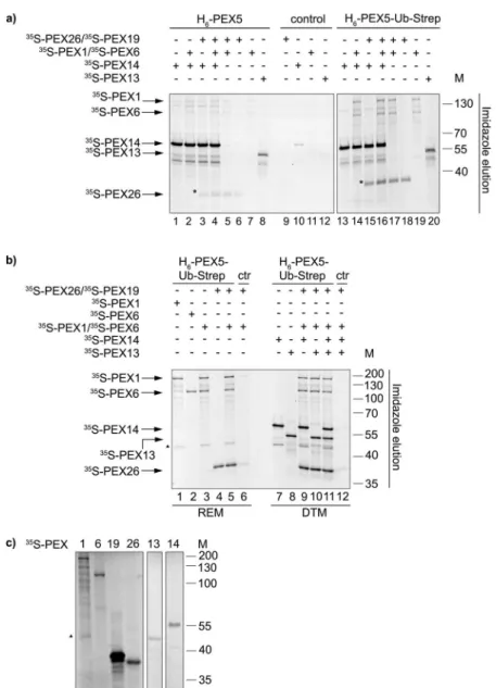
Export of DTM-embedded monoubiquitinated PEX5 is an ATP-dependent step catalysed by the receptor export module, which comprises PEX1, PEX6 and PEX26. Aiming to better understand the role of ubiquitin in this process, a series of in vitro pull-down assays were performed. We used magnetic Ni-beads containing purified H6-PEX5, H6-PEX5-Ub-Strep or no recombinantprotein (negative control) as baits. In vitro synthesised 35S-PEX1 and 35S-PEX6 (individually or pre-mixed) and
Figure 3. H6-PEX5 and H6-PEX5-Ub-Strep are functional in the PTS2 import into peroxisomes in vitro.
Post-nuclear supernatant (PNS) from mouse liver was incubated with the in vitro 35S-synthesised proteins PEX5,
PEX7 and pre-thiolase, and recombinant H6-PEX5 and H6-PEX5-Ub-Strep for 30 min at 37 °C or at 0 °C. After
the import reaction, the samples were treated with proteinase K, precipitated and analysed by SDS-PAGE, blotting and autoradiography. The arrows to the left indicate 35S-PEX5, 35S-PEX7, and 35S-pre-thiolase. The
processed, mature 35S-mat-thiolase is indicated by an asterisk (*). The Ponceau S-stained membrane served as
loading control. The numbers to the right indicate the molecular mass (M) of proteins in kDa. The uncropped version of the blot is presented in Supplementary Fig. S6. The import reaction is representative for two experiments.
35S-PEX26/35S-PEX19 (note that PEX26 was co-synthesised with PEX19 in order to ensure that it remains soluble;
see materials and methods for details) were investigated as preys. Some of these assays also contained 35S-PEX14
and 35S-PEX13, two components of the DTM. The aim was to cover the possibility that the interactions between
PEX5 and the REM components might be dependent on DTM proteins. The results of these experiments are shown in Fig. 4 and Supplementary Fig. S5a. The samples used for Fig. 4a and Supplementary Fig. S5a were incu-bated with ATP and cytosol from ΔPEX5 fibroblasts to provide putative co-factors that might be required for some protein-protein interactions.
Figure 4. H6-PEX5 and H6-PEX5-Ub-Strep interact with the peroxisomal export machinery and with PEX13
and PEX14. (a) Pull-down assay with magnetic Ni-beads that were coupled with purified H6-PEX5, H6
-PEX5-Ub-Strep or used uncoupled (control). In vitro synthesised 35S-PEX1/35S-PEX6, 35S-PEX26/35S-PEX19, 35S-PEX14 and 35S-PEX13 were added to the coupled beads and incubated with cytosol from human ΔPEX5
fibroblasts and ATP. The fractions were eluted with imidazole. The bound fractions (50%) were analysed by SDS-PAGE/autoradiography. Both gels originate from one experiment. The gels were processed in parallel. The results are representative for three individual experiments. (*): The asterisk indicates 35S-Pex26. (b) Pull-down
assay with magnetic Ni-beads that were coupled with H6-PEX5-Ub-Strep or used uncoupled (control). In vitro
synthesised 35S-PEX26/35S-PEX19 and REM proteins (35S-PEX1, 35S-PEX6) or DTM proteins (35S-PEX14, 35S-PEX13) were added to the beads and incubated with buffer A and ATPγS. The samples were eluted with
imidazole. The bound fractions (50%) were analysed by SDS-PAGE/autoradiography. The experiments were performed twice. (c) In vitro TNT reaction of 35S-labelled PEX1, PEX6, PEX19, PEX26, PEX13 and PEX14
analysed by SDS-PAGE/autoradiography. The TNT reactions for PEX13 and PEX14 are from different gels. The numbers to the right indicate the molecular mass (M) of proteins in kDa (a–c). The full-length gels are presented in Supplementary Fig. S6. (▲): The triangle to the left in (b,c) represents a translation product of PEX1 that binds to H6-PEX5-Ub-Strep.
Both, H6-PEX5 and H6-PEX5-Ub-Strep pulled-down 35S-PEX1 and 35S-PEX6 (Fig. 4a, lanes 7 and 19,
respectively and Supplementary Fig. S5a). However, the amounts of radiolabelled PEX1 and PEX6 recovered with H6-PEX5-Ub-Strep were clearly larger than those obtained with H6-PEX5. A similar result was found
with 35S-PEX26. Note that although large amounts of 35S-PEX19 were present in these assays (to maintain 35S-PEX26 in solution) almost no PEX19 was recovered in the pull-down samples (Fig. 4a compared to 4c, see
also Supplementary Fig. S5a, lanes 1 and 5). These results strongly suggest distinct interactions of H6-PEX5 and
H6-PEX5-Ub-Strep with the peroxisomal export proteins PEX1, PEX6 and PEX26.
In addition, both, H6-PEX5 (Fig. 4a, lanes 1 and 8, respectively) as well as H6-PEX5-Ub-Strep (Fig. 4a,
lanes 13 and 20, respectively) have the capability to bind to the peroxisomal docking proteins 35S-PEX13 and 35S-PEX14. Interestingly, we consistently found that slightly more 35S-PEX26 was recovered with the H
6-PEX5
and H6-PEX5-Ub-Strep beads when these assays were made in the presence of 35S-PEX14 and 35S-PEX1/35S-PEX6
(Fig. 4a, compare lanes 4 and 16 with lanes 6 and 18, respectively). Additional binding assays performed in binding buffer instead of cytosol from ΔPEX5 fibroblasts and in the presence of ATPγS instead of ATP yielded similar results (Fig. 4b). The eluates of the samples in Fig. 4a,b were additionally blotted for PEX5 to investigate the binding to the Ni-beads (see Supplementary Fig. 5b,c). We regularly noticed that less H6-PEX5-Ub-Strep
compared to H6-PEX5 could be coupled to the Ni-beads (see Supplementary Fig. 5b). The results above
sug-gest that 35S-PEX1/35S-PEX6, 35S-PEX13, 35S-PEX14 and 35S-PEX26 interact independently with H
6-PEX5 and
H6-PEX5-Ub-Strep.
The interaction between PEX26 and the DTM proteins.
To better characterize the interactions involving PEX26 and other components of the peroxisomal import machinery, additional pull-down assays were performed, but this time using as a bait in vitro synthesised myc-tagged 35S-PEX26. As shown in Fig. 5, only lowamounts of radiolabelled prey proteins (PEX1, PEX6, PEX13 and PEX14) were recovered in these assays. The fact that only nanograms of bait are used in these experiments (in contrast with typical pull-down assays in which micrograms of recombinant bait proteins are generally used) may explain this result. For radiolabelled PEX1, and probably also for PEX6, the amounts recovered with PEX26-myc beads do not differ much from those appearing in the negative control with myc-tagged phythanoyl-CoA-hydroxylase (PHYH-myc) (Fig. 5, compare lanes 1 and 2 with lanes 6 and 7, respectively). However, a clear enrichment of radiolabelled PEX14 at PEX26-myc beads compared to beads loaded with the negative control was observed (Fig. 5, lanes 4 and 9). A possible PEX13 bind-ing seems to be weak. Thus, these results suggest that PEX14 interacts with PEX26, as described previously34,49. Figure 5. Interaction of PEX26 with the DTM proteins. Pull-down assay with PEX26-myc and the myc-tagged phythanoyl-CoA-hydroxylase (PHYH-myc) (control) with the REM and DTM proteins. (a) In vitro synthesised proteins (TNT) 35S-PEX26-myc/35S-PEX19, 35S-PEX1, 35S-PEX6, 35S-PEX14, 35S-PEX13 and 35S-PHYH-myc
were added to magnetic myc-Trap
®
-beads and incubated with buffer C and ATP. The samples were eluted with myc-peptide. The bound fractions (50%) were analysed by SDS-PAGE/autoradiography. (b) In vitro TNT reaction of 35S-PEX26-myc/35S-PEX19, 35S-PEX1, 35S-PEX6, 35S-PEX14, 35S-PEX13 and 35S-PHYH-mycanalysed by SDS-PAGE/autoradiography. The numbers to the right indicate the molecular mass (M) of proteins in kDa. The full-length gels are presented in Supplementary Fig. S6. The experiment is representative for four replicates.
PEX6 and PEX26 bind to H
6-PEX5-Ub-Strep.
Given the crucial role of ubiquitin in the dislocationof PEX5 from the DTM, we next asked whether PEX1, PEX6 and PEX26 can bind ubiquitin itself. For this purpose, magnetic Strep-beads containing Strep-Ub-alkyne, H6-PEX5-Ub-Strep or H6-BRD4-GFP-Strep
(referred to as Strep-GFP) were used in pull-down assays with in vitro synthesised 35S-PEX1, 35S-PEX6, and 35S-PEX26/35S-PEX19, or a mixture of these proteins. Both, radiolabelled PEX6 and PEX26 bound much better to
the H6-PEX5-Ub-Strep-containing beads than to those containing Strep-Ub-alkyne (Fig. 6, compare lanes 6 and 7
with lanes 2 and 3, respectively), while almost no binding occurred to the control with Strep-GFP-coupled beads. The interaction of PEX1, PEX6, and PEX26 together with Strep-Ub-alkyne is weak, compared to the stronger binding to H6-PEX5-Ub-Strep (see Fig. 6 lanes 4 and 8).
Discussion
To get more insights into the extraction of monoubiquitinated PEX5 from the DTM by the REM components we generated PEX5 that was chemically ubiquitinated (H6-PEX5-Ub-Strep, see Fig. 1). In vitro analysis revealed that
H6-PEX5-Ub-Strep binds PEX7/PTS2 proteins (Fig. 2a and Supplementary Fig. 3) and interacts with the DTM
components PEX14 and PEX13 (Fig. 4). In addition, this protein is functional in PTS2 protein import as shown by complementation studies in ΔPEX5 fibroblasts with endogenous thiolase (Fig. 2c) and by the in vitro import assay (Fig. 3). Thus, we have no evidence that the binding of H6-PEX5-Ub-Strep to PEX7/PTS2 proteins or to
DTM proteins is affected by the ubiquitination. This also indicates that the deubiquitination is not a mandatory step neither for the binding to cargo nor for the binding to PEX13 and PEX14.
However, both the PTS1 protein pull-down assays and the complementation studies in ΔPEX5 fibroblasts indi-cate that H6-PEX5-Ub-Strep is not able to bind to PTS1 proteins and thus cannot complement the import defect of
PTS1 proteins in ΔPEX5 fibroblasts (Supplementary Fig. 4). We performed additional experiments that suggested that the oxidative conditions used in the CuAAC reaction affect the PTS1-binding domain of PEX5 (there are 5 cysteines in this domain) inducing small conformational changes. Indeed, H6-PEX5 subjected to this treatment
(H6-PEX5 “click”) as a control did not interact with PTS1 proteins in an in vitro pull-down experiment. This was
shown using the PTS1 protein pre-SCP2 as an example (Supplementary Fig. 4c). We also recognized that the elec-troporation of purified H6-PEX5-Ub-Strep into ΔPEX5 fibroblasts resulted in a significant reduction of the amount
of this protein after the first and the second day (Fig. 2d). Albeit H6-PEX5-Ub-Strep is functional in recovering the
PTS2 protein import it is conceivable that the half-life of H6-PEX5-Ub-Strep might be shorter than that of H6-PEX5.
The chemically added ubiquitin cannot be cleaved from PEX5, in contrast to the wild type modification. With one ubiquitin permanently attached, it is possible that PEX5 is poly-ubiquitinated leading to an increased proteaso-mal degradation. We do not have any data whether H6-PEX5-Ub-Strep is used for several rounds of peroxisomal
transport like it is assumed for the endogenous PEX528,50–52 or just once. In the complementation studies in ΔPEX5 fibroblasts (Fig. 2) H6-PEX5-Ub-Strep was added in a vast excess to compensate for the short half-life of this protein.
Although the REM proteins were thought to be involved in the export of monoubiquitinated PEX5, until very recently neither the binding of Ub-PEX5 to the PEX1/PEX6 complex nor to PEX26 had been reported32,34. Our results show a binding of H6-PEX5 to the docking/REM components that is weak but clearly increased when
PEX5 is ubiquitinated. The interactions were independent of each other (Fig. 4 and Supplementary Fig. S5). While this manuscript was under review similar findings, i.e. the result that the AAA ATPases Pex1p and Pex6p interact with N-terminally linked ubiquitin-Pex5p fusion protein were reported for the yeast proteins53.
Our in vitro binding assays suggest that in particular PEX6 and PEX26 proteins bind only slightly to H6-PEX5
(Fig. 4a and Supplementary Fig. S5a) and do not, or bind only slightly to Strep-Ub-alkyne (Fig. 6). However, both PEX6 and PEX26 independently display a substantial binding to ubiquitinated PEX5 (Fig. 4 and Supplementary Fig. S5a). A similar binding behaviour of PEX1 to H6-PEX5-Ub-Strep was obtained, if the latter was pulled
Figure 6. PEX6 and PEX26 bind to H6-PEX5-Ub-Strep. Pull-down assay with magnetic Strep-Tactin
®
XT-beads that were coupled with purified Strep-Ub-alkyne, H6-PEX5-Ub-Strep or H6-BRD4-GFP-Strep (referred
to as Strep-GFP; control). In vitro synthesised 35S-PEX26/35S-PEX19, 35S-PEX1, 35S-PEX6 (TNT) were added to
the beads and incubated with buffer C and ATP. The samples were eluted with buffer C containing biotin. The eluates (50%) were analysed by SDS-PAGE/autoradiography. The numbers to the right indicate the molecular mass (M) of proteins in kDa. The full-length gels are presented in Supplementary Fig. S6. The experiment is representative for three replicates. (*): The asterisk indicates a translation product of PEX1 that binds especially to H6-PEX5-Ub-Strep and migrates almost to the same position as PEX6.
quantitative proteomic studies with PEX14 indicating that yeast PEX15 is a component of the transient pore16. Thus, PEX26 might act as a bridge between embedded (monoubiquitinated) PEX5, the DTM and PEX1/PEX6 complex. In recent studies, Miyata et al. identified AWP1/ZFAND6 as a PEX5 export factor that recognizes PEX6 and monoubiquitinated PEX5 and enhances the export of PEX5 from the peroxisomal membrane54, but did not bind the PEX1/PEX6 complex. Our interaction studies demonstrate that the binding of ubiquitinated PEX5 and PEX6 does not require additional cytosol. However small amounts of rabbit reticulocyte lysate are present in our pull-down assays and thus we cannot exclude that traces of rabbit AWP1 or other factors are present and needed for the interactions. According to very recent data the PEX1/PEX6 complex extracts Ub-PEX5 from the peroxiso-mal DTM using a processive threading mechanism31,40. However, exactly how the PEX1/PEX6 complex engages the DTM-embedded Ub-PEX5 protein and what is the mechanistic role of ubiquitin in all this process remains completely unknown. Clearly, an answer to these questions will require reconstituting the extraction reaction
in vitro using pure recombinant proteins. The chemical synthesis of the bona fide substrate of the PEX1/PEX6
unfoldase described here is thus the first step towards this goal.
Methods
Cloning.
For protein production, N-terminal hexahistidinyl-tagged hsPEX5L was cloned intoNcoI/NotI sites of pET9dNHis6 (a kind gift of G. Stier, Heidelberg, for a brief description see55) creating
pVS1 (kindly provided by W. Schliebs, Bochum). Briefly, the NcoI/BglII fragment from pGD10611 contain-ing the cDNA for PEX5L was first ligated into the NcoI/BamHI sites of pET21d+, cut with NcoI/NotI and finally cloned into pet9dNHis6 creating pVS1. For site-directed mutagenesis of pVS1 the forward primer
(GD496) (5′-GTGGAGGCCGAATAGGGGGGTGCCACC-3′ and the reverse primer (GD467) (5′-GTTGGCACC CCCCTATTCGGCCTCCAC-3′) were used to generate the cysteine-amber stop codon mutations at posi-tion 11 resulting in H6-PEX5C11AzF (pVS06). To generate Strep-UbΔGG-GyrA-CBD (pVS09; referred to
as Strep-Ub-GyrA-CBD) an N-terminal Strep-tag
®
II and a TEV-cleavage site was amplified by oligonucle-otide hybridization using the forward primer (GD504) (5′-TATGTCCGCCTGGAGCCACCCGCAGTT CGAAAAAGAAAACCTGTATTTTCAGGGCCA-3′) and the reverse primer (GD505) (5′-TATGGCCCTG AAAATACAGGTTTTCTTTTTCGAACTGCGGGTGGCTCCAGGCGGACA-3′) and cloned into theNdeI/NdeI sites of pNW0442. The ubiquitin used in this work is derived from S. cerevisiae.
Full-length hsPEX13 was amplified from cDNA using the forward primer (GD144) (5′-CAGGATCCGGTACC TTACCATGGCGTCCCAGCCGCCAC-3′) and the reverse primer (GD145) (5′-GGTCTAGATCAAAG ACCTTGCTTTTCTCCATC-3′) and cloned into the BamHI/XbaI sites of pcDNA3.1/Zeo (pCK19). The PEX1 construct pBM57 contains the coding region of full-length human hsPEX1 including 411 bp of the 3′untranslated region in the BamHI/XhoI sites of pcDNA3.1/Zeo. The PEX26-myc plasmid (pJD10) encodes hsPEX26 with a C-terminal myc-(epitope) tag in the EcoRI/NotI sites of pcDNA3.1/Zeo.
Expression in E. coli.
H6-PEX5 was expressed in E. coli BL21 (DE3) in 2 L of YEP (10 g peptone, 10 g NaCland 5 g yeast extract per 1 mL) containing appropriate antibiotics. Cultures were grown to an OD600 = 0.4–0.6 at
37 °C, induced with IPTG (1 mM) and further grown for 18 h at 18 °C.
H6-PEX5AzF was co-expressed with pEVOL-pAzF, a vector encoding the orthogonal pAzFRS/tRNACUA pair56
(kindly provided by P.G. Schultz, Addgene plasmid 31186) in E. coli BL21 (DE3) in 2 L of M9-minimal media containing appropriate antibiotics as described before43. Cultures were grown to an OD
600 = 0.3–0.4 at 37 °C. The
unnatural amino acid p-azidophenylalanine (AzF; Bachem) was added to a final concentration of 1 mM. Protein expression was induced with IPTG (1 mM) and 0.02% arabinose for 12 h at 28 °C.
Strep-Ub-GyrA-CBD was expressed in E. coli BL21 (DE3) in 8 L of YEP containing appropriate antibiotics.
Cells were grown to an OD600 = 0.4–0.6 at 37 °C, induced with IPTG (0.4 mM) and further incubated for 4 h at
28 °C.
H
6-PEX5/H
6-PEX5AZF purification.
Pelleted cells were resuspended in HisA buffer (50 mM NaH2PO4,300 mM NaCl, 10 mM imidazole, pH 8) (5–7 mL/g pellet) and lysed using an EmulsiFlex
®
-C3 system (Avestin). After centrifugation (25,000 g, 1 h, 4 °C) the filtered supernatant was loaded onto a 1 mL HisTrap™
HP column (GE Healthcare) for one hour. The proteins were eluted using a linear gradient of HisB buffer (50 mM NaH2PO4,300 mM NaCl, 500 mM imidazole, pH 8) and dialysed against click buffer (20 mM HEPES, 150 mM NaCl, pH 8). H6-PEX5 was further purified on a Superdex
™
200 HiLoad™
16/60 column (GE Healthcare) using click buffer.(10 mM final concentration). After dialysis against HisA buffer, H6-PEX5-Ub-Strep was first purified using a
1 mL HisTrap
™
HP column as described above. The eluted protein was directly loaded onto a 1 mL Strep-Tactin®
Superflow®
high capacity cartridge (IBA) using StrepA buffer (20 mM Tris, 150 mM NaCl, 1 mM EDTA, pH 8). H6-PEX5-Ub-Strep was eluted using a linear gradient of StrepB buffer (20 mM Tris, 150 mM NaCl, 1 mM EDTA,5 mM desthiobiotin, pH 8). Purified H6-PEX5-Ub-Strep was dialysed against click buffer. The success of the click
reaction and purification of H6-PEX5-Ub-Strep was confirmed by SDS-PAGE.
Capillary electroporation of human fibroblasts.
PEX5-deficient human fibroblast cells (PDB005T)45 were referred to as ΔPEX5 fibroblasts. Cells were cultured in Dulbecco’s modified Eagle medium (DMEM; Sigma) supplemented with 10% fetal calf serum, 2 mM glutamine and 50 mg/L gentamicin at 37 °C and 8.5% CO2.Capillary electroporation57 was carried out with a Neon
™
Transfection System (Life Technologies). Cells were washed twice with Hank’s Balanced Salt Solution (HBSS) and resuspended in electroporation buffer (250 mM sucrose, 1 mM MgCl2 in Dulbecco’s-PBS)58. Each 10 µL electroporation reaction containing 2 × 105 ΔPEX5fibroblasts and purified H6-PEX5 (4 µM), Strep-Ub-alkyne (4 µM) or H6-PEX5-Ub-Strep (8 µM) was pulsed at
1050 V (30 ms). To monitor the PTS1 import we added 66 ng pEGFP-PTS1 (pEB22.11, coding for EGFP with the C-terminal sequence RPPLHSKL) as reporter plasmid. After electroporation cells were resuspended in pre-warmed supplemented DMEM and sedimented at 200 g for 5 min to remove recombinant proteins. Cells were then seeded either into 6-well-plates prepared with coverslips for indirect immunofluorescence microscopy or into 25 cm2 culture flasks for immunoblot detection.
Cytosol extraction from ΔPEX5 fibroblasts.
For cytosol extraction four to seven 175 cm2 culture flasksof 90% confluent ΔPEX5 fibroblasts were harvested and resuspended in cytosol extraction buffer (20 mM HEPES, 150 mM NaCl, 5 mM imidazole, 20% glycerol, pH 7.4, protease inhibitor cocktail (PI) (Sigma; 1:200)). To isolate the post-nuclear supernatant (PNS), cells were lysed using a pre-cooled ball-bearing-homogenizer59, centrifuged (700 g, 10 min, 4 °C) and the pellet resuspended in cytosol extraction buffer. The centrifugation was repeated and the resulting two supernatants (PNS) were pooled and pelleted (100,000 g, 1 h, 2 °C) to separate the cytosol from the organelles. The cytosolic fraction was stored in aliquots at −80 °C.
Immunoblot detection.
For an SDS-PAGE gel either 20 µg of total protein after electroporation or 1.1% of bound and 2.8% of unbound fractions after pull-down binding assays were separated and transferred onto a nitrocellulose membrane (Roth) using a semidry blotting system (Biorad). α-PEX5 (GDA2, rabbit polyclonal) was diluted 1:5,000 in TBS-TX buffer (100 mM Tris, 100 mM NaCl, pH 7.4, 0.1% Triton X®
100). To generate these PEX5 antibody pVS1 was expressed in E. coli BL21 (DE3) and recombinant protein was purified under native conditions as described20. The α-MAPK (α-mitogen activated protein kinase) antibody (QIAexpress®
Tag 100, Qiagen, against the epitope of MAP kinase 2 (EETARFQPGYRS); mouse monoclonal) was diluted 1:2,000 in TBS-Tw buffer (100 mM Tris, 100 mM NaCl, pH 7.4, 0.02% Tween®
20). After using the enhanced chemilu-minescence Western Blot substrate (Thermo Scientific) the blot was exposed to film (Hyperfilm; Amersham) for different times and developed.Immunofluorescence microscopy.
Cells on coverslips were stained for immunofluorescence microscopy as described before60. As primary antibodies α-thiolase (ACAA1) (rabbit polyclonal, dilution 1:100; a kind gift of Nancy E. Braverman), α-PMP70 (ABCD3) (sheep 1:100; a kind gift of Stephen J. Gould), α- PEX1461 (rabbit polyclonal 1:400) or α-AFP (mouse monoclonal (3E6), 1:100) (QBiogen) were used. The secondary antibodies (donkey) α-mouse or α-rabbit conjugated to Alexa-488 or Alexa-594 were purchased from Life Technologies and were used in a dilution of 1:300 and 1:200, respectively.The stained cells were analysed using a Zeiss Axiovert 200 M fluorescence microscope equipped with a Plan-Apochromat 63x/1.4 oil objective and AxioVision 4.8 software.
In vitro synthesis of proteins and autoradiography.
The in vitro transcription and translation (TNT) experiments were performed with the TNT®
Quick Coupled Transcription/Translation System (Promega) according to manufacturer’s instruction. The translation products from the rabbit reticulocyte lysate (RRL) were labelled with 35S-methionine (35 mCi/mL; Hartmann Analytic). The amounts of protein synthesisedin our reactions were not determined but yields of 2–6 ng of protein/microliter of lysate are common accord-ing to the manufacturers information. The reactions were carried out for two hours at 30 °C. Generation of PEX26-myc (referred to PEX26) was performed together with the chaperone PEX1962 (pCS3). For this, both
40 µL per reaction) were coupled with 7 µg of purified H6-PEX5 and 14 µg H6-PEX5-Ub-Strep or without protein
(control) in buffer A (cytosol extraction buffer with 0.02% Tween
®
20) for 45 min at 4 °C. Protein concentra-tions of purified fracconcentra-tions were estimated at 280 nm using the respective extinction coefficients (Expasy). As this was difficult for H6-PEX5-Ub-Strep, due to the unusual linkage, the amounts coupled to the beads wereadjusted to obtain equivalent band intensities on a Coomassie-stained SDS-PAGE gel. The coupled Ni-beads were incubated with the radiolabelled translation products (10 µL reaction samples of PEX1461, 20 µL of PEX1, PEX6, PEX19/PEX26, PEX13, pre-thiolase-myc66 (referred to as thiolase), myc-PEX714 (referred to as PEX7; pEB13.10), PTS2-CAT6, PTS2-GFP64 (pJM205), PTS1-GFP (pSM102)64 and pre-SCP265 and 40 µL of PEX1/PEX6) either in buffer A or in ΔPEX5 fibroblast cytosol supplemented with 2 mM ATP (Sigma) or 2 mM ATPγS (Sigma) and 4 mM MgCl2 (150 µL total incubation volume) for 2 h at 30 °C. If necessary, the reactions were filled up with RRL
to have the same amount of RRL in each sample. After incubation, unbound fractions were removed and the Ni-beads were washed four times prior to elution with 30 µL buffer B (20 mM HEPES, 150 mM NaCl, 200 mM imidazole, 20% glycerol, pH 7.4, PI (1:200) and 0.02% Tween
®
20) for 10 min.Pull-down binding assays with myc-beads.
Magnetic myc-beads (myc-Trap®
_MA; Chromotek; 20 µL per reaction) were used according to manufacturer’s instruction. The radiolabelled translation products (4 µL PHYH-myc67, 10 µL PEX14, 20 µL of PEX1, PEX6 and PEX13 and 40 µL of PEX26-myc/PEX19) were incu-bated with myc-beads in buffer C (20 mM HEPES, 150 mM NaCl, 20% glycerol, pH 7.4, PI (1:200) and 0.02% Tween®
20) containing 2 mM ATP and 4 mM MgCl2 as described above under ‘Pull-down binding assays withNi-beads’. The elution was performed with 30 µL buffer C containing 5 µg myc-peptide (Chromotek).
Pull-down binding assays with Strep-beads.
Magnetic Strep-beads (MagStrep “type3” XT beads; IBA; 40 µL per reaction) were coupled with purified 4.6 µg Strep-Ub-alkyne and 14 µg H6-PEX5-Ub-Strepor 14 µg H6-BRD4-GFP-Strep (control) in buffer C for 45 minutes at 4 °C. The unrelated control protein
H6-BRD4-GFP-Strep (containing the bromodomain4(1)) was expressed from pET28a-BRD4(1)-TagGFP in E.
coli BL21 (DE3) and purified by NTA-chromatography. The radiolabelled translation products (20 µL reaction samples of PEX1, PEX6, PEX19/PEX26) were incubated with Strep-beads in buffer C supplemented with 2 mM ATP and 4 mM MgCl2, as described above under ‘Pull-down binding assays with Ni-beads’. The bound proteins
were eluted with 30 µL buffer C containing 50 mM biotin.
In vitro import assay.
The principles of the PTS2-centered import assay including the preparation of mouse liver PNS (post nuclear supernatant) have been described before47. Briefly, 500 µg of PNS were incubated in 100 µL import buffer (250 mM sucrose, 50 mM KCl, 20 mM MOPS-KOH (pH 7.4), 3 mM MgCl2) supplemented with3 mM ATP and the TNT 35S-lysate mix for 30 min at 37 °C or at 0 °C (control). The TNT 35S-lysate mix contained
0.37 µL of the PTS2 protein 35S-pre-thiolase, 0.67 µL 35S-PEX7 and 0.5 µL 35S-PEX511 (pGD106), respectively. In addition, H6-PEX5 (0.96 µM) or H6-PEX5-Ub-Strep (0.84 µM and 1.69 µM) were added in some reactions.
Proteinase K (PK; Sigma) was added in PK buffer (20 mM MOPS-KOH (pH 7.2)) to a final concentration of 400 µg/mL, incubated for 40 min at 0 °C and stopped with PMSF (500 µg/mL final concentration). The samples were filled up to 1 mL in SEMK buffer (250 mM sucrose, 1 mM EDTA (pH 8.0), 20 mM MOPS-KOH (pH 7.2), 80 mM KCl, and centrifuged (11,300 g, 15 min, 4 °C). The pellet was precipitated with 1 ml TCA (10%) washed with acetone, separated by SDS-PAGE and transferred onto a nitrocellulose membrane (Roth) using a semidry blotting system (Biorad). For the SDS gel 60% of the samples and 0.075 µL of the TNT reactions were used. The dried membrane was exposed to autoradiography for six to eight days.
Data Availability
No datasets were generated or analysed during the current study.
References
1. Lazarow, P. B. & Fujiki, Y. Biogenesis of peroxisomes. Annu Rev Cell Biol 1, 489–530 (1985).
2. Brocard, C. & Hartig, A. Peroxisome targeting signal 1: is it really a simple tripeptide? Biochim Biophys Acta 1763, 1565–1573 (2006). 3. Gould, S. J., Keller, G. A., Hosken, N., Wilkinson, J. & Subramani, S. A conserved tripeptide sorts proteins to peroxisomes. J Cell Biol
108, 1657–1664 (1989).
4. Gatto, G. J. Jr., Geisbrecht, B. V., Gould, S. J. & Berg, J. M. Peroxisomal targeting signal-1 recognition by the TPR domains of human PEX5. Nat Struct Biol 7, 1091–1095 (2000).
5. Fodor, K. et al. Ligand-induced compaction of the PEX5 receptor-binding cavity impacts protein import efficiency into peroxisomes.
21703–21714 (2000).
14. Dodt, G., Warren, D., Becker, E., Rehling, P. & Gould, S. J. Domain mapping of human PEX5 reveals functional and structural similarities to Saccharomyces cerevisiae Pex18p and Pex21p. J Biol Chem 276, 41769–41781 (2001).
15. Azevedo, J. E. & Schliebs, W. Pex14p, more than just a docking protein. Biochim Biophys Acta 1763, 1574–1584 (2006).
16. Oeljeklaus, S. et al. Identification of core components and transient interactors of the peroxisomal importomer by dual-track stable isotope labeling with amino acids in cell culture analysis. J Proteome Res 11, 2567–2580 (2012).
17. Carvalho, A. F. et al. The N-terminal half of the peroxisomal cycling receptor Pex5p is a natively unfolded domain. J Mol Biol 356, 864–875 (2006).
18. Rayapuram, N. & Subramani, S. The importomer–a peroxisomal membrane complex involved in protein translocation into the peroxisome matrix. Biochim Biophys Acta 1763, 1613–1619 (2006).
19. Saidowsky, J. et al. The di-aromatic pentapeptide repeats of the human peroxisome import receptor PEX5 are separate high affinity binding sites for the peroxisomal membrane protein PEX14. J Biol Chem 276, 34524–34529 (2001).
20. Schliebs, W. et al. Recombinant human peroxisomal targeting signal receptor PEX5. Structural basis for interaction of PEX5 with PEX14. J Biol Chem 274, 5666–5673 (1999).
21. Shiozawa, K., Konarev, P. V., Neufeld, C., Wilmanns, M. & Svergun, D. I. Solution structure of human Pex5.Pex14.PTS1 protein complexes obtained by small angle X-ray scattering. J Biol Chem 284, 25334–25342 (2009).
22. Emmanouilidis, L., Gopalswamy, M., Passon, D. M., Wilmanns, M. & Sattler, M. Structural biology of the import pathways of peroxisomal matrix proteins. Biochim Biophys Acta 1863, 804–813 (2016).
23. Dias, A. F. et al. The peroxisomal matrix protein translocon is a large cavity-forming protein assembly into which PEX5 protein enters to release its cargo. J Biol Chem 292, 15287–15300 (2017).
24. Mukai, S. & Fujiki, Y. Molecular mechanisms of import of peroxisome-targeting signal type 2 (PTS2) proteins by PTS2 receptor Pex7p and PTS1 receptor Pex5pL. J Biol Chem 281, 37311–37320 (2006).
25. Williams, C., van den Berg, M., Sprenger, R. R. & Distel, B. A conserved cysteine is essential for Pex4p-dependent ubiquitination of the peroxisomal import receptor Pex5p. J Biol Chem 282, 22534–22543 (2007).
26. Platta, H. W. et al. Regulation of peroxisomal matrix protein import by ubiquitination. Biochim Biophys Acta 1863, 838–849 (2016). 27. Francisco, T. et al. Ubiquitin in the peroxisomal protein import pathway. Biochimie 98, 29–35 (2014).
28. Miyata, N. & Fujiki, Y. Shuttling mechanism of peroxisome targeting signal type 1 receptor Pex5: ATP-independent import and ATP-dependent export. Mol Cell Biol 25, 10822–10832 (2005).
29. Platta, H. W., Grunau, S., Rosenkranz, K., Girzalsky, W. & Erdmann, R. Functional role of the AAA peroxins in dislocation of the cycling PTS1 receptor back to the cytosol. Nat Cell Biol 7, 817–822 (2005).
30. Birschmann, I. et al. Pex15p of Saccharomyces cerevisiae provides a molecular basis for recruitment of the AAA peroxin Pex6p to peroxisomal membranes. Mol Biol Cell 14, 2226–2236 (2003).
31. Pedrosa, A. G. et al. Peroxisomal monoubiquitinated PEX5 interacts with the AAA ATPases PEX1 and PEX6 and is unfolded during its dislocation into the cytosol. J Biol Chem 293, 11553–11563 (2018).
32. Matsumoto, N., Tamura, S. & Fujiki, Y. The pathogenic peroxin Pex26p recruits the Pex1p-Pex6p AAA ATPase complexes to peroxisomes. Nat Cell Biol 5, 454–460 (2003).
33. Weller, S. et al. Alternative splicing suggests extended function of PEX26 in peroxisome biogenesis. Am J Hum Genet 76, 987–1007 (2005).
34. Tamura, S., Matsumoto, N., Takeba, R. & Fujiki, Y. AAA peroxins and their recruiter Pex26p modulate the interactions of peroxins involved in peroxisomal protein import. J Biol Chem 289, 24336–24346 (2014).
35. Ciniawsky, S. et al. Molecular snapshots of the Pex1/6 AAA + complex in action. Nat Commun 6, 7331 (2015).
36. Blok, N. B. et al. Unique double-ring structure of the peroxisomal Pex1/Pex6 ATPase complex revealed by cryo-electron microscopy.
Proc Natl Acad Sci USA 112, E4017–4025 (2015).
37. Gardner, B. M., Chowdhury, S., Lander, G. C. & Martin, A. The Pex1/Pex6 complex is a heterohexameric AAA + motor with alternating and highly coordinated subunits. J Mol Biol 427, 1375–1388 (2015).
38. Tan, D., Blok, N. B., Rapoport, T. A. & Walz, T. Structures of the double-ring AAA ATPase Pex1-Pex6 involved in peroxisome biogenesis. FEBS J 283, 986–992 (2016).
39. Grimm, I., Saffian, D., Girzalsky, W. & Erdmann, R. Nucleotide-dependent assembly of the peroxisomal receptor export complex.
Sci Rep 6, 19838 (2016).
40. Gardner, B. M. et al. The peroxisomal AAA-ATPase Pex1/Pex6 unfolds substrates by processive threading. Nat Commun 9, 135 (2018).
41. Grou, C. P. et al. Identification of ubiquitin-specific protease 9X (USP9X) as a deubiquitinase acting on ubiquitin-peroxin 5 (PEX5) thioester conjugate. J Biol Chem 287, 12815–12827 (2012).
42. Weikart, N. D. & Mootz, H. D. Generation of site-specific and enzymatically stable conjugates of recombinant proteins with ubiquitin-like modifiers by the Cu(I)-catalyzed azide-alkyne cycloaddition. Chembiochem 11, 774–777 (2010).
43. Sommer, S., Weikart, N. D., Brockmeyer, A., Janning, P. & Mootz, H. D. Expanded click conjugation of recombinant proteins with ubiquitin-like modifiers reveals altered substrate preference of SUMO2-modified Ubc9. Angew Chem Int Ed Engl 50, 9888–9892 (2011). 44. Weikart, N. D., Sommer, S. & Mootz, H. D. Click synthesis of ubiquitin dimer analogs to interrogate linkage-specific UBA domain
binding. Chem Commun (Camb) 48, 296–298 (2012).
45. Dodt, G. et al. Mutations in the PTS1 receptor gene, PXR1, define complementation group 2 of the peroxisome biogenesis disorders.
Nat Genet 9, 115–125 (1995).
46. Slawecki, M. L. et al. Identification of three distinct peroxisomal protein import defects in patients with peroxisome biogenesis disorders. J Cell Sci 108, 1817–1829 (1995).
47. Rodrigues, T. A. et al. A cell-free organelle-based in vitro system for studying the peroxisomal protein import machinery. Nat Protoc
57. Kim, J. A. et al. A novel electroporation method using a capillary and wire-type electrode. Biosens Bioelectron 23, 1353–1360 (2008). 58. Brees, C. & Fransen, M. A cost-effective approach to microporate mammalian cells with the Neon Transfection System. Anal
Biochem 466, 49–50 (2014).
59. Balch, W. E. & Rothman, J. E. Characterization of protein transport between successive compartments of the Golgi apparatus: asymmetric properties of donor and acceptor activities in a cell-free system. Arch Biochem Biophys 240, 413–425 (1985). 60. Schmidt, F. et al. The role of conserved PEX3 regions in PEX19-binding and peroxisome biogenesis. Traffic 13, 1244–1260 (2012). 61. Will, G. K. et al. Identification and characterization of the human orthologue of yeast Pex14p. Mol Cell Biol 19, 2265–2277 (1999). 62. Soukupova, M., Sprenger, C., Gorgas, K., Kunau, W. H. & Dodt, G. Identification and characterization of the human peroxin PEX3.
Eur J Cell Biol 78, 357–374 (1999).
63. Yahraus, T. et al. Theperoxisome biogenesis disorder group 4 gene, PXAAA1, encodes a cytoplasmic ATPase required for stability of the PTS1 receptor. EMBO J 15, 2914–2923 (1996).
64. Kalish, J. E. et al. Characterization of a novel component of the peroxisomal protein import apparatus using fluorescent peroxisomal proteins. EMBO J 15, 3275–3285 (1996).
65. Stanley, W. A. & Wilmanns, M. Dynamic architecture of the peroxisomal import receptor Pex5p. Biochim Biophys Acta 1763, 1592–1598 (2006).
66. Braverman, N. et al. PEX7 Gene Structure, Alternative Transcripts, and Evidence for a Founder Haplotype for the Frequent RCDP Allele, L292ter. Genomics 63(2), 181–192 (2000).
67. Mihalik, S. J. et al. Identification of PAHX, a Refsum disease gene. Nature Genetics 17(2), 185–189 (1997).
Acknowledgements
We are grateful to Stephen J. Gould (Johns Hopkins University, Baltimore), Nancy E. Braverman (McGill University, Montreal), Wolfgang Schliebs (Ruhr University, Bochum) and Daniel Passon (EMBL, Hamburg) for providing plasmids and antibodies. Work in J.E.A. lab is funded by FEDER (Fundo Europeu de Desenvolvimento Regional), through COMPETE 2020 – Operacional Programme for Competitiveness and Internationalization (POCI), Portugal 2020, and by Portuguese funds through Fundação para a Ciência e Tecnologia (FCT)/Ministério da Ciência, Tecnologia e Inovação in the framework of the projects “Institute for Research and Innovation in Health Sciences” (POCI-01-0145-FEDER-007274) and “The molecular mechanisms of peroxisome biogenesis” (PTDC/BEXBCM/2311/2014), and through Norte 2020 – Programa Operacional Regional do Norte, under the application of the “Porto Neurosciences and Neurologic Disease Research Initiative at i3S” (NORTE-01-0145-FEDER-000008). We acknowledge support by the Open Access Publishing Fund of the University of Tübingen and the Deutsche Forschungsgemeinschaft for publishing costs.
Author Contributions
V.H., S.S., P.F., J.B., and N.v.T. performed the experiments. V.H. and J.B. prepared the figures. V.H., H.D.M., D.S., J.E.A. and G.D. designed the study. V.H., J.E.A. and G.D. wrote the manuscript. All authors reviewed the manuscript.
Additional Information
Supplementary information accompanies this paper at https://doi.org/10.1038/s41598-018-34200-5. Competing Interests: The authors declare no competing interests.
Publisher’s note: Springer Nature remains neutral with regard to jurisdictional claims in published maps and institutional affiliations.
Open Access This article is licensed under a Creative Commons Attribution 4.0 International License, which permits use, sharing, adaptation, distribution and reproduction in any medium or format, as long as you give appropriate credit to the original author(s) and the source, provide a link to the Cre-ative Commons license, and indicate if changes were made. The images or other third party material in this article are included in the article’s Creative Commons license, unless indicated otherwise in a credit line to the material. If material is not included in the article’s Creative Commons license and your intended use is not per-mitted by statutory regulation or exceeds the perper-mitted use, you will need to obtain permission directly from the copyright holder. To view a copy of this license, visit http://creativecommons.org/licenses/by/4.0/.