Avaliação não invasiva da pressão venosa central por ecocardiografia em cuidados intensivos
– Particularidades nos doentes com dilatação do ventrículo direito e exacerbação de doença pulmonar crónica
Non invasive evaluation of central venous pressure using echocardiography in the intensive care
– Specific features of patients with right ventricular
enlargement and chronic exacerbated pulmonary disease Recebido para publicação/received for publication: 06.07.19
Aceite para publicação/accepted for publication: 06.08.29 Paulo Marcelino1
Alexandra Borba2
Ana Paula Fernandes1
Susan Marum1
Nuno Germano3
Mário G Lopes4
1 Assistente Hospitalar de Medicina Interna, subespecialista de Medicina Intensiva / Hospital Assistant, Internal Medicine, sub-speciality Intensive Medicine 2 Interna do Internato Complementar de Pneumologia / Intern, Complementary Internship, Pulmonology
3 Assistente Eventual de Medicina Interna / Future Assistant, Internal Medicine
4 Professor Agregado da Faculdade de Medicina de Lisboa, chefe de Serviço de Cardiologia, director do Serviço de Cardiologia do Hospital de Santa Maria, Lisboa / / Institute Professor, Lisbon University School of Medicine, Head, Cardiology Unit, Director, Cardiology Unit, Hospital de Santa Maria, Lisbon
Proveniência / Study undertaken at: Hospital de Curry Cabral, Unidade de Cuidados Intensivos e Centro de Cardiologia da Universidade de Lisboa (CCUL) / Intensive Care Unit, Hospital de Curry Cabral and Cardiology Unit, Universidade de Lisboa (CCUL)
Correspondência/Correspondence to: Paulo Marcelino
Hospital de Curry Cabral, Unidade de Cuidados Intensivos Rua da Beneficência, 8
1069-166 Lisboa, Portugal e-mail : [email protected]
Resumo
Objectivos: Determinar a possibilidade de avaliação não invasiva da pressão venosa central (PVC) através da análise da veia cava inferior (VCI), obtida por eco-cardiografia transtorácica (ETT).
Desenho: Estudo prospectivo com 3 anos de duração.
Local: Unidade de Cuidados Intensivos Polivalente (UCIP) de 16 camas.
Métodos: Estudados doentes admitidos numa UCIP nos quais se avaliou a PVC em simultâneo com exame ETT que, para além da visualização da VCI, consistiu na obten-ção da dimensão das cavidades cardíacas e funobten-ção sistólica
Abstract
Objectives: To determine the possibility of non-in-vasive estimation of central venous pressure (CVP) through inferior vena cava (IVC) analysis, using trans-thoracic echocardiography (TTE).
Design: A prospective 3-year study.
Setting: A 16-bed medical/surgical Intensive Care Unit (ICU).
Methods: Patients admitted to the ICU were enrol-led. CVP measurement and TTE (determining cardiac chambers dimension and left ventricular shortening fraction) with IVC analysis (maximum dimension and
do ventrículo esquerdo. Para a correlação foram utilizados testes estatísticos paramétricos e não paramétricos.
Resultados: Admitidos 560 doentes com registo simul-tâneo de PVC e ETT e incluídos 477 doentes em que foi possível visualizar a VCI, com idade média de 62,6 ± ±17,3 anos, média de internamento de 11,9 ± 18,7 dias, um índice APACHE II médio de 23,9 ± 8,9 e SAPS II médio de 55,7 ± 20,4. Por análise de regressão linear verificou-se uma relação entre a PVC e a dimensão má-xima da VCI (p=0,013), o índice da VCI (p=0,001) e a presença de ventilação mecânica (p=0,002). A correla-ção linear entre a PVC e a dimensão máxima da VCI e respectivo índice foi de 0,34 e 0,44. Por teste de qui-qua-drado, verificou-se uma relação estatisticamente signifi-cativa entre os seguintes intervalos de valores: índice da VCI <25% e PVC> 13mmHg; índice da VCI entre 26 e 50% e PVC entre 8 e 12mmHg; índice da VCI> 51% e PVC> 7mmHg; dimensão máxima da VCI> 20mmHg e PVC> 13 mmHg; dimensão máxima da VCI> 10mm e PVC> 7mmHg. Nos doentes com dilatação do ventrí-culo direito (VD) observou-se uma relação mais fraca entre a PVC <7mmHg e a dimensão máxima da VCI <10mm; nos doentes admitidos por exacerbação de doença pulmonar crónica verificou-se uma correlação fraca en-tre a PVC <7mmHg e o índice da VCI> 50%. A dimen-são máxima da VCI, mas não o seu índice, correlacio-nou-se com a dilatação do VD e AD.
Conclusões: A análise da VCI por ETT revelou-se útil na avaliação qualitativa da PVC em doentes admi-tidos numa UCIP. Em doentes com dilatação do VD e admitidos por exacerbação de doença pulmonar cró-nica, os métodos avaliados não foram fidedignos para valores baixos de PVC. A dilatação da VCI traduz melhor a cronicidade da doença, enquanto o índice da VCI reflecte melhor o estado de volemia.
Rev Port Pneumol 2006; XII (6): 637-658
Palavras-chave: Pressão venosa central, ecocardiogra-fia, cuidados intensivos, doença pulmonar crónica.
IVC index) were performed simultaneously. Parame-tric and non-parameParame-tric statistical analysis was perfor-med to establish correlations between variables.
Results: 560 patients were admitted to the study, in-cluding 477 in whom IVC was analysed, aging 62.2 ± 17.3 years, a mean ICU stay 11.9 ± 18.7 days, a APA-CHE II score 23.9 ± 8.9 and a SAPS II score 55.7 ± 20.4. Through linear regression analysis CVP was in-fluenced by IVC index (p=0.001), IVC maximum di-mension (p=0.013) and presence of mechanical ven-tilation (p=0.002). A statistically significant correlation was found between the following parameters: an IVC index< 25% and a CVP >13mmHg; an IVC index and a CVP 26%-50%; an IVC index >51% and CVP< 7mmHg; an IVC maximum dimension > 20mm and a CVP >13mmHg; an IVC maximum dimension <10mmHg and CVP< 7mmHg. Patients with right ventricle enlargement presented a lack of agreement between IVC maximum dimension and CVP> 7mmHg was observed, and in patients with chronic respiratory failure (who presented a high prevalence of right ventricular enlargement) a lack of agreement between IVC index >50% and CVP< 7mmHg was also observed.
Conclusions: IVC analysis is a possible way to non-invasively estimate CVP in a medical/surgical ICU. However, patients with right ventricular enlargement and admitted with chronic respiratory failure present a lack of agreement between IVC parameters and low values of CVP. IVC dimension is a marker of chronic disease and IVC index correlated better with CVP.
Rev Port Pneumol 2006; XII (6): 637-658
Key-words: Echocardiography, intensive care, central venous pressure, inferior vena cava, chronic respira-tory failure.
Introdução
A aferição do estado de volemia é um passo importante na avaliação de qualquer doente, em particular em cuidados intensivos (UCI). De entre os parâmetros habitualmente utili-zados na aferição da volemia destaca-se tal-vez a pressão venosa central (PVC), pela fre-quência com que é utilizada, quer em UCI quer noutros locais. Embora durante algum tempo preterida face à utilização da pressão de encravamento da artéria pulmonar1,2,
re-centemente diversos ensaios clínicos refe-rem-se a este parâmetro como indicador de volemia, atribuindo-lhe mesmo valores-alvo durante a fluidoterapia3,4, ou documentando
o mesmo valor prognóstico relativamente a outros parâmetros invasivos5.
O exame ecocardiográfico pode ser utiliza-do como técnica não invasiva de obtenção de parâmetros hemodinâmicos6, entre os
quais a PVC. A relação das características da veia cava inferior (VCI) e a PVC foram esta-belecidas há mais de 20 anos7,8. Estes
traba-lhos originais verificaram que doenças que afectam o coração direito podem ser identi-ficadas pelas repercussões nesta estrutura vascular. Por outro lado, os trabalhos realiza-dos em doentes sujeitos a hemodiálise per-mitiram estabelecer uma ligação entre as ca-racterísticas da VCI e a volemia intravascular, possibilitando a avaliação do “peso seco” por variáveis derivadas da análise da VCI9-12.
A análise da cinética respiratória da VCI per-mite uma análise aproximada do retorno ve-noso e da resistência encontrada pelo san-gue ao entrar na aurícula direita, havendo mesmo alguns autores que lhe chamam
Star-ling resistor13.
Em cuidados intensivos a importância deste parâmetro tem sido subestimada. Um estu-do de Jue e col14 não conseguiu
correlacio-Introduction
Measuring volemia is a crucial step in as-sessing the state of health of a patient, particularly a patient in an intensive care unit (ICU). Central venous pressure (CVP) is one of the methods frequently used to measure volemia, both in ICU and in other locations. It lost ground to the use of pulmonary artery occlusion pres-sure for a while1,2, but several recent
dif-ferent clinical trials have referred to it as a test indicative of volemia. They attribute the same target-values to it as to fluid therapy3,4, or show this technique has the
same weight in prognosis as other inva-sive procedures5.
Echocardiography can be used as a non-invasive technique to obtain
haemody-namic parameters6, one of which is CVP.
The relationship between the inferior vena cava (IVC) and the CVP was estab-lished over 20 years ago7,8. These original
studies show that diseases which affect the right heart can be identified by the repercussions in this vascular structure. On the other hand, the work undertaken on haemodialysis patients has allowed a link to be forged between IVC characte-ristics and intravascular volemia. This has made it possible to measure the dr y weight by variables derived from the ana-lysis of the IVC9-12.
Analysing the IVC’s respiratory kinetics allows a close analysis to be made of the venous return and the resistance met by the blood entering the right auricular. Some authors call this the “starling re-sistor”13.
The importance of this parameter has been underestimated in intensive care units. In their study, Jue et al 14 were unable to
corre-A aferição do estado de volemia é um passo importante na avaliação de qualquer doente O exame ecocardiográfico pode ser utilizado como técnica não invasiva de obtenção de parâmetros hemodinâmicos
nar as variáveis da VCI com a PVC num con-junto de 49 doentes ventilados, lançando le-gítima dúvida sobre esta avaliação neste con-texto particular.
No presente trabalho colocámos como hi-pótese a possibilidade de aferir a PVC de forma não invasiva por ecocardiografia trans-torácica (ETT) numa UCIP, através da aná-lise da VCI. Pretendeu-se analisar a influên-cia de outros parâmetros ecocardiográficos e clínicos na PVC, de forma a avaliar o im-pacto da ETT no contexto da avaliação he-modinâmica não invasiva.
Material e métodos Doentes
Foram incluídos os doentes com medições da PVC medida por cateterização venosa central em locais adequados (cateteres veno-sos centrais jugulares ou subclávios, n=560) e sujeitos a exames ecocardiográficos nas primeiras 24 horas de admissão. O estudo iniciou-se em Agosto de 2002 e terminou em Dezembro de 2005.
Para caracterização clínica e demográfica dos doentes foram considerados os seguintes parâmetros: Idade, sexo, tempo de interna-mento na UCI, mortalidade, pressão arterial média, frequência cardíaca e índices de gra-vidade APACHE II e SAPS II e os grupos de diagnóstico mais representativos. Os diag-nósticos dos doentes foram definidos pros-pectivamente, respeitando as patologias refe-rentes a órgãos e sistemas de órgãos ou com número de doentes representativo.
Ecocardiogafia
As variáveis ecocardiográficas considera-das para análise foram: Determinação da dimensão do ventrículo esquerdo (VE),
late the ICV variables with the CVP in a po-pulation of 49 patients on ventilation, casting a real doubt on this evaluation in this speci-fic context.
In this work we postulate the hypothesis of measuring the CVP non-invasively by trans-thoracic echocardiography (TTE) in a Por-tuguese intensive care unit via IVC analysis. We aimed to analyse the impact of other echocardiographic and clinical parameters on the IVC as a way of evaluating the impact of TTE as part of a non-invasive haemody-namic evaluation.
Methods Patients
The study population comprised patients whose CVP was measured via central venous catheters which are suitably located (jugular or subclavian central venous catheters n=560). The subjects had also undergone an echocardicac exam in the first 24 hours fol-lowing admission. The study ran from Au-gust 2002-December 2005.
The following parameters were used to cha-racterise the patients’ clinical and demo-graphic profile: age, gender, length of ICU hospitalisation, mortality, median arterial pressure, cardiac rate and APACHE II and SAPS II severity indexes and the most repre-sentative diagnostic groups. The patients’ diagnostics were defined prospectively, fol-lowing the pathologies referent to the organs and organ systems or with a representative patient number.
Echocardiography
The echocardiograph variables taken into account for analysis were: measurement of the diameter of the left ventricle (LV), Colocámos como
hipótese a possibilidade de aferir a PVC de forma não invasiva por ecocardiografia transtorácica
aurícula esquerda e ventrículo direito (VD) em incidência paraesternal eixo lon-go; determinação da função sistólica do ventrículo esquerdo, avaliada de acordo com a fracção de encurtamento em modo M, em incidência paraesternal eixo lon-go, com o cursor ao nível das pontas da válvula mitral, avaliação da distância e-septo; determinação do gradiente de re-gurgitação tricúspide em incidência api-cal 4 câmaras utilizando o Doppler contínuo; avaliação da VCI em incidên-cia subcostal, a cerca de 2 cm da aurícula direita, com determinação da dimensão máxima e dimensão mínima e índice da VCI (dimensão máxima – dimensão mí-nima x 100/dimensão máxima), em modo M e ajustada por avaliação bimensional; determinação da relação E/A do fluxo transvalvular mitral nos doentes em rit-mo sinusal, utilizando o Doppler pulsa-do, com o cursor localizado na câmara de entrada do VE e o tempo de relaxamento isovolumétrico (TRIV), igualmente por Doppler pulsado, com o cursor entre a válvula mitral e o tracto de saída do VE, de modo a captar o fluxo de entrada e de saída e determinando o tempo entre o fim do fluxo de saída do VE e o início da onda E do fluxo transvalvular mitral.
Nas seguintes condições considerou-se dila-tação das cavidades cardíacas: VE com di-mensão telediastólica> a 32mm/m2, aurícula
esquerda> 22mm/m2 e VD> 14mm/m2.
A área corporal foi calculada pela fórmula de Jacobson15. Uma fracção de
encurtamen-to do VE <30% foi considerada como dis-função sistólica. A presença de alteração das dimensões das cavidades ou da fracção de encurtamento do VE foi valorizada como alteração cardíaca.
left auricular and right ventricle (RV) in parasternal long-axis view; determining the systolic function of the left ventricle, evaluated in accordance with the M-mode shortening fraction, in parasternal long--axis view, with the cursor at the level of the mitral valve points, evaluation of the left septal distance; determination of the tricuspid regurgitation gradient in apical 4-chamber incidence using continuous Doppler; evaluation of the IVC in sub-costal incidence, approx. 2 cm from the right auricular, with measurement of the maximum and minimum diameter and IVC index (maximum diameter – mini-mum diameter x 100/maximini-mum diame-ter), in M mode and adjusted for bimen-sional evaluation; calculating the relationship between the E/A of the mitral transval-vular flow for the patients with sinusal rhythm, using Doppler pulse, with the cursor at the level of the LV entrance chamber and the isovolumetric relaxation time (TRIV), by Doppler pulse, with the cursor between the mitral valve and the LV exit tract to capture the entrance and exit flow and measure the time between the entrance and exit flow of the LV and the beginning of the E wave of the mi-tral transvalvular flow.
The following are taken as enlargement of the cardiac cavities: LV with
teledias-tolic diameter > 32mm/m2, left
auricu-lar> 22mm/m2 and RV> 14mm/m2. The
corporal areas was calculated using the
Jacobson formula15. A shortening
frac-tion of the LV <30% was taken as systo-lic dysfunction. Alteration in the diame-ters of the cavities or the shortening fraction of the LV was taken as cardiac modification.
Medições invasivas
A PVC foi medida com o doente em de-cúbito dorsal logo após a realização do ecocardiograma transtorácico e registada de forma independente de forma que o ecocardiografista não tivesse conhecimen-to do seu valor. Para o efeiconhecimen-to foram utili-zados monitores AGILENT 2001, atra-vés de um transdutor de pressão ligado ao lúmen distal do cateter, previamente aferido à pressão zero atmosférica. A PAM e a FC foram registadas no mesmo moni-tor no início da realização do exame eco-cardiográfico.
Análise estatística
A estatística descritiva consistiu na obtenção de médias e desvio-padrão para cada uma das variáveis.
As diferenças entre os grupos das variáveis estudadas foram analisadas com o teste t de
student, recorrendo ao teste de
Mann-Whi-they sempre que a distribuição normal não foi encontrada.
As variáveis ecocardiográficas estudadas (di-mensões das cavidades, presença de disfun-ção sistólica, dimensão máxima e índice da veia cava inferior) foram estudadas de acor-do com a sua influência no valor de PVC. Para estabelecer a influência sobre a PVC, realizou-se uma análise de regressão linear com a variável numérica PVC como variável dependente na modalidade stepwise backward. A análise de correlação entre as variáveis e a PVC foi efectuada através de testes de cor-relação linear e do teste não paramétrico de qui-quadrado.
Foi considerado significativo um valor de p> 0,05 para um intervalo de confiança de 95% e um valor de r> 0,85 na correlação de Pearson.
Invasive evaluations
CVP was measured with a patient prone on his/her back immediately following the transthoracic echocardiogram. It was registered separately from the echocar-diogram so that the echocardiologist did not know the CVP result. AGILENT 2001 monitors were used, via a pressure transducer connected to the distal lumen of the catheter which had been previously set to zero atmospheric pressure. The PAM and the FC were registered on the same monitor at the beginning of the echocardiograph exam.
Statistical analysis
The statistical description was the obtaining of the medians and standard deviation for each of the variables.
The Student ‘T’ test was used to analyse the differences between the groups of variables studied. The Mann-Whitney test was used whenever a non-normal distribution oc-curred.
The echocardiograph variables studied (ca-vity diameters, presence of systolic dysfunc-tion, maximum diameter and index of the inferior vena cava) were studied according to their influence on the CVP. To establish the influence on the CVP, a linear regression with a numerically variable CVP as a varia-ble dependant on the stepwise backward procedure was used.
Linear correction tests and the chi-squared non-parametric test were used to analyse the correlations between the variables and the CVP.
A value of p> 0.05 for a 95% confidence interval and a value of r> 0.85 in the Pear-son correlation were considered statistically significant.
Resultados Doentes
Do total de doentes admissíveis (n= 560) fo-ram incluídos no estudo 477, em que a VCI foi visualizada (impossibilidade de visualiza-ção de 14,8%). Dos doentes em que a veia cava inferior não foi visualizada, 69 eram do foro cirúrgico, 29 dos quais em pós-opera-tório de transplante hepático, e 24 do foro médico.
Os doentes incluídos apresentaram uma ida-de média ida-de 62,2 anos, 11,9 dias ida-de interna-mento, índice APACHE II de 23,6, SAPS II de 55,9 e mortalidade de 30,1 %. Cento e trinta e três doentes encontravam-se em tilação espontânea e 344 mecanicamente ven-tilados no momento da obtenção das variá-veis (Quadro I).
O principal grupo diagnóstico está represen-tado pelos doentes com infecção ou sépsis, seguido dos doentes com patologia
respira-Results Patients
From the total number of admissible patients (n= 560) 477 took part in the study, having their IVC visualised. It was impossible to visualise in 14.8%. Of the patients whose inferior vena cava was not visualised, 69 had undergone surgery with 29 of these being liver transplant post-op, and 24 were under medical care.
The patients had a median age of 62.2 years of age, had been admitted to hospital for an average of 11.9 days, had APACHE II inde-xes of 23.6, SAPS II indeinde-xes of 55.9 and a mortality rate of 30.1 %. One hundred and thirty three patients had spontaneous ventila-tion and 344 were on mechanical ventilaventila-tion when the variables were measured (Table I). The main group diagnosed was made up of patients with infections or sepsis. The next group was patients with respiratory
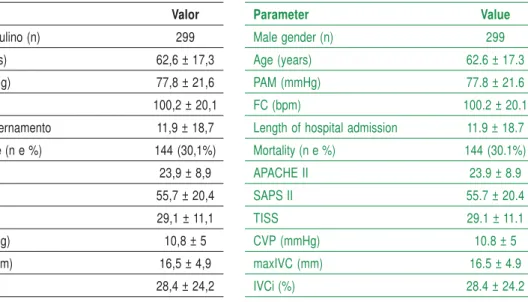
patho-Quadro I – Características globais dos doentes estuda-dos (n=477) Parâmetro Valor Sexo masculino (n) 299 Idade (anos) 62,6 ± 17,3 PAM (mmHg) 77,8 ± 21,6 FC (bpm) 100,2 ± 20,1 Dias de internamento 11,9 ± 18,7 Mortalidade (n e %) 144 (30,1%) APACHE II 23,9 ± 8,9 SAPS II 55,7 ± 20,4 TISS 29,1 ± 11,1 PVC (mmHg) 10,8 ± 5 VCImax (mm) 16,5 ± 4,9 VCIi (%) 28,4 ± 24,2
Table I – Global characteristics of the patient population
(n=477) Parameter Value Male gender (n) 299 Age (years) 62.6 ± 17.3 PAM (mmHg) 77.8 ± 21.6 FC (bpm) 100.2 ± 20.1
Length of hospital admission 11.9 ± 18.7
Mortality (n e %) 144 (30.1%) APACHE II 23.9 ± 8.9 SAPS II 55.7 ± 20.4 TISS 29.1 ± 11.1 CVP (mmHg) 10.8 ± 5 maxIVC (mm) 16.5 ± 4.9 IVCi (%) 28.4 ± 24.2
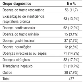
Quadro II – Distribuição de acordo com os principais gru-pos diagnósticos
Grupo diagnóstico N e %
Doença do tracto respiratório 56 (11,7) Exacerbação de insuficiência
63 (13,2%) respiratória crónica
Doença cardiovascular 62 (12,9%) Doença do tracto urinário 15 (3,1%) Doença gastrintestinal 37 (7,7%) Doença neurológica 12 (2,5%) Doenças infecciosas ou sépsis 71 (14,9%) Doenças cirúrgicas 82 (17,2%) Transplante hepático 51 (10,7%)
Outros 38 (7,9%)
Total 477
Table II – Distribution of the agreement with the main
diagnosed groups
Diagnosed group N e %
Respiratory tract disease 56 (11.7)
Worsening of chronic respiratory 63 (13.2%)
insufficiency
Cardiovascular disease 62 (12.9%)
Urinary tract disease 15 (3.1%)
Gastrointestinal disease 37 (7.7%)
Neurological disease 12 (2.5%)
Infectious disease or sepis 71 (14.9%)
Surgical diseases 82 (17.2%)
Liver transplant 51 (10.7%)
Other 38 (7.9%)
Total 477
tória, em particular os admitidos por exacer-bação de doença pulmonar crónica (Quadro II), situação clínica por nós definida previa-mente16,17.
Do total de doentes, registou-se pelo menos 1 alteração cardíaca em 209 (44,1%): 157 com depressão da função sistólica do VE, 67 com dilatação do VE, 106 com dilatação da AE, 78 com dilatação do VD.
Correlações entre variáveis ecocardiográficas e a PVC
A análise de regressão linear com as variá-veis ecocardiográficas estudadas, admitin-do a PVC como variável dependente, re-velou uma relação com significado estatístico com a dimensão máxima da VCI (p=0,013) e respectivo índice (p=0,001). A análise de correlação linear entre a di-mensão máxima da VCI e a PVC revelou valores de r de 0,338 e 0,441 respectiva-mente (Figs. 1 e 2).
logy, in particular patients admitted to hos-pital due to worsening of chronic pulmo-nary disease (Table II), clinical situation pre-viously established by us16,17.
Out of the total patient population, there was at least 1 cardiac change registered out of 209 (44.1%): 157 with depression of the systolic function of the LV, 106 with enlargement of the LA and 78 with enlargement of the RV. Correlation between echocardiograph variables and the CVP
Analysing the linear regression with the echo-cardiographic variables studied, considering the CVP as a dependant variable, showed a statistically significant relation between the maximum diameter of the ICV (p=0.013) and the respective index (p=0.001). Analysing the linear correlation between the maximum dia-meter of the ICV and the ICV index with the CVP showed r values of 0.338 and 0.441 res-pectively (Figs. 1 and 2).
Fig. 1 – Gráfico de dispersão da correlação entre o índice da VCI e a PVC
Índice da VCI
Série 1 Linear (Série 1)
PVC
Fig. 1 – Graph of the dispersal of the correlation between the IVC index and the CVP
IVC Index
Series 1 Linear Series 1
Fig. 2 – Gráfico de dispersão da correlação entre a dimensão máxima da VCI e a PVC
DImensão máxima da VCI
Série 1 Linear (Série 1)
PVC
Fig. 2 – Graph of the dispersal of the correlation between the maximum diameter of the IVC and the CVP
Maximum diameter of the IVC
Series 1 Linear Series 1
A presença de ventilação mecânica influen-ciou igualmente a PVC (p=0,002) e obtive-ram-se correlações fortes mas sem significa-do estatístico entre a PVC e a dimensão significa-do VD (p=0,071) e da AD (p=0,099).
Correlação entre a PVC e o índice da VCI
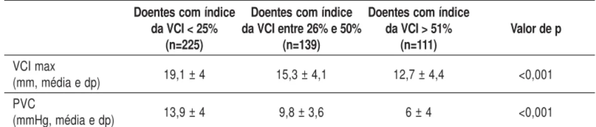
Para efeitos de análise comparativa por teste não paramétrico de qui-quadrado, a variável PVC foi categorizada em 3 variáveis: Inferior a 7mmHg, entre 8 e 12mmHg e superior a 13mmHg; o índice da VCI foi dividido em 3 variáveis categóricas: De 0% a 25%, de 26% a 50% e superior a 51%. A análise do con-junto dos doentes mostrou que 111 doentes (23,2%) apresentava um índice inferior a 25%, 139 (29,1%) um índice entre 26 e 50% e 225 (47,2%) um índice superior a 51%. No Quadro III mostra-se a análise de frequên-cia entre os valores categorizados de PVC o e índice da VCI.
Mechanical ventilation also influenced the CVP (p=0.002) and allowed for stronger – but not statistically significant – correlations between the CVP and the diameter of the RV (p=0.071) and the RA (p=0.099). Correlation between the CVP and the ICV index
To use the comparative analysis of the non-parametric chi-squared test, the variable CVP was categorised under 3 variables: below 7mmHg, between 8 and 12mmHg and above 13mmHg and the IVC index was divided into 3 categorical variables: from 0% – 25%, from 26% – 50% and over 51%. Analysing the set of patients showed that 111 patients (23.2%) had an index below 25%, 139 (29.1%) had an index between 26 and 50% and 225 (47.2%) had an index over 51%. Table III shows the analysis of the frequency between the cate-gorisable CVP values and the IVC index.
Quadro III – Valores médios de diversos parâmetros de acordo com o índice da veia cava inferior Doentes com índice Doentes com índice Doentes com índice
da VCI < 25% da VCI entre 26% e 50% da VCI > 51% Valor de p
(n=225) (n=139) (n=111) VCI max 19,1 ± 4 15,3 ± 4,1 12,7 ± 4,4 <0,001 (mm, média e dp) PVC 13,9 ± 4 9,8 ± 3,6 6 ± 4 <0,001 (mmHg, média e dp)
Legenda: VCI – veia cava inferior; VCImax – dimensão máxima da veia cava inferior; PVC – pressão venosa central; mmHg – milímetros de
mercúrio.
Table III – Median values of the various parameters in agreement with the inferior venous cava index Patients with Patients with Patients with
IVC index < 25% IVC index < 25% IVC index > 51% p value (n=225) (n=139) (n=111) Max IVC 19.1 ± 4 15.3 ± 4.1 12.7 ± 4.4 <0.001 (mm, mean and sd) CVP 13.9 ± 4 9.8 ± 3,6 6 ± 4 <0.001 (mmHg, mean and sd)
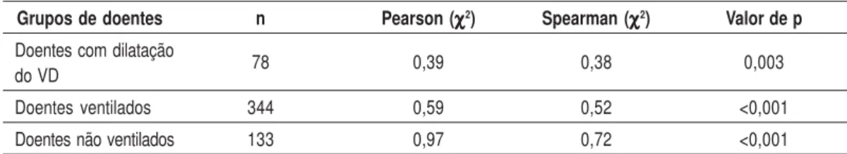
Na análise das duas variáveis categóricas, PVC e índice da VCI, pelo teste de qui-quadrado, obteve-se um valor de p <0,001; para um va-lor de r do teste de correlação de Pearson foi de 0,565, a correlação de Spearman de 0,592. Esta correlação é apresentada em forma de gráfico de dispersão na Fig. 3 para a generali-dade dos doentes. Nesta figura observa-se que alguns doentes com índices da VCI elevados apresentam PVC igualmente elevadas. Dada a influência da ventilação mecânica e da dimensão do VD na PVC verificaram-se as correlações entre estes 2 parâmetros nes-tes 2 grupos de doennes-tes (Quadro IV). Verifi-cou-se que a correlação nos doentes ventila-dos foi menor do que nos doentes não ventilados e que foi substancialmente me-nor nos doentes com dilatação do VD.
In analysing the two variable categories, the CVP and the IVC index, using the chi-squared test gave a value <0.001. For a value of r the Pearson correlation test was 0.565 and the Spearman correlation was 0.592. This correlation is shown for the majority of the patients in the dispersion graph in Fig. 3. This figure shows that some patients with high IVC indexes had equally high CVPs.
Mechanical ventilation and the diameter of the RV influence the CVP. The correlations between these 2 parameters in these 2 groups of patients are shown in Table IV. It is seen that the correlation of the ventilated patients was less than in the non-ventilated patients and was markedly less in the patients with RV enlargement.
Quadro IV – Comparação entre as variáveis de análise da veia cava inferior e a pressão venosa central nos diferentes grupos de doentes estudados
Grupos de doentes n Pearson (χχχχχ2) Spearman (χχχχχ2) Valor de p
Doentes com dilatação
78 0,39 0,38 0,003
do VD
Doentes ventilados 344 0,59 0,52 <0,001
Doentes não ventilados 133 0,97 0,72 <0,001 Legenda: VCImax – dimensão máxima da veia cava inferior; VCIi – índice da veia cava inferior; Pearson e Spearman referem-se às
correlações do teste de qui-quadrado.
Table IV – Comparison between the analysis variables of the inferior vena cava and the central venous pressure in the
different patient groups studied
Patient groups n Pearson (χχχχχ2) Spearman (÷2) P value
Patients with RV
78 0.39 0.38 0.003
enlargement
Ventilated patients 344 0.59 0.52 <0.001
Non-ventilated patients 133 0.97 0.72 <0.001
Legend: maxIVC – maximum diameter of inferior vena cava; iVCI – inferior vena cava index; Pearson and Spearman are the chi-squared
Fig. 3 – Gráfico de dispersão da PVC relativamente ao índice da VCI na totalidade dos doentes estudados
PVC
Doentes com índice da VCI < 25%
Doentes com índice da VCI entre 26% e 50%
Doentes com índice da VCI > 51%
Índice da VCI
Fig. 3 – Graph of the dispersal of CVP relative to the IVC index in the total patient population
CVP
Patients with IVC index < 25%
Patients with IVC index between 26% and 50% Patients with IVC index > 51%
Verificou-se igualmente a correlação entre o índice da VCI e a PVC nos doentes admitidos por exacerbação de doença pulmonar crónica, que apresentaram uma elevada prevalência de dilatação do VD (35 de 63 doentes, 54%). Neste grupo de doentes apenas 5 apresentaram valo-res de PVC <7mmHg e 5 índice da VCI> 50%. Apesar de poucos doentes se apresentarem na faixa mais baixa de PVC (n=5) ou na mais ele-vada de índice da VCI (n=5), nota-se uma gran-de dispersão gran-de valores neste intervalo (Fig. 4), denotando uma ausência de correlação entre estes dois parâmetros nesta faixa de valores (p não significativo).
Correlação entre a PVC e a dimensão máxima da VCI
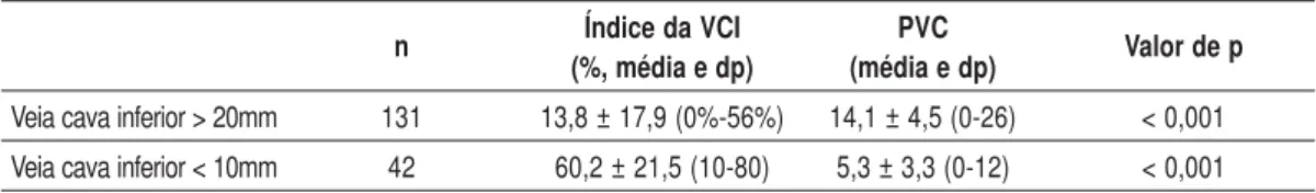
Por teste não paramétrico de qui-quadrado, ob-servou-se uma correlação estatisticamente signi-ficativa entre uma VCI dilatada (dimensão má-xima > 20mm) e valores de PVC > 13mmHg, e uma VCI com dimensão inferior a 10mm e PVC < 7mmHg (Quadro V).
A correlation was also seen between the IVC index and the CVP in the patients admitted to hospital for worsening of chronic pulmo-nary disease. These had a greater rate of RV enlargement (35 out of the 63 patients, 54%). Out of this group of patients, only 5 had CVP values of <7mmHg and 5 had an IVC index of > 50%. While only a few patients fell into the lowest CVP band (n=5) or into the highest IVC index band (n=5), there was a great dispersion of values in this interval (Fig. 4), denoting an absence of correlation between these 2 parameters in this band of values (p non-significant).
Correlation between the CVP and the maximum diameter of the IVC
The non-parametric chi-squared test showed a statistically significant correlation between IVC enlargement (maximum diameter> 20mm) and CVP values> 13mmHg, and an IVC with a diameter less than 10mm and CVP < 7mmHg (Table V).
Quadro V – Comparação entre os valores de pressão venosa central e dimensão máxima da veia cava inferior na globalidade dos doentes
n Índice da VCI PVC Valor de p
(%, média e dp) (média e dp)
Veia cava inferior > 20mm 131 13,8 ± 17,9 (0%-56%) 14,1 ± 4,5 (0-26) < 0,001 Veia cava inferior < 10mm 42 60,2 ± 21,5 (10-80) 5,3 ± 3,3 (0-12) < 0,001
Table V – Comparison between the central venous pressure values and maximum diameter of the inferior vena cava in
the majority of patients
n IVC index CVP P value (%, mean and sd) (mean and sd)
Inferior vena cava> 20mm 131 13.8 ± 17.9(0%-56%) 14.1 ± 4.5 (0-26) <0.001
Fig. 4 – Gráfico de tipo error bar da correlação entre intervalos de índice da VCI e a PVC nos doentes admitidos por exacerbação de insuficiência respiratória crónica
Legenda: Índice 1 – doentes com índice da veia cava inferior < 25%; índice 2 – doentes com índice da veia cava inferior entre 26 e
50%; índice 3 – doentes com índice da veia cava inferior> 51%
PVC1
Índice 1
Fig. 4 – Graph of the error bar type of the correlation between the intervals of the IVC index and the CVP in patients
admitted to hospital for worsening of chronic respiratory insufficiency disease
Legend: Index 1 – patients with inferior vena cava index < 25%; index 2 – patients with inferior vena cava index between 26 and
50%; index 3 – patients with inferior vena cava index > 51%
CVP1
Table VI – Analysis of the maximum diameter of the IVC and the CVP in patients with right ventricle enlargement
n IVC > 20mm IVC < 10mm CVP > 13mmHg CVP < 7mmHg (n and %) (n and %) (n and %) (n and %)
Patients with
78 45 (57.7%) 7 (8.9%) 48 (61.5%) 4 (5.1%)
RV enlargement
Quadro VI – Análise da dimensão máxima da VCI e a PVC em doentes com dilatação do ventrículo direito
n VCI > 20mm VCI < 10mm PVC > 13mmHg PVC < 7mmHg
(n e %) (n e %) (n e %) (n e %)
Doentes com
78 45 (57,7%) 7 (8,9%) 48 (61,5%) 4 (5,1%) dilatação do VD
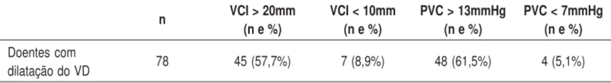
Verificou-se que esta correlação se mantinha no conjunto de doentes ventilados e nos doentes com exacerbação de doença pulmonar crónica. Contudo, observou-se que os doentes com dila-tação do VD apresentaram uma frequência de valores de dimensão da VCI <10mm mais baixa (n=7, 8,1%), enquanto 57,7% (n=45) apresen-taram uma VCI > 20mm. Quatro destes doen-tes apresentaram PVC < 7mmHg (Quadro VI).
Outras correlações
Por análise de regressão linear com o índice da VCI e a dimensão máxima da VCI como variáveis dependentes, verificou-se que a di-mensão máxima se correlacionou com a dila-tação do VD e com o índice da VCI (p <0,001 e p=0,03, respectivamente), mas não se veri-ficou qualquer relação entre o índice da VCI e os parâmetros ecocardiográficos referidos.
Discussão
Estudos prévios e principais achados
Não encontrámos na literatura estudo seme-lhante realizado numa UCIP e com um nú-mero semelhante de doentes. Mesmo o
estu-It can be seen that this correlation is steady in the ventilated patient group and in the patients with worsening of chronic pulmo-nary disease. In addition, it was seen that the patients with RV enlargement had a frequen-cy of ICV diameter values <10mm lower (n=7.8.1%) while 57.7% (n=45) had an IVC of > 20mm. Four of these patients had CVP of <7mmHg (Table VI).
Other correlations
To analyse the linear regression with the IVC index and the maximum diameter of the IVC as dependant variables, it is seen that the maxi-mum diameter correlates with the enlarged RV and with the IVC index (p <0.001 and p=0.03 respectively). No relation was seen between the IVC index and the abovementioned echocar-diograph parameters, however.
Discussion
Earlier studies and main findings We could find no comparable earlier study carried out in a PICU and with a similar number of patients. Even the Jue et al study
do de Jue e col incluiu apenas 49 doentes ven-tilados, nos quais não foram avaliadas a influ-ência de determinados parâmetros ecocardi-ográficos, em particular a dilatação do VD ou a presença de doença pulmonar crónica. Observámos uma fraca correlação linear entre estas variáveis, já descrita na literatu-ra18, mas uma possibilidade de aferir
qualita-tivamente a PVC por ETT, em especial nos seus valores extremos.
Deparámos com duas dificuldades principais relativamente à obtenção de dados. A primei-ra tem a ver com a dificuldade na visualização da VCI, que na sua maioria se deveu à presen-ça de pensos abdominais nos doentes cirúrgi-cos, inviabilizando um conjunto de exames. Ainda nestes doentes, a presença de líquido intra-abdominal de natureza diversa (inflama-tória ou não) e a interposição de ansas intesti-nais dificultaram a visualização de estruturas intrabdominais, entre as quais a veia cava in-ferior. Contudo, o sucesso alcançado parece significativo, pois mais de 80% dos doentes puderam ser avaliados. Este sucesso parece--nos ser de valorizar na perspectiva de uma possível utilização sistemática da avaliação da VCI neste grupo particular de doentes. Por outro lado, em alguns doentes, observou-se um conflito assinalável entre os dados da PVC e da VCI, uma vez que em alguns obtivemos va-lores baixos de PVC e vava-lores elevados de índi-ce da VCI. Este conflito não comprometeu a correlação geral, que se manteve significativa para a generalidade dos doentes, em especial por aná-lise estatística com métodos não paramétricos, reforçando aliás o carácter essencialmente qua-litativo da informação obtida por ETT. Pode-mos apontar diversas causas para este facto. Apesar do cuidado observado com a visualiza-ção do posicionamento dos cateteres centrais utilizados para determinação da PVC e do
cui-population only had 49 ventilated patients and of these, the influence of determined echocardiograph parameters, in particular RV enlargement or the presence of chronic pul-monary disease, were not evaluated. We saw a weak linear correlation between these variables, as has already been seen in earlier studies18, but there was the chance
of measuring the CVP qualitatively by TTE, particularly its most extreme values. There were two main difficulties inherent in obtaining these data. The first had to do with the difficulty of visualising the IVC, which was largely due to the surgery patients having abdominal plasters which made it impossi-ble to carry out certain exams. Intrabdomi-nal liquid (inflammatory or non-inflam-matory) in these surgery patients and the interposition of intestinal ansas made it hard to visualise the intrabdominal structures, one of which is the inferior vena cava. This not-withstanding, the success achieved seems sig-nificant in that it was possible to evaluate over 80% of these patients. We feel this suc-cess makes it possible to evaluate the ICV of this particular group of patients in a sys-temic way.
On the other hand, a marked conflict was seen in some patients between the CVP and IVC data, in that we obtained low CVP values and high ICV index values in some patients. This division did not compromise the general cor-relation which remained significant for the majority of patients, in particular for a statis-tical analysis with non-parametric methods. This served to strengthen the essentially qual-itative character of the information gleaned through TTE. There are several causes for this. Despite the care taken with visualising the positioning of the central catheters used for determining the CVP and the care taken
dado na medição deste parâmetro, não é possí-vel aferir o local exacto da medição, ao contrário do que é possível por ETT. Por outro lado, sen-do a PVC um parâmetro de pressão, pode não relacionar-se de forma perfeita com a volemia19.
Na determinação dos parâmetros da VCI, sen-do um vaso distensível, participam não só a volémia do doente, mas também repercute a pressão da circulação pulmonar. Se em condi-ções normais esta é uma circulação de baixa pressão, em condições patológicas esta relação pode alterar-se, e a VCI nestes casos reflecte mais a sobrecarga de pressão do que a volemia. Observámos que, quer a PVC quer os parâ-metros de análise da VCI, se modificaram no mesmo sentido (ou seja, a uma maior PVC corresponde um menor índice da VCI) nos doentes com maior probabilidade de apre-sentar alterações da pressão da circulação pulmonar (doentes ventilados ou com dila-tação do VD). De qualquer das formas, a presença de uma pressão aumentada na cir-culação pulmonar afectou a qualidade da correlação entre estes dois parâmetros.
Avaliação dos dados na perspectiva clínica
Verificou-se que os doentes com dilatação do VD mostraram uma relação mais fraca na apreciação qualitativa da PVC através da dimensão máxima da VCI. Este dado vem de encontro às descrições da literatura e po-demos inferir que nestas condições a análise da VCI reflecte preferencialmente a conção de sobrecarga crónica das cavidades di-reitas. Como a população de doentes com exacerbação de doença pulmonar crónica é especialmente afectada pela dilatação do VD (54% no nosso estudo), não é de estranhar que neste grupo de doentes as conclusões sejam similares.
in measuring this parameter, it was not possi-ble to evaluate the exact location of the measu-rement, unlike that which is made possible by TTE. On the other hand, as CVP is a pres-sure parameter it cannot be perfectly related with volemia19. In determining the IVC
pa-rameters, as the IVC is a vein which distends, it is not only a part of the patient’s volemia, but also rebounds pressure to the pulmonary circulation. If under normal conditions this is the low pressure circulation, under patho-logical conditions this relationship can change and in these cases it is the IVC rather than the volemia which better mirrors the overload of pressure.
We saw that both the CVP and the parame-ters used to measure the IVC changed in the same way (or rather a greater CVP corre-sponds to a lower IVC index) in the patients more likely to show changes in the pulmo-nary circulation pressure, i.e. ventilated pa-tients or papa-tients with RV enlargement. Whatever the case, elevated pulmonary cir-culation pressure impacts on the correlation quality of these 2 parameters.
A clinical perspective evaluation of the data
Patients with RV enlargement show a weaker relationship in the qualitative appreciation of the CVP through the maximum diameter of the IVC. These data come from earlier stu-dies made and from them we can infer that under these conditions an analysis of the IVC preferentially reflects the chronic overload condition of the right cavities. As the pa-tient population with worsened chronic pul-monary disease is essentially affected by RV enlargement (54% in our study), it is not strange that the conclusion should be similar in this group of patients.
Contudo, esta sobrecarga pode igualmente reflectir-se na própria PVC, como se verifica por análise de regressão linear, na qual a di-latação do VD (e da AD) influenciou forte-mente a PVC, embora sem atingir significa-do estatístico. Este dasignifica-do, aliasignifica-do ao facto de que apenas 5,1% (4 de 78 doentes) com dila-tação do VD apresentou valores de PVC in-feriores a 7mmHg, pode sugerir-nos que es-tes dois parâmetros, entre si correlacionados, são pouco úteis para detectar situações de baixa pressão, sugestivas de hipovolemia. Uma situação de sobrecarga crónica fisioló-gica da circulação pulmonar foi verificada em atletas de alta competição. Num estudo feito em atletas e nadadores de competição, sujei-tos a sobrecarga direita crónica pelo esforço físico e respectiva sobrecarga na circulação pulmonar, os autores descreveram uma di-mensão máxima da VCI aumentada mas com valores do respectivo índice superiores a 50%, próximo dos valores normais descritos para indivíduos normais11,20. Sugere-se, assim, que
a sobrecarga crónica altera fundamentalmen-te a dimensão máxima da VCI e o índice da VCI reflecte mais o estado actual de vole-mia, com uma melhor aproximação por aná-lise de correlação ao valor de PVC. Esta afir-mação é bem fundamentada no estudo presente, onde se verificou uma correlação entre a dimensão máxima da VCI e a dilata-ção das cavidades direitas, o mesmo não se verificando para o índice da VCI.
Mas convém destacar que a análise da VCI mantém o seu valor para identificar valores elevados de PVC nestes doentes. Este fac-to parece-nos importante, uma vez que a detecção de situações de hipovolemia é im-portante para diagnósticos diferenciais ou para preparar os doentes durante o desma-me ventilatório.
Additionally, this overload can equally be reflected in the CVP itself, as the linear regression analysis shows, in which the RV (and the RA) enlargement impacts strongly on the CVP, although it does not reach sta-tistical significance. This fact, taken togeth-er with the fact that only 5.1% (4 out of the 78 patients) with RV enlargement show CVP values below 7mmHg, could suggest that these 2 parameters, correlated in them-selves, are not very useful for detecting low pressure situations suggestive of hypo-volemia.
A chronic physiological overload of the pul-monary circulation has been seen in highly trained athletes. The authors of a study into competition athletes and swimmers subject to chronic right overload and respective pulmonary circulation overload caused by physical effort describe an increased maxi-mum IVC diameter but with the respective index values above 50%, close to normal values seen in normal individuals11,20. This
suggests that a chronic overload fundamen-tally alters the maximum diameter of the IVC and the IVC index reflects better the actual state of volemia, with a close approximation by correlation analysis to the CVP index. Our study corroborates this idea. Our study shows a corelation between the maximum diameter of the IVC and the enlargment of the right cavities. This is not true of the IVC index.
It is important to highlight that the analy-sis of the IVC maintains its value of iden-tifying the raised CVP values of these pa-tients. This fact strikes us as important in that detecting incidences of hypovolemia is crucial for differential diagnoses or for preparing patients during weaning from ventilation.
A presença de ventilação mecânica altera a fisiologia respiratória, modificando a cor-relação de pressões intratorácicas. No con-junto de doentes não ventilados, verificá-mos que as dimensões da VCI foram idênticas às dos doentes ventilados, mas o índice da VCI foi maior e a PVC mais baixa (em média 20%). O grau de correlação en-tre valores categóricos de PVC e do índice da VCI foram superiores nos doentes não ventilados. Para analisar estes dados temos de ter em conta que muitos dos doentes ventilados apresentavam concomitantemen-te as alconcomitantemen-terações cardíacas identificadas, em particular os doentes com dilatação do VD. Por outro lado, verifica-se igualmente que apenas 9 destes doentes apresentaram va-lores de PVC inferiores a 7mmHg, pelo que se pode aplicar o raciocínio exposto sobre a possibilidade de diagnosticar situações tí-picas de baixa pressão nestes doentes com estes métodos (invasivo e não invasivo). A presença concomitante de ventilação me-cânica e dilatação do VD não foi explora-da, mas pode revelar-se uma associação na qual estes dados podem ser ainda mais exa-cerbados. Os doentes ventilados com exacer-bação de insuficiência respiratória crónica podem ser especialmente visados.
Os dados de Doppler podem ser correlacio-nados com diversos parâmetros de preen-chimento intravascular. No nosso estudo, a relação E/A mitral e o TRIV não foram pa-râmetros correlacionáveis com a PVC. Con-vém assinalar que estes parâmetros são obti-dos ao nível do VE, e a PVC é um parâmetro de enchimento do ventrículo direito. A utili-dade prática destes parâmetros pode ser ques-tionada, pois alterações de ritmo e frequência cardíaca podem modificá-los de forma sig-nificativa ou inviabilizar a sua determinação.
The presence of mechanical ventilation changes the respiratory physiology, alter-ing the correlation of intrathoracic pres-sures. In the non-ventilated patient group, we saw that the IVC diameters were iden-tical to those of the ventilated patients, but the IVC index was greater and the CVP lower (on average 20%). The degree of correlation between the CVP categorical variables and the IVC index was higher in the non-ventilated patients. To analyse these data, we have to bear in mind that many ventilated patients have concomitant cardiac alterations identified, in particular the patients with RV enlargement. Con-versely, equally only 9 of these patients had CVP values lower than 7mmHg, meaning that the abovementioned rationale about the possibility of diagnosing typical low pressure situations in these patients with these methods (invasively and non-inva-sively) can be applied. The concomitant presence of mechanical ventilation and RV enlargement has not been explored, but it may reveal an association in which these data could be further heightened. The ven-tilated patients with worsening of chronic respiratory insufficiency could be particu-larly targeted.
The Doppler data can be correlated with the various intravascular filling parameters. In our study, the mitral E/A relationship and the TRIV were not correlational parameters with the CVP. It should be stressed that these parameters are obtained at the level of the LV and the CVP is a filling parameter of the right ventricle. The practical use of these parameters can be questioned, as alterations in the cardiac rhythm and rate can change significantly or make their measurement invalid.
Clinical implications and conclusion Using TTE to analyse the IVC (IVC index and maximum diameter) proves useful in the non-invasive evaluation of CVP in patients admitted to a PICU.
The parameter which better correlated with the CVP was the ICV índex, especially in non-ventilated patients.
We noted that a RV enlargement and in the patients admitted for worsening of chronic pulmonary disease, the correlations between the IVC index and the CVP were altered by the low CVP values. The existence of pathol-ogies which impact on chronic overload of the right cavities can change the diagnostic viability of these methods in detecting inci-dences of hypovolemia.
If in the PICU in which the study was car-ried out 63 (13.2%) patients had worsening of chronic pulmonary disease, there could be a much more significant rate of these patients and RV enlargement in ICUs for respiratory patients.
Conversely, analysing IVC can identify states of major volemia in all the patients, includ-ing the patients with worseninclud-ing of chronic pulmonary disease and RV enlargement, al-lowing in this way the detection of incidences of hypervolemia, important in critical situa-tions such as ventilatory weaning.
Implicações clínicas e conclusão
A análise da VCI por ETT (índice da VCI e dimensão máxima) revelou-se útil na avalia-ção não invasiva da PVC em doentes admiti-dos numa UCIP.
O parâmetro que melhor se correlacionou com a PVC foi o índice da VCI, em especial nos doentes não ventilados.
Notámos que a presença de um VD dilata-do e, nos dilata-doentes admitidilata-dos por exacerba-ção de doença pulmonar crónica, as correla-ções entre o índice da VCI e a PVC foram alteradas para valores baixos de PVC. A exis-tência de patologias que condicionam sobre-carga crónica nas cavidades direitas pode al-terar a fiabilidade diagnóstica destes métodos para detectar situações de hipovolemia. Se, na UCIP onde decorreu o estudo, 63 (13,2%) doentes apresentavam exacerbação de doen-ça pulmonar crónica, nas UCI dedicadas a doentes respiratórios pode existir uma pre-valência mais significativa destes doentes e de dilatação do VD.
Por outro lado, a análise da VCI pode identifi-car estados de maior volemia em todos os doen-tes, incluindo nos doentes com exacerbação de doença pulmonar crónica e dilatação do VD, permitindo assim detectar situações de hiper-volemia, importantes para situações críticas, como, por exemplo, desmame ventilatório.
Bibliografia/Bibliography
1. Forrester J, Diamond G, Mc Hugh TJ, et al. Filling pressures in the right and left sides of the heart in acute myocardial infarction: a reapraisal of central venous pres-sure. N Eng J Med 1971; 285:190-193.
2. Magder S. More respect for CVP. Intensive Care Med 1998; 24:651-653.
3. Paul L Marino. Hemodynamic monitoring. Tissue oxygenation. In: Paul L Marino, editor. The ICU Book. William & Wilkins, 1997: 190.
4. Rivers E, Bryant Nguyen, Havstad S, et al. Early goal-oriented therapy in the treatment of severe sepsis and septic shock. N Eng J Med 2001; 345:1368-1377. 5. The ARDS Clinical Trials Network. Pulmonary-artery versus central venous catheter to guide treatment of acute lung injury. N Eng J Med 2006;(NEJMMoa061895). 6. Marcelino P, Marum S, Fernandes AP, Ribeiro JP. Non invasive evaluation of Central Venous Pressure by echo-cardiography. Rev Port Cardiol 2002; 21:125-133.
O parâmetro que melhor se correlacionou com a PVC foi o índice da VCI
A análise da VCI pode identificar estados de maior volemia em todos os doentes
7. Kircher B, Himelman RB, Schiller NB. Noninvasive esti-mation of right atrial pressure from the inspiratory colapse of the inferior vena cava. Am J Cardiol 1990; 66:493-496. 8. Moreno F, Hagan AD, Holmen JR, Pryor TA, Stri-ckland RD, Castle H. Evaluation of Size and Dynamics of Inferior Vena Cava as an Index of Right Sided Cardiac Function. Am J Cardiol 1984; 53:579-585.
9. Ando Y, Yanagiba S, Asano Y. The inferior vena cava diameter as a marker of dry weight in chronic hemo-dialyzed patients. Artif Organs 1995; 19:1237-1242. 10. Mandelbaum A, Ritz E. Vena cava diameter measu-rement for estimation of dry weight in haemodialysis patients. Nephrol Dial Transplant 1996; 11:S24-S27. 11. Minutiello L. Non-invasive evaluation of central ve-nous pressure derived from respiratory variations in the diameter of the inferior vena cava. Minerva Cardioan-giol 1993; 41:433-437.
12. Yanagiba S, Ando Y, Kusano E, Asano Y. Utility of the inferior vena cava diameter as a marker of dry weight in no-noliguric hemodialyzed patients. ASAIO J 2001; 47:528-532. 13. Jardin F, Vieillard-Baron A. Ultrasonographic exa-mination of the venae cavae. Intensive Care Med 2006; 32:203-206.
14. Jue J, Chung W, Schiller NB. Does inferior vena cava size predict right atrial pressure in patients receiving
me-chanical ventilation? J Am Soc Echocardiogr 1992; 5:613--619.
15. Jacobson B. Medicine and Clinical Engeneering. En-glewood Cliffs, New Jersey: Prentice-Hall, 1977. 16. Marcelino P, Germano N, Grilo A, Flora L, Marum S, Fernandes AP et al. Linfopénia em doentes submeti-dos a ventilação mecânica por exacrbaçã de insuficiência respiratória crónica: estudo prospectivo. Rev Port Pneu-mol 2004; 10:373-381.
17. Marcelino P, Germano N, Nunes AP, Flora L, Molei-ro A, Marum S et al. Determinantes cardíacas do tempo de ventilação mecânica e mortalidade de doentes com insuficiência respiratória crónica exacerbada. A impor-tância dos parâmetros ecocardiográficos. Rev Port Pneu-mol 2006; 12:131-146.
18. Baumann UA, Marquis C, Stoupis C, Willenberg TA, TakalaJ, Jacob SM. Estimation of central venous pressu-re by ultrasound. Resuscitation 2005; 64:193-199. 19. Marcelino P, Frade F, Marum S, Fernandes AP, Ri-beiro JP, Lopes MG. Cardiac Doppler variation with vo-lume status changes in General Intensive Care. Rev Port Cardiol 2004; 23:183-196.
20. Minutiello L. Value of the vena cava index in healthy young subjects. Echocardiography study. Minerva Car-dioangiol 1994; 42:229-232.