Abstract
Objective: To identify non-invasive predictors of esophageal varices in children and adolescents with chronic liver disease or extrahepatic portal venous obstruction (EHPVO).
Methods: 53 patients younger than 20 years with chronic liver disease or EHPVO and no history of bleeding or prophylactic treatment of esophageal varices (EV) were assessed. They were divided into 2 groups: group I (35 with chronic liver disease) and group II (18 with EHPVO). Their blood count, international normalized ratio (INR), albumin, bilirubin, abdominal ultrasonography and upper endoscopy results were taken. A splenic index was determined by dividing the patients’ spleen dimension by its uppermost limit according to their age. The variables were compared to EV presence or not. Univariate (chi-square test, Fischer’s exact test and Wilcoxon exact test) and multivariate (logistic regression) analyses were performed. A receiver operating characteristic (ROC) curve was constructed and the area under the ROC curve was calculated.
Results: EV were observed in 48.5% of group I patients and in 83.3% of group II patients. Low platelet count (p = 0.0015), splenomegaly (p = 0.0003) and splenic index (p = 0.0007) were statistically signiicant predictors of EV among group I patients. The multivariate analysis showed low platelet count (odds = 21.7) as an independent predictor of EV in patients with chronic liver disease.
Conclusion: Platelet count, splenic index and platelet-splenic index ratio were predictors of EV in children and adolescents with chronic liver disease. There were no EV predictors among group II patients.
J Pediatr (Rio J). 2012;88(4):341-6: Esophageal varices, extra-hepatic portal venous obstruction, cirrhosis, portal hypertension, child, adolescent.
O
riginala
rticleCopyright © by Sociedade Brasileira de Pediatria
341 Introduction
Taking into account the high prevalence of esophageal varices (EV) in adults with cirrhosis and high morbidity and mortality related to upper gastrointestinal bleeding, there is a consensus to perform esophagogastroduodenoscopy
(EGD) during diagnosis and every 1-2 years depending on the severity of the liver disease.1,2
However, endoscopic screening in children and adolescents diagnosed with cirrhosis results in up to 50%
Non-invasive predictors of esophageous varices
in children and adolescents with chronic liver disease
or extrahepatic portal venous obstruction
Roberta V. Alcantara,1 Roberto M. Yamada,2 Adriana M. A. De Tommaso,1
Maria Angela Bellomo-Brandão,1 Gabriel Hessel3
1. PhD, Saúde da Criança e do Adolescente. Faculdade de Ciências Médicas, Universidade Estadual de Campinas (UNICAMP), Campinas, SP, Brazil. 2. PhD. Adjunct professor, Universidade Federal de São Carlos (UFSCar), São Carlos, SP, Brazil.
3. Associate professor, Departamento de Pediatria, Faculdade de Ciências Médicas, UNICAMP, Campinas, SP, Brazil.
No conflicts of interest declared concerning the publication of this article.
Suggested citation: Alcantara RV, Yamada RM, De Tommaso AM, Bellomo-Brandão MA, Hessel G. Non-invasive predictors of esophageous varices in children and adolescents with chronic liver disease or extrahepatic portal venous obstruction. J Pediatr (Rio J). 2012;88(4):341-6.
Manuscript submitted Jan 10 2012, accepted for publication Apr 25 2012.
normal endoscopies.3,4 In addition to patients undergoing an invasive procedure unnecessarily, general anesthesia is necessary to perform the procedure in most children. One also has to consider the overpopulation in medical services and the high cost to the health system.
To restrict EGD to those patients who have ultrasonographic or laboratory indicators able to predict the presence of varices would result in a better risk/ beneit ratio for the endoscopic study.
In recent years, several studies have attempted to address this issue by correlating the presence of EV and various clinical, laboratory and/or ultrasonographic parameters.3-14 Among these, there are only two studies on the pediatric age group.3,4
Fagundes et al.3 found that splenomegaly, discovered on physical examination, and hypoalbuminemia were indicators of EV among 85 chronic liver disease patients. Gana et al.4 assessed the size of the spleen by ultrasonography (US) and also found that splenomegaly was an indicator of EV among the children and adolescents in the study. In addition, they created an index which includes the number of platelets, the spleen size and the serum albumin level.
In this study, the main objective was to identify non-invasive predictors for EV in children and adolescents with chronic liver disease or extrahepatic portal venous obstruction (EHPVO).
Methods
This cross-sectional study included patients treated in the outpatient clinic for Pediatric Hepatology of Hospital de Clínicas da Universidade Estadual de Campinas (Unicamp), Brazil, a tertiary university hospital, from January 2007 to September 2011. It was conducted following the principles of the Declaration of Helsinki and approved by the Research Ethics Committee of Unicamp’s School of Medical Sciences (Faculdade de Ciências Médicas), opinion n. 141/2007. Informed consent was obtained from parents or caregivers.
The inclusion criteria were: patients with up to 20 years of age in follow-up because of chronic liver disease or EHPVO who agreed to participate, not having personal history of acute gastrointestinal bleeding or sclerotherapy and ligation of EV, not in use of beta-blockers, not having a history of portosystemic shunt. The exclusion criterion was the carrying out of EGD in the 3 months prior or subsequent to the performance of laboratory tests and abdominal US.
53 patients who met the inclusion criteria were included in the study. Of these, 31 were female. The patients were divided into two groups. In group I 35 patients with chronic liver disease were included, and in group II, 18 patients with EHPVO. The Child-Pugh score15 was calculated for patients in group I.
Laboratory tests (complete blood count, coagulation, serum albumin and total bilirubin) were performed in the Clinical Laboratory (Laboratório de Análises Clínicas) of Unicamp’s Hospital de Clínicas and collected up to 3 months after EGD completion.
The diagnosis of chronic liver disease was based on the clinical, laboratory and ultrasound assessments. Liver biopsy was performed in 88.5% of patients (31/35). EHPVO diagnosis was made using abdominal Doppler US.
Blood count assessed: hemoglobin, total leukocyte and platelet counts. We used the reference values for hemoglobin and white blood cells according to age group16. Thrombocytopenia was characterized when there were fewer than 150,000 platelets/mm3. The international normalized ratio (INR) < 1.25 was considered normal. Hypoalbuminemia was deined as serum albumin < 3.5 g/dL.
All patients underwent abdominal US at Unicamp’s Center for Diagnosis of Digestive Tract Diseases (Centro de Diagnóstico de Doenças do Aparelho Digestivo - Gastrocentro), using a Toshiba Power Vision 6000 device and a convex linear transducer of 3.75 MHz. Tests were performed by two experienced physicians who had no information on the result of the patient’s EGD. The assessment included the longitudinal dimension of the spleen, which was expressed in millimeters. In order to standardize he data the longitudinal dimension of each patient’s spleen was divided by the maximum size as the reference value for age group,17 so that it was determined how many times larger the spleen was. The value obtained was called the splenic index (SI) and SI > 1.0 was deined as splenomegaly.
Considering that there may be several reasons that explain thrombocytopenia in patients with liver disease, it was chosen to determine a platelet/spleen ratio by dividing the number of platelets by the SI. The result was then divided by 1000 to facilitate viewing of the data. Furthermore, the number of platelets in patients taking azathioprine (patients with autoimmune hepatitis) was compared to other patients in group I.
EGD was performed in all patients by two experienced pediatric endoscopists who had no information on the results of abdominal US. The presence of EV, gastric varices (GV) and portal hypertensive gastropathy (PHG) was assessed, as well as their characteristics.18-20 The examinations were performed in the operating center of Hospital de Clínicas da Unicamp and in Gastrocentro, under sedation or general anesthesia, with endoscopes Pentax FG27x and Fuginon EG590WR.
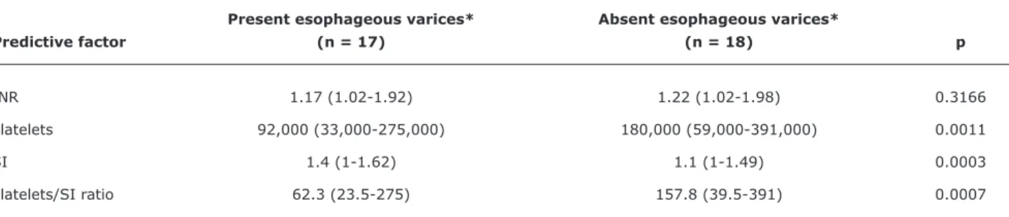
Present esophageous varices* Absent esophageous varices*
Predictive factor (n = 17) (n = 18) p
INR 1.17 (1.02-1.92) 1.22 (1.02-1.98) 0.3166
Platelets 92,000 (33,000-275,000) 180,000 (59,000-391,000) 0.0011
SI 1.4 (1-1.62) 1.1 (1-1.49) 0.0003
Platelets/SI ratio 62.3 (23.5-275) 157.8 (39.5-391) 0.0007
Table 1 - Univariate analysis of the following predictive factors for the presence of esophageal varices in patients with chronic liver disease: internaional normalized ratio, platelets, splenic index and platelets/splenic index ratio
INR = international normalized ratio; n = number of patients; SI = splenic index. * Median (minimum-maximum) values are expressed.
Wilcoxon test was used.
from the variables with p < 0.1 in the univariate analysis.21 The accuracy was determined from the area under the ROC curve using the computer program SPSS 17.0. Sensitivity, speciicity, positive predictive value (PPV) and negative predictive value (NPV) of the variables indicative of the presence of EV were determined.22
Statistical analysis was performed using the SAS 9.1 computer program. A signiicance level of 5% was adopted.
Results
The mean age at completion of EGD was 9 years in group I (1-20 years, median = 11 years) and six years in group II (11 months to 19 years, median = 5 years).
Group I included 19 patients with autoimmune hepatitis, 10 with biliary atresia, three with cholangitis, one with cystic ibrosis, one with histiocytosis and one with idiopathic cirrhosis.
Among patients in group I, 82.4% were classiied as Pugh A, 14.7% as Pugh B, and 2.9% as Child-Pugh C.
There was no difference in the number of platelets when compared to patients with autoimmune hepatitis (in azathioprine) and other patients with chronic liver disease (p = 0.7711).
Five patients in group I and two patients in group II showed splenic size within normal limits for age.
No EV was found in 18 patients from group I and three from group II.
A univariate analysis of categorical variables (anemia, leukopenia, thrombocytopenia, hypoalbuminemia, INR > 1.25 and splenomegaly) was performed, to determine the presence or absence of EV. Thrombocytopenia was the only variable that showed a statistically signiicant difference between patients in group I for the presence
or absence of EV (p = 0.0015). There was no difference when the same parameters were assessed in group II.
When comparing the continuous variables to the presence of EV, there was signiicant difference in the number of platelets, splenomegaly and platelet/SI ratio in group I (Table 1). There was no difference when the same parameters were assessed in group II.
After logistic regression, the variable number of platelets was identiied as an indicator of EV among patients in group I (cutoff < 119,000; odds ratio = 21.7; 95%CI 3.7-126.9, p = 0.0006). Sensitivity, speciicity, PPV and NPV of the variables are shown in Table 2.
The same variables, categorical and continuous, were assessed comparing patients with or without GV. A statistical difference was found between the following variables for group I patients: platelet count, SI and platenets/SI ration (Table 3). Among group II patients, the presence of thrombocytopenia was statistically signiicant among those with GV (p = 0.0216).
When compared to the presence or absence of PHG, categorical variables thrombocytopenia (p = 0.0286) and INR > 1.25 (p = 0.0233) were signiicant. Continuous variables (platelet count, INR and platelet/SI ratio) were indicative of PHG among group I patients (Table 4). Univariate analysis of these variables for the presence of PHG showed no statistical difference among group II patients.
Discussion
Present PHG* Absent PHG*
Predictive factor (n = 8) (n = 27) p
INR 1.53 (1.08-1.98) 1.20 (1.02-1.45) 0.0278
Platelets 102,000 (50,000-145,000) 144,000 (33,000-391,000) 0.0332
SI 1.35 (1.15-1.60) 1.15 (1-1.62) 0.0491
Platelets/SI ratio 75.5 (31.2-109) 125.2 (23.5-391) 0.0369
Table 4 - Univariate analysis of the following predictive factors for the presence of portal hypertensive gastropathy in group I patients: international normalized ration, platelets, splenic index and platelets/splenic index ratio
INR = international normalized ratio; n = number of patients; PHG = portal hypertensive gastropathy; SI = splenic index. * Median (minimum-maximum) values are expressed.
Wilcoxon test was used.
Predictive factor Cutoff Sensitivity Especiicity PPV NPV Accuracy
Platelets < 119,000 76.4% 88.2% 86.6% 78.9% 83%
(95%CI) (74-78.9) (86.3-90) (84.7-88.6) (76.6-81.3) (68.7-97.4)
SI ≥ 1.18 88.2% 72.2% 75% 86.6% 85.9%
(95%CI) (86.4-90) (69.7-74.7) (72.5-77.7) (84.7-88.5) (73-98.9)
Platelets/splenic index ratio < 88 88.2% 70.5% 75% 85.7% 84.1%
(95%CI) (86.3-90) (67.9-73.2) (72.5-77.5) (83.7-87.7) (70.1-98.1)
Present gastric varices* Absent gastric varices*
Predictive factor (n = 7) (n = 28) p
INR 1.22 (1.08-1.92) 1.20 (1.02-1.98) 0.5685
Platelets 92,000 (50,000-145,000) 144,000 (33,000-391,000) 0.0369
SI 1.54 (1.21-1.62) 1.15 (1-1.52) 0.0041
Platelets/SI ratio 59.3 (31.2-109) 125 (23.5-391) 0.0192
Table 2 - Sensitivity, speciicity, positive predictive value, negative predictive value and accuracy of predictive factors (platenets, splenic index and platenets/splenic index ratio) for the presence of esphageous varices among group I patients (n = 35)
95%CI = 95% confidence interval; NPV = negative predictive value; PPV = positive predictive value; SI = splenic index.
Table 3 - Univariate analysis of the following predictive factors for the presence of gastric varices in group I patients: international normalized ration, platelets, splenic index and platelets/splenic index ratio
INR = international normalized ratio; n = number of patients; SI = splenic index. * Median (minimum-maximum) values are expressed.
Wilcoxon test was used.
On the other hand, EVs are not present in 30-50% of children with cirrhosis,3,4,24 which implies performing an invasive and expensive procedure in many patients unnecessarily. In the present study no EV was observed in 51.4% of patients with chronic liver disease. It is noteworthy that most of these were classiied as Child-Pugh A.
Factors predictive for the presence of EV in cirrhotic adults have been studied in order to restrict EGDs to patients with risk for EV.5-14
References
1. de Franchis R. Evolving consensus in portal hypertension. Report of the Baveno IV consensus workshop on methodology of diagnosis and therapy in portal hypertension. J Hepatol. 2005;43:167-76.
2. Bittencourt PL, Farias AQ, Strauss E, Mattos AA; Pannel of the 1st Brazilian Consensus of Variceal Bleeding, Brazilian Society of Hepatology. Variceal bleeding: consensus meeting report from the Brazilian Society of Hepatology. Arq Gastroenterol. 2010;47:202-16.
3. Fagundes ED, Ferreira AR, Roquete ML, Penna FJ, Goulart EM, Figueiredo Filho PP, et al. Clinical and laboratory predictors of esophageal varices in children and adolescents with portal hypertension syndrome. J Pediatr Gastroenterol Nutr. 2008;46:178-83.
4. Gana JC, Turner D, Mieli-Vergani G, Davenport M, Miloh T, Avitzur Y, et al. A clinical prediction rule and platelet count predict esophageal varices in children. Gastroenterology. 2011;141:2009-16.
5. Zaman A, Hapke R, Flora K, Rosen HR, Benner K. Factors predicting the presence of esophageal or gastric varices in patients with advanced liver disease. Am J Gastroenterol. 1999;94:3292-6.
6. Chalasani N, Imperiale TF, Ismail A, Sood G, Carey M, Wilcox CM, et al. Predictors of large esophageal varices in patients with cirrhosis. Am J Gastroenterol. 1999;94:3285-91.
7. Ng FH, Wong SY, Loo CK, Lam KM, Lai CW, Cheng CS. Prediction of oesophagogastric varices in patients with liver cirrhosis. J Gastroenterol Hepatol. 1999;14:785-90.
8. Pilette C, Oberti F, Aubé C, Rousselet MC, Bedossa P, Gallois Y, et al. Non-invasive diagnosis of esophageal varices in chronic liver diseases. J Hepatol. 1999;31:867-73.
This result indirectly relects the presence of portal hypertension by hypersplenism found in most patients with splenomegaly. Moreover, this variable remained even after multivariate analysis: group I patients with less than 119,000 platelets were more likely to have EV, showing high speciicity (88.2%), PPV (86.6%), NPV (78.9%), and accuracy (83%).
Studies in children3,4 and adults5-11,13,14 had a similar result. Fagundes et al.3 studied 85 children and adolescents with chronic liver disease and found that patients with less than 120,000 platelets were more likely to have EV. Likewise, Gana et al.4 also found a lower platelet count in patients with EV.
Although there are reports in the literature that approximately 2% of patients taking azathioprine had thrombocytopenia,25 a comparison was performed between the number of platelets of patients taking the drug and the other patients with chronic liver disease. There was no statistical difference. Therefore, the assessment of this variable was maintained for patients with autoimmune hepatitis.
The presence of splenomegaly was signiicant among patients with chronic liver disease who had EV (p = 0.0003), a result also found by other authors.3,4,6,11,13,14 In this
case series, SI ≥ 1.18 showed good accuracy (85.9%) and NPV of 86.6%. It is possible that the splenic size can discriminate patients as to the necessity of performing EGD, because among the 15 patients with SI ≤ 1.18, only two had EV, in both of them small-caliber, with no sign of red (low risk of bleeding, without the need for primary prophylaxis).
When assessing the platelets/SI ratio two important variables were associated in an attempt to reduce interference from other mechanisms causing thrombocytopenia and enlarged spleen, and after the univariate analysis, the variable proved predictive of the presence of EV in group I.
A similar assessment was made by Giannini et al.12 in adults and recently validated.26 The authors propose that the ratio of platelet number and diameter of the spleen (in millimeters) be computed and that values of less than 909 are indicative of EGD in these patients. The cutoff used showed a sensitivity of 91.5%, speciicity 67%, PPV of 76.6% and NPV of 87%.
Gana et al.4 also included the ratio platelets/spleen between the variables studied in 108 children (98 and 10 with liver EHPVO). They divided the number of platelets by the spleen’s z score and obtained a sensitivity of 83%, speciicity of 53% and PPV and NPV of 79% and 58% respectively.
The comparison of the platelets/spleen ratio between this and the other studies is not possible because different methodologies were used. In adults,12 it is possible to divide
the number of platelets by the size of the spleen, what cannot be done in children, as there are different reference values for each age group. Likewise, a comparison to the results of Gana et al.4 is not possible, since reference values for splenic dimension were not used similarly. The measurement performed in the present study is easily reproducible.
The presence of EV was observed in 83.3% of patients with EHPVO in the present case series. In other studies, EVs were observed in 85-100% of these patients.3,4,27,28 These results suggest that these children are referred late for specialized medical services.
In relation to GV and PHG among patients with chronic liver disease, a similar behavior to the analysis of EV was found. This reinforces the hypothesis that these variables are correlated with portal hypertension.
It is possible that the number of patients in group II was insuficient for a better assessment of variables, especially because most patients had EV. Moreover, the most frequent presentation of EHPVO is acute gastrointestinal bleeding,29,30 hampering the increase in cases.
9. Schepis F, Cammà C, Niceforo D, Magnano A, Pallio S, Cinquegrani M, et al. Which patients with cirrhosis should undergo endoscopic screening for esophageal varices detection? Hepatology. 2001;33:333-8.
10. Zaman A, Becker T, Lapidus J, Benner K. Risk factors for the presence of varices in cirrhotic patients without a history of variceal hemorrhage. Arch Intern Med. 2001;161:2564-70.
11. Madhotra R, Mulcahy HE, Willner I, Reuben A. Prediction of esophageal varices in patients with cirrhosis. J Clin Gastroenterol. 2002;34:81-5.
12. Giannini E, Botta F, Borro P, Risso D, Romagnoli P, Fasoli A, et al.
Platelet count/spleen diameter ratio: proposal and validation of a non-invasive parameter to predict the presence of oesophageal varices in patients with liver cirrhosis. Gut. 2003;52:1200-5.
13. Sharma SK, Aggarwal R. Prediction of large esophageal varices in patients with cirrhosis of the liver using clinical, laboratory and imaging parameters. J Gastroenterol Hepatol. 2007;22:1909-15.
14. Cherian JV, Deepak N, Ponnusamy RP, Somasundaram A, Jayanthi V. Non-invasive predictors of esophageal varices. Saudi J Gastroenterol. 2011;17:64-8.
15. Pugh RN, Murray-Lyon IM, Dawson JL, Pietroni MC, Williams R.
Transection of the oesophagus for bleeding oesophageal varices.
Br J Surg. 1973;60:646-9.
16. Nathan DG, Oski FA. Nathan and Oski’s Hematology of Infancy and Childhood. 6th ed. Philadelphia, PA: WB Saunders; 2003. 17. Konuş OL, Ozdemir A, Akkaya A, Erbaş G, Celik H, Işik S. Normal
liver, spleen, and kidney dimensions in neonates, infants, and children: evaluation with sonography. AJR Am J Roentgenol. 1998;171:1693-8.
18. Idezuki Y. General rules for recording endoscopic indings of
esophagogastric varices (1991). Japanese Society for Portal Hypertension. World J Surg. 1995;19:420-3.
19. Sarin SK, Mishra SR. Endoscopic therapy for gastric varices. Clin Liver Dis. 2010;14:263-79.
20. Cubillas R, Rockey DC. Portal hypertensive gastropathy: a review.
Liver Int. 2010;30:1094-102.
21. Conover WJ. Practical nonparametric statistics. 3rd ed. New York: John Wiley & Sons; 1999.
22. Agresti A. Categorical data analysis. 2nd ed. New York: Wiley-Interscience; 2002.
23. de Franchis R; Baveno V Faculty. Revising consensus in portal hypertension: report of the Baveno V consensus workshop on methodology of diagnosis and therapy in portal hypertension. J Hepatol. 2010;53:762-8.
24. Duché M, Ducot B, Tournay E, Fabre M, Cohen J, Jacquemin E, et al. Prognostic value of endoscopy in children with biliary atresia at risk for early development of varices and bleeding.
Gastroenterology. 2010;139:1952-60.
25. Connell WR, Kamm MA, Ritchie JK, Lennard-Jones JE. Bone marrow
toxicity caused by azathioprine in inlammatory bowel disease:
27 years of experience. Gut. 1993;34:1081-5.
26. Giannini EG, Zaman A, Kreil A, Floreani A, Dulbecco P, Testa E, et al.
Platelet count/spleen diameter ratio for the noninvasive diagnosis of esophageal varices: results of a multicenter, prospective, validation study. Am J Gastroenterol. 2006;101:2511-9.
27. Alvarez F, Bernard O, Brunelle F, Hadchouel P, Odièvre M, Alagille D. Portal obstruction in children. I. Clinical investigation and hemorrhage risk. J Pediatr. 1983;103:696-702.
28. Abd El-Hamid N, Taylor RM, Marinello D, Mufti GJ, Patel R, Mieli-Vergani G, et al. Aetiology and management of extrahepatic portal vein obstruction in children: King’s College Hospital experience.
J Pediatr Gastroenterol Nutr. 2008;47:630-4.
29. Cohen J, Edelman RR, Chopra S. Portal vein thrombosis: a review. Am J Med. 1992;92:173-82.
30. Gürakan F, Eren M, Koçak N, Yüce A, Ozen H, Temizel IN, et al.
Extrahepatic portal vein thrombosis in children: etiology and long-term follow-up. J Clin Gastroenterol. 2004;38:368-72.
Correspondence:
Roberta Vacari de Alcantara Departamento de Pediatria,
Faculdade de Ciências Médicas da UNICAMP
Rua Tessália Vieira de Camargo, n. 126, Barão Geraldo CEP 13083-887 - Campinas, SP - Brazil