Faculdade de Medicina de Lisboa
Pathogenesis of Gammaherpesviruses
Based upon Structural Data
Sofia Isabel Arriaga Mimoso Cerqueira
Supervisor: Prof. Doutor João Pedro Monteiro e Louro Machado de Simas
Thesis specially elaborated for the degree of Doctor of Philosophy in
Biomedical Sciences (Specialization in Microbiology and Parasitology)
As opiniões expressas nesta publicação são da exclusiva
responsabilidade do seu autor.
Faculdade de Medicina de Lisboa
Pathogenesis of Gammaherpesviruses
Based upon Structural Data
Sofia Isabel Arriaga Mimoso Cerqueira
Supervisor: Prof. Doutor João Pedro Monteiro e Louro Machado de Simas
Thesis specially elaborated for the degree of Doctor of Philosophy in
Biomedical Sciences (Specialization in Microbiology and Parasitology)
Júri:
Presidente: Doutor José Augusto Gamito Melo Cristino, Professor Catedrático e Presidente do Conselho Científico da Faculdade de Medicina da Universidade de Lisboa
Vogais:
Doutora Maria Arménia Abreu Fonseca de Carvalho Teixeira Carrondo, Professora Catedrática do Instituto de Tecnologia Química e Biológica da Universidade Nova de Lisboa
Doutor Adriano José Alves de Oliveira Henriques, Professor Associado do Instituto de Tecnologia Química e Biológica da Universidade Nova de Lisboa
Doutor Luís Ricardo Simões da Silva Graça, Professor Associado com Agregação da Faculdade de Medicina da Universidade de Lisboa
Doutor Nuno Fernando Duarte Cordeiro Correia dos Santos, Professor Associado com Agregação da Faculdade de Medicina da Universidade de Lisboa
Doutor Mário Nuno Ramos d’Almeida Ramirez, Professor Associado com Agregação da Faculdade de Medicina da Universidade de Lisboa
Doutor João Pedro Monteiro e Louro Machado de Simas, Professor Associado da Faculdade de Medicina da Universidade de Lisboa (Orientador)
The author was recipient of a scholarship from Fundação para a Ciência e a Tecnologia
(SFRH/BD/80152/2011)
A impressão desta tese foi aprovada pelo Conselho Científico da
Faculdade de Medicina de Lisboa em reunião de 17 de Novembro
de 2015.
PREFACE
The current thesis presents data obtained during my PhD research project, developed at Instituto de Medicina Molecular (iMM Lisboa) in the period of January 2012 to April 2015, under the supervision of Professor Doutor João Pedro Simas (Faculdade de Medicina, Universidade de Lisboa). My PhD work was inserted into the project «Pathogenesis of Kaposi’s sarcoma herpesvirus LANA», in the scope of Harvard Medical School-Portugal (HMS-PT) Program in Translational Research and Information, from Fundação para a Ciência e a Tecnologia (FCT). This was a project involving three research teams: Prof. Kenneth M. Kaye Laboratory (Harvard Medical School, USA), Prof. Mª Arménia Carrondo and Dr Colin E. McVey Laboratory (Instituto de Tecnologia Química e Biológica, Universidade Nova de Lisboa, Portugal), and Prof. J. Pedro Simas Laboratory (Instituto de Medicina Molecular, Faculdade de Medicina da Universidade de Lisboa, Portugal), the coordinator of the project. The work presented in chapter 5 of this thesis was performed in strict collaboration with Dr Min Tan from Prof. K.M. Kaye Lab and results presented in Figure 5.4 were obtained by him. In addition, the work performed in chapter 6 was developed in collaboration with Dr Imre Berger (European Molecular Biology Laboratory, Grenoble, France), who received me in his laboratory for two weeks during June 2012, where the results presented in Figures 6.3 and 6.4 were generated by me.
This thesis is organized in eight chapters, which are preceded by a summary written in Portuguese and an abstract. Before the description of the results, an introductory review of the subject is provided in chapter 1, followed by the aims of the work. In chapters 2, 3, 4, 5 and 6 the original data obtained during this research project are presented and discussed. Final considerations, which integrate the results presented in previous chapters as well as future directions, are presented in chapter 7. Finally, chapter 8 concerns the description of the materials and methodologies employed to carry out the presented work. The publications that resulted from the research carried out throughout the duration of this project are included in appendix 1 and 2.
Data presented in this dissertation were purely the result of work carried out by me, except for results presented in Figure 5.4, and it is clearly acknowledged in the text whenever data or reagents produced by others were utilized. This work has not been submitted for any degree at this or any university.
PUBLICATIONS
Appendix 1
Correia, B.*, Cerqueira, S.A.*, Beauchemin, C., Pires de Miranda, M., Li, S., Ponnusamy, R., Rodrigues, L., Schneider, T.R., Carrondo, M.A., Kaye, K.M., Simas, J.P., and McVey, C.E. (2013). Crystal structure of the gamma-2 herpesvirus LANA DNA binding domain identifies charged surface residues which impact viral latency. PLoS Pathog. 9, e1003673. *shared first authorship
Construction of recombinant viruses and all animal experiments presented in this publication (Figure 5 and Table 4) were performed by me.
This article was published back to back with two other articles from independent teams, led by Prof. Thomas F. Schulz (Hellert et al., 2013) and Prof. Paul M. Lieberman (Domsic et al., 2013).
Appendix 2
Cerqueira, S.A.*, Tan, M.*, Li, S., Juillard, F., McVey, C.E., Kaye, K.M., and Simas, J.P. LANA E3 ubiquitin-ligase activity impacts gammaherpesvirus latent infection. In preparation. *shared first authorship
Data presented in Figures 4, 5 and 7, and Tables 1 and 2 were obtained by me. The MuHV-4 ORF50-deficient recombinant viruses and the cell lines latently infected with these viruses used in Figure 8, panel D were generated by me.
These publications result from the work performed by the consortium in the scope of the HMS-PT Program.
ACKNOWLEDGEMENTS
Em primeiro lugar, quero agradecer ao meu supervisor, Prof. Doutor J. Pedro Simas, pela oportunidade de realizar o meu doutoramento no seu laboratório A sua orientação científica, conselhos, ensinamentos e o interesse demonstrado ao longo do meu doutoramento foram fundamentais. Agradeço-lhe ainda a possibilidade de integrar uma equipa de investigação multidisciplinar, no âmbito do programa «HMS-PT Program in Translational Research and Information» da FCT, que em muito contribuiu para a minha formação científica.
Quero também agradecer à Faculdade de Medicina de Lisboa por me ter aceitado enquanto estudante de doutoramento, bem como ao Instituto de Medicina Molecular por me ter proporcionado as condições apropriadas para o desenvolvimento do meu projecto. Agradeço ainda aos membros do meu comité de tese, Prof. Doutor João Ferreira e Doutor Colin E. McVey, pelas críticas construtivas.
Um agradecimento especial a todos os membros presentes e passados da Unidade de Patogénese Viral pelo apoio, conselhos e amizade. Em particular, agradeço à Lénia Rodrigues, pelos conhecimentos transmitidos, orientação, proteção e, sobretudo, pela grande amizade demonstrada. Obrigada à Marta Miranda e à Sofia Marques por partilharem comigo a sua experiência e conhecimento, à Cristina pelos muitos ensinamentos, pela inspiração e pelos bons momentos no final dos dias trabalhosos, à Diana pela ajuda e enorme cumplicidade, à Inês pela boa disposição, à Ana Lopes por todas as encomendas e confidências, à Teresa por ser “a minha professora preferida”, à Filipa pela rapidez com que me ensinou tanto, à Marta Alenquer e ao Jérémie.
Agradeço também aos investigadores e funcionários do instituto que, pela constante ajuda e disponibilidade, contribuíram para este trabalho. Em particular, agradeço aos membros da Unidade de Citometria de Fluxo e da Unidade de Histologia pelo apoio prestado, e ainda aos membros da Unidade de Reumatologia, pela boa vizinhança e companheirismo.
I would like to thank all the members of HMS-PT team, for great scientific discussion and for the good times we spent. Special thanks should go to Prof. Kenneth M. Kaye, for his invaluable collaboration and for sharing with us his scientific expertise in the field, Dr Min Tan, for his participation in complementing my PhD project, Dr Colin E. McVey, for guiding me through the fascinating world of protein structure, and Dr Rajesh Ponnusamy, for being so helpful in providing me the figures showing mLANA structural features. Further thanks go to Dr Imre Berger, who kindly received me in his laboratory, and to all the members of his team, particularly Fred and Alice.
Por fim, agradeço à minha família e aos meus amigos todo o apoio e motivação. Ao David, por estar sempre ao meu lado e me aceitar como sou. E à minha mãe, pelo amor incondicional que tem suportado a minha vida.
RESUMO
A principal característica dos herpesvírus é a sua capacidade de estabelecerem infeções latentes que persistem durante toda a vida do hospedeiro. Os gamaherpesvírus, em particular, são importantes agentes com potencial oncogénico que estabelecem latência em linfócitos, especialmente células B. Para persistirem no hospedeiro, os gamaherpesvírus exploram o ciclo de vida normal da célula B, induzindo a proliferação de células B latentemente infetadas em centros germinativos e a sua posterior diferenciação em células B de memória de longa duração. Durante a latência, o genoma viral é mantido sob a forma de um epissoma circular extracromossomal no núcleo da célula hospedeira e a expressão de proteínas virais é limitada. Assim, é necessário que os epissomas virais repliquem em sincronia com a divisão celular e que sejam segregados para os núcleos-filhos após a mitose, de modo a persistirem em células em proliferação. Este processo é controlado por proteínas de manutenção do epissoma, codificadas pelos gamaherpesvírus. Os gama-2-herpesvírus, como o vírus associado ao sarcoma de Kaposi (KSHV) ou o herpesvirus de murganho-4 (MuHV-4), expressam uma proteína associada à latência (LANA) que medeia a persistência do epissoma, através da ligação a repetições terminais (TRs) presentes no genoma viral. LANA regula também a transcrição celular através da sua atividade de ubiquitina-ligase E3. A estrutura cristalina do domínio de ligação ao DNA (DBD) de LANA foi recentemente resolvida, revelando várias características estruturais que se encontram associadas a funções específicas da proteína. O estudo dos mecanismos subjacentes às funções de LANA é de extrema importância, uma vez que esta proteína é essencial para o estabelecimento e manutenção da infeção latente in
vivo. Contudo, a contribuição individual de cada uma das funções atribuídas a LANA no decorrer
da infeção natural do hospedeiro encontra-se ainda pouco caracterizada.
A estrita especificidade de hospedeiro dos gamaherpesvírus humanos, o vírus Epstein-Barr (EBV) e o vírus KSHV, tem limitado os estudos in vivo. O gamaherpesvírus MuHV-4 é um parasita natural de roedores selvagens, geneticamente relacionado com os gamaherpesvírus humanos e que, à semelhança destes, infeta latentemente células B, induzindo a sua proliferação em centros germinativos e posterior diferenciação em células B de memória, como estratégia para persistir durante toda a vida do hospedeiro. Além disso, a proteína LANA de MuHV-4 (mLANA) apresenta homologia aminoacídica, estrutural e funcional com a proteína LANA de KSHV (kLANA), representando assim um modelo para o estudo da patogénese de LANA in vivo, usando um modelo de infeção animal. Esta tese teve como principal objetivo avaliar o papel de determinadas funções de LANA no contexto da infeção latente in vivo por gamaherpesvírus, recorrendo à infeção de ratinhos de laboratório com o vírus MuHV-4 como modelo experimental. Com este propósito, foram construídos vírus MuHV-4 recombinantes contendo mutações baseadas na estrutura do DBD de mLANA, de modo a interferir com interfaces específicas da proteína. A capacidade destes recombinantes estabelecerem e manterem uma infeção latente foi analisada após infeção intranasal de ratinhos.
A estrutura cristalina do DBD de mLANA permitiu identificar uma interface de ligação ao DNA, a qual possibilita a interação de mLANA com as sequências TRs do DNA viral. Para avaliar a importância desta interação no contexto da infeção latente in vivo, construiu-se um vírus recombinante contendo mLANA incapaz de se ligar às TRs, devido a mutações específicas na interface de ligação ao DNA. Seguidamente, procedeu-se à infeção intranasal de ratinhos com o vírus recombinante e analisou-se o comportamento do mesmo durante o estabelecimento e manutenção da latência. Os resultados obtidos demonstraram que a ligação de mLANA às TRs do DNA viral é essencial para a expansão da latência em células B do centro germinativo, bem como para a persistência do vírus no hospedeiro. Estas experiências validam assim a importância da ligação de mLANA ao DNA viral durante a infeção latente in vivo, indicando ainda que a incapacidade do vírus amplificar a latência em células B dos centros germinativos tem consequências severas na manutenção da infeção latente a longo termo, o que suporta o modelo proposto para a infeção por gamaherpesvírus.
No lado oposto à interface de ligação ao DNA de mLANA encontra-se uma região extensa de resíduos carregados positivamente, designada região positiva dorsal. Para investigar o papel funcional desta região na infeção latente in vivo, foram construídos vírus MuHV-4 recombinantes contendo mutações na periferia ou na zona central da região positiva dorsal de mLANA. Os vírus foram administrados intranasalmente a ratinhos e o efeito das mutações introduzidas em mLANA no estabelecimento e na manutenção da latência foi avaliado. Os resultados obtidos demonstraram que a região positiva dorsal, particularmente a zona periférica, é necessária para a eficiente expansão da latência em células B do centro germinativo. As mutações na zona central tiveram um impacto menor na latência. Embora estudos anteriores tenham demonstrado que a região positiva dorsal de mLANA está envolvida na ligação à proteína celular Brd4, os resultados obtidos neste estudo não permitiram estabelecer uma relação entre os fenótipos observados e a perda de ligação a Brd4. Deste modo, o mecanismo através do qual a região positiva dorsal de mLANA exerce a sua função permanece desconhecido.
Estudos anteriores realizados no nosso laboratório demonstraram que mLANA promove a formação de um complexo de ubiquitina-ligase através do recrutamento das proteínas celulares ElonguinaB/C e Culina5, de modo a promover a poli-ubiquitinação e consequente regulação de dois fatores de transcrição celulares: o fator nuclear kappa B (NF-κB) e Myc. A interação de mLANA com a ElonguinaB/C e com a Culina5 é mediada pelo motivo supressor da sinalização por citocinas (SOCS-box), presente em mLANA, o qual se localiza perpendicularmente à interface de ligação ao DNA. Para esclarecer a relevância biológica da atividade de ubiquitina-ligase de mLANA, foram construídos vírus MuHV-4 recombinantes contendo mutações na SOCS-box de mLANA. Estas mutações foram introduzidas tendo em conta a estrutura do DBD, de modo a inibir a função de ubiquitina-ligase de mLANA sem interferir com a sua função de manutenção do epissoma viral. Os vírus recombinantes foram administrados intranasalmente a ratinhos e o seu fenótipo foi depois analisado durante o estabelecimento e manutenção da latência. As mutações introduzidas resultaram numa atenuação dos níveis de latência no baço, demonstrando que a atividade de ubiquitina-ligase de mLANA contribui para a amplificação da latência viral in vivo.
Embora não tenha sido possível inibir completamente a atividade de ubiquitina-ligase de mLANA, o facto de uma inibição parcial desta função ter sido suficiente para reduzir os níveis de latência sugere que a SOCS-box poderá ser um alvo farmacológico para inibir a atividade de ubiquitina-ligase de mLANA e, consequentemente, controlar a infeção viral.
Durante o estudo do impacto da atividade de ubiquitina-ligase de mLANA in vivo, foi identificado um vírus recombinante capaz de expandir a latência em ratinhos apesar de conter mutações em mLANA que comprometiam severamente a ligação às TRs do DNA viral, eliminando assim a persistência do epissoma. Tendo em conta que, conforme demonstrado no presente estudo, a ligação de mLANA às TRs é fundamental para o estabelecimento e manutenção da latência in vivo, equacionou-se a hipótese de que este mutante, mLANAV199A/L202A, seria capaz de
mediar a manutenção do epissoma, mas o ensaio usado para testar esta função não seria suficientemente sensível, devido à presença de um número de cópias de TRs inferior ao número de cópias presente no genoma viral completo. Para determinar se o mutante mLANAV199A/L202A
seria capaz de mediar a persistência do epissoma no contexto do genoma viral completo, estabeleceram-se linhas celulares latentemente infetadas com um vírus MuHV-4 deficiente em ORF50, o qual é incapaz de replicação lítica, contendo as mutações V199A/L202A em mLANA (vORF50-mLANAV199A/L202A). A presença de epissomas foi posteriormente analisada em gel
Gardella. Os resultados obtidos revelaram a presença de epissomas em todas as linhas celulares infetadas com vORF50-mLANA
V199A/L202A, demonstrando que mLANA é capaz de mediar a
persistência do epissoma na presença de mutações que reduzem consideravelmente a sua ligação às TRs, dependendo do número de cópias de TRs existentes nos epissomas. Estes resultados sugerem que a ligação de mLANA a múltiplas cópias de TRs poderá compensar perdas consideráveis na capacidade de ligação ao DNA, de modo a manter eficazmente o epissoma.
Além dos estudos de patogénese anteriormente referidos, no âmbito desta tese testou-se também a possibilidade de expressão de mLANA na sua forma completa em células de inseto, usando baculovírus. Os resultados mostraram que bons níveis de expressão de mLANA podem ser obtidos recorrendo a este sistema de expressão. Esta descoberta poderá contribuir para a futura resolução da estrutura cristalina de mLANA na forma completa, uma vez que apenas a estrutura do seu DBD C-terminal é conhecida, facilitando assim o estudo de outras funções de mLANA in vivo, com base em dados estruturais.
Globalmente, os resultados apresentados nesta tese validam o papel essencial de LANA na infeção latente por gamaherpesvírus, evidenciando ainda a vantagem da realização de estudos de patogénese baseados em informação estrutural. Assim, este estudo sugere que a inibição farmacológica de determinadas funções de LANA, usando como alvo características estruturais associadas a funções específicas da proteína, poderá ser uma estratégia para controlar a infeção latente por gamaherpesvírus e as patologias relacionadas.
Palavras-chave: gamaherpesvírus; proteína associada à latência; manutenção do epissoma; região positiva dorsal; ubiquitina-ligase
ABSTRACT
The hallmark of herpesviruses is the establishment of lifelong latent infections in specific cell types. Gammaherpesviruses, a herpesvirus subfamilywith causative roles in several malignancies, drive proliferation of latently infected B cells in germinal centres (GCs) to expand latency and to achieve long-term persistence in memory B cells. During latency, the viral genome is maintained as a non-integrated circular episome within the host cell nucleus, and viral protein expression is highly restricted. To persist in proliferating cells, viral episomes must replicate in step with normal cell division and segregate to newly formed nuclei after mitosis. This process is mediated by gammaherpesvirus episome maintenance proteins. In particular, gamma-2-herpesviruses encode a latency-associated nuclear antigen (LANA), which has been shown to mediate episome persistence and is also known as a cellular transcription modulator, including through E3 ubiquitin-ligase activity. Remarkably, the crystal structure of LANA DNA binding domain has been solved and revealed several structural features that are associated with specific LANA functions.
The aim of this thesis was to address the role of particular LANA functions in the context of gammaherpesvirus latent infection in vivo, using infection of laboratory mouse with murid herpesvirus-4 (MuHV-4) as an experimental model. To this end, MuHV-4 recombinant viruses harbouring structure-based mutations targeting specific MuHV-4 LANA (mLANA) interfaces were engineered, and the ability of these recombinants to establish and maintain latent infection was analysed upon intranasal infection of mice. These experiments demonstrated that mLANA binding to terminal repeat (TR) elements in viral DNA is essential for latency expansion in GC B cells and persistence in the host. Interestingly, mLANA was capable of mediating episome persistence despite mutations that resulted in considerably reduced TR DNA binding, depending on TR elements number present in episomes. This work also showed that the novel mLANA dorsal positive patch, opposite to the DNA binding interface, is required for efficient latency expansion in GC B cells. In addition, recombinants bearing mutations in mLANA suppressor of cytokine signalling (SOCS)-box, which lies within a loop protruding perpendicular to the DNA binding interface, exhibited an attenuation of latency levels in spleen, demonstrating that mLANA E3 ubiquitin-ligase activity contributes to latency amplification.
Overall, these findings validate LANA as an essential player in gammaherpesvirus latent infection and reveal the advantage of performing pathogenesis studies guided by structural data. Hence, this study constitutes a primer for pharmacological inhibition of LANA functions, through targeting of specific structural features, as a putative strategy to control gammaherpesvirus latent infection and associated pathologies.
Keywords: gammaherpesvirus; latency-associated nuclear antigen; episome maintenance; dorsal positive patch; E3 ubiquitin-ligase
ABBREVIATIONS
AcNPV Autographa californica nuclear polyhedrosis virus
AIDS acquired immunodeficiency syndrome
AP alkaline phosphatase APS ammonium persulfate ATP adenosine triphosphate BAC bacterial artificial chromosome
BAFF B cell-activating factor
BAFF-R BAFF receptor
BCR B cell receptor
BET bromodomain and extra-terminal
BHK baby hamster kidney
BLAST Basic Local Alignment Search Tool
bp base pair
CI confidence interval cpe cytopathic effect
CRL5 Cullin5-RING E3 ubiquitin-ligase
CSR class switch recombination
CTL cytotoxic T lymphocyte
DBD DNA binding domain DIG digoxigenin
DMEM Dulbecco’s modified Eagle’s medium
DNA deoxyribonucleic acid
dNTP deoxynucleotide
dpa day of proliferation arrest dpi days post-infection DTT dithiothreitol
EBER EBV-encoded RNA
EBNA EBV nuclear antigen
EBV Epstein-Barr virus EC5S ElonginBC/Cullin5/SOCS
EC5SkLANA ElonginBC/Cullin5/SOCS-kLANA
EC5SmLANA ElonginBC/Cullin5/SOCS-mLANA
EDTA ethylenediaminetetraacetic acid
eGFP enhanced green fluorescent protein
EMBL European Molecular Biology Laboratory
ERK1 extracellular signal-regulated kinase 1
FBS foetal bovine serum FC flow cytometry
FCT Fundação para a Ciência e a Tecnologia GC germinal centre
GFP green fluorescent protein
GMEM Glasgow's modified Eagle's medium
GPT guanosine phosphoribosyl transferase
GSK-3 glycogen synthase kinase-3
h hour
HCMV human cytomegalovirus
HECT homology to E6AP C-terminus
HEK human embryonic kidney
HHV human herpesvirus
HIV human immunodeficiency virus
HMW high molecular weight
HMS-PT Harvard Medical School-Portugal
HRP horseradish peroxidase HSV herpes simplex virus
hTERT human telomerase reverse transcriptase
IgG class G immunoglobulin
IL-8 interleukin-8
IPTG isopropyl-β-D-thiogalactopyranoside
ITQB Instituto de Tecnologia Química e Biológica
κB kappa B
kDa kilodaltons
kLANA KSHV latency-associated nuclear antigen
KS Kaposi’s sarcoma
KSHV Kaposi’s sarcoma-associated herpesvirus
LANA latency-associated nuclear antigen
LB Luria Bertani LBS LANA binding site
LMP latent membrane protein
Luc luciferase
MCD multicentric Castleman’s disease
MCM mini-chromosomal maintenance protein
MEF mouse embryonic fibroblast
MHC major histocompatibility complex
MHV-68 murine gammaherpesvirus-68
min minute
mLANA MuHV-4 latency-associated nuclear antigen
mLBS MuHV-4 LANA binding site
MPA mycophenolic acid MOI multiplicity of infection
MuHV-4 murid herpesvirus-4
MZ marginal zone
NBT nitroblue tetrazolium chloride
NCBI National Centre for Biotechnology Information
NF-κB nuclear factor-kappa B
NK natural killer nt nucleotide NTA nitrilotriacetic acid OD optical density ON overnight
ORC origin recognition complex
ORF open reading frame PBS phosphate-buffered saline
PBS-T PBS-Tween
PCR polymerase chain reaction
PEL primary effusion lymphoma
PFU plaque forming unit
polyA polyadenylation
RBL red blood cell lysis RBR RING-between-RING
RFC replication factor C
RING really interesting new gene
RNA ribonucleic acid rpm revolutions per minute RT room temperature
SAXS small-angle X-ray scattering
SDS sodium dodecyl sulfate
SDS-PAGE SDS-polyacrylamide gel electrophoresis
Sf Spodoptera frugiperda
SHM somatic hypermutation
SHPM single-hit Poison Model
SL soluble lysate
SOCS suppressor of cytokine signalling
SSC saline sodium citrate TAE Tris-acetate-EDTA
TE Tris-EDTA
TEMED tetramethylethylenediamine
TNF tumour necrosis factor TPB tryptose phosphate broth TR terminal repeat
tRNA transferRNA
UV ultraviolet
vFLIP viral Fas-associated death domain-like interleukin-1β-converting enzyme-inhibitory
protein
VHL von Hippel-Lindau
vIRF-3 viral interferon regulatory factor-3
VZV varicella-zoster virus
WB Western-blot
WCE whole cell extract
WT wild-type
TABLE OF CONTENTS
PREFACE ... iii PUBLICATIONS ... v ACKNOWLEDGEMENTS ... vii RESUMO ... ix ABSTRACT ... xiii ABREVIATIONS ... xv TABLE OF CONTENTS ... xix INDEX OF FIGURES ... xxv INDEX OF TABLES ...xxvii1. Introduction ... 1 1.1. Herpesvirus ... 3 1.1.1. General properties ... 3 1.1.2. The subfamily Gammaherpesvirinae... 4 1.2. Epstein-Barr virus (EBV) ... 5 1.2.1. Germinal centre (GC) model of EBV infection ... 6 1.3. Kaposi’s sarcoma-associated herpesvirus (KSHV) ...10 1.3.1. KSHV latency ...10 1.4. Murid herpesvirus-4 (MuHV-4) ...12 1.4.1. MuHV-4 model of infection ...12 1.4.2. MuHV-4 latency ...14 1.5. Latency-associated nuclear antigen (LANA) ...17 1.5.1. KSHV LANA (kLANA) ...17 1.5.2. MuHV-4 LANA (mLANA) ...20 1.6. Crystal structure of the DNA binding domain (DBD) of gammaherpesvirus episome maintenance proteins ...23 1.6.1. Crystal structure of EBNA1 DBD ...23 1.6.2. Crystal structure of kLANA DBD ...24 1.6.3. Crystal structure of mLANA DBD ...25 1.7. E3 ubiquitin-ligase complexes assembled by LANA proteins ...27 1.8. Aims ...30
2. mLANA binding to TR DNA is essential for gammaherpesvirus latency expansion in GC B cells and persistence in the host ...31 2.1. Introduction ...33 2.2. Results ...35
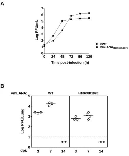
2.2.1. Generation of a MuHV-4 recombinant virus harbouring mutations on mLANA ventral face ... 35 2.2.2. vmLANAH186D/K187E recombinant virus displays normal lytic replication kinetics in vitro and in vivo ... 36
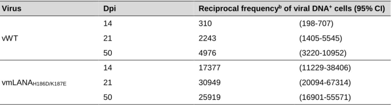
2.2.3. mLANA binding to viral TR DNA is required for the establishment and maintenance of MuHV-4 latency ... 38 2.2.4. mLANA binding to viral TR DNA is essential for MuHV-4 latency expansion in GC B cells ... 41 2.2.5. Loss of mLANA-mediated TR DNA binding abolishes MuHV-4 colonization of splenic follicles ... 43 2.2.6. Phenotypic changes in vmLANAH186D/K187E recombinant virus are intrinsic to ORF73
locus and not the consequence of mutations elsewhere in MuHV-4 genome ... 44 2.3. Discussion ... 45
3. mLANA dorsal positive patch is required for efficient expansion of gammaherpesvirus latency in GC B cells... 47 3.1. Introduction ... 49 3.2. Results ... 51 3.2.1. Generation of a MuHV-4 recombinant virus harbouring mutations on mLANA dorsal positive patch ... 51 3.2.2. Recombinant viruses containing mutations on mLANA dorsal positive patch display normal lytic replication kinetics in vitro and in vivo ... 52 3.2.3. The positive patch periphery of mLANA dorsal face is required for efficient expansion of MuHV-4 latency ... 54 3.2.4. The positive patch periphery of mLANA dorsal face is required for efficient MuHV-4 colonization of GC B cells ... 56 3.2.5. The positive patch periphery of mLANA dorsal face is required for efficient MuHV-4 latency amplification in splenic follicles ... 58 3.2.6. Phenotypic changes in dorsal recombinant viruses are intrinsic to ORF73 locus and not the consequence of mutations elsewhere in MuHV-4 genome ... 59 3.3. Discussion ... 60
4. mLANA E3 ubiquitin-ligase activity impacts on gammaherpesvirus latency amplification ... 63 4.1. Introduction ... 65 4.2. Results ... 67 4.2.1. mLANAV199A, mLANAV199A/L202A and mLANAP203A/P206A mutants exhibit impaired
ability to inhibit NF-κB and to activate Myc transcriptional activities ... 67 4.2.2. mLANAV199A, mLANAV199A/L202A and mLANAP203A/P206A mutants exhibit diminished
4.2.3. mLANAV199A, mLANAV199A/L202A and mLANAP203A/P206A mutants weakly associate
with ElonginC and Cullin5 ...71 4.2.4. Generation of MuHV-4 recombinant viruses harbouring mutations in mLANA SOCS-box ...73 4.2.5. Impairment of mLANA E3 ubiquitin-ligase activity impacts on MuHV-4 latency amplification ...74 4.3. Discussion ...76
5. DNA binding-deficient mLANA mediates episome persistence in the context of multiple TR elements ...79 5.1. Introduction ...81 5.2. Results ...82 5.2.1. Generation of MuHV-4 ORF50-deficient recombinant viruses harbouring mutations in mLANA SOCS-box ...82 5.2.2. Generation of cell lines latently infected with MuHV-4 ORF50-deficient recombinant viruses ...83 5.2.3. mLANAV199A/L202A mutant efficiently mediates episome persistence in a whole-virus
context ...84 5.3. Discussion ...86
6. Full-length mLANA is expressed in insect cells using baculovirus MultiBac system ...87 6.1. Introduction ...89 6.2. Results ...90 6.2.1. Construction of transfer plasmids for integration of ORF73 gene into baculovirus genome ...90 6.2.2. Generation of recombinant baculoviruses expressing full-length mLANA ...91 6.2.3. Small-scale expression of full-length mLANA ...93 6.3. Discussion ...95
7. Final Considerations ...97
8. Materials and Methods ...105 8.1. Materials ...107 8.1.1. General reagents ...107 8.1.2. Antibodies ...107 8.1.2.1. Primary antibodies ...107 8.1.2.2. Secondary antibodies ...108 8.1.3. Cell lines ...108 8.1.4. Bacterial strains ...109 8.1.5. Plasmids ...110
8.1.5.1. Plasmids for transient expression in mammalian cells ... 110 8.1.5.2. Plasmids for construction of MuHV-4 recombinant viruses ... 113 8.1.5.3. Plasmids for in situ hybridization ... 114 8.1.5.4. Plasmids for baculovirus studies ... 114 8.1.6. Viruses ... 115 8.1.6.1. MuHV-4 viruses ... 115 8.1.6.2. Baculoviruses ... 117 8.1.7. Mice ... 118 8.2. Methods ... 118 8.2.1. Isolation and analysis of DNA... 118 8.2.1.1. High molecular weight (HMW) cellular/viral DNA extractions ... 118 8.2.1.2. Plasmid DNA isolation ... 118 8.2.1.3. Quantification of DNA ... 120 8.2.1.4. Restriction endonuclease digestion ... 120 8.2.1.5. Analysis and isolation of DNA by gel electrophoresis ... 121 8.2.1.6. DNA sequencing ... 121 8.2.2. Polymerase Chain Reaction (PCR) ... 121 8.2.3. Cloning procedures ... 123 8.2.3.1. Cloning of inserts into pBamG vector ... 123 8.2.3.2. Subcloning of inserts into the shuttle vector ... 123 8.2.3.3. Subcloning of GST-His-mLANA and His-mLANA inserts into pPBac plasmid ... 124 8.2.3.4. DNA ligation ... 124 8.2.3.5. Bacterial transformation... 124 8.2.4. Cell cultures and transfections ... 126 8.2.4.1. Media and culture conditions ... 126 8.2.4.2. Transfections ... 126 8.2.5. Construction of MuHV-4 recombinant viruses ... 127 8.2.5.1. Shuttle vector cloning ... 127 8.2.5.2. BAC mutagenesis in E. coli ... 127 8.2.5.3. Virus reconstitution ... 128 8.2.5.4. Removal of BAC sequences ... 128 8.2.6. Construction of recombinant baculoviruses ... 129 8.2.6.1. Codon optimization of ORF73 gene ... 129 8.2.6.2. Subcloning of GST-His-mLANA and His-mLANA inserts into pPBac plasmid ... 129 8.2.6.3. Generation of recombinant EMBacY plasmids ... 129 8.2.6.4. Baculovirus reconstitution ... 130 8.2.7. Animal experiments ... 130 8.2.7.1. Ethics statement ... 130
8.2.7.2. Mice infection ...130 8.2.8. MuHV-4 virus assays...131 8.2.8.1. Viral infection of cell cultures ...131 8.2.8.2. Virus working stocks ...131 8.2.8.3. In vitro multi-step growth curves...131 8.2.8.4. Plaque assay (suspension assay) ...132 8.2.8.5. Infectious centre assay ...132 8.2.9. Flow cytometry ...133 8.2.9.1. Staining of splenocytes ...133 8.2.9.2. Purification of germinal centre (GC) B cell populations ...133 8.2.10. Limiting dilution analysis of infected splenocytes ...133 8.2.10.1. Statistical analysis of limiting dilution assay ...134 8.2.10.2. Real-time PCR ...134 8.2.11. In situ hybridization ...135 8.2.11.1. Generation of digoxigenin (DIG) UTP-labelled riboprobes ...135 8.2.11.2. Preparation of tissue for in situ hybridization ...135 8.2.11.3. In situ hybridization ...135 8.2.12. Episome maintenance assays ...136 8.2.13. Protein methods ...137 8.2.13.1. Reporter gene assays ...137 8.2.13.2. Immunoprecipitations ...137 8.2.13.3. Poly-ubiquitination assays ...138 8.2.13.4. Sodium dodecyl sulfate-polyacrylamide gel electrophoresis (SDS-PAGE)
...138 8.2.13.5. Transfer of proteins into nitrocellulose membranes ...139 8.2.13.6. Western-blot ...139 8.2.14. mLANA expression in insect cells using baculovirus (feasibility study) ...139 8.2.14.1. Processing of samples for SDS-PAGE and YFP fluorescence measurement ...139 8.2.14.2. Coomassie Blue staining ...140 8.2.14.3. Baculovirus amplification and small-scale protein expression ...140
9. References ...141
INDEX OF FIGURES
Figure 1.1. Normal B cell response and the parallel with the GC model of EBV persistence ... 7 Figure 1.2. Model of MuHV-4 infection ...13
Figure 1.3. Schematic diagram of KSHV LANA (kLANA) and MuHV-4 LANA (mLANA) ...20
Figure 1.4. The ubiquitination cascade ...28 Figure 1.5. E3 ubiquitin-ligase complex assembled by mLANA (EC5SmLANA) ...29
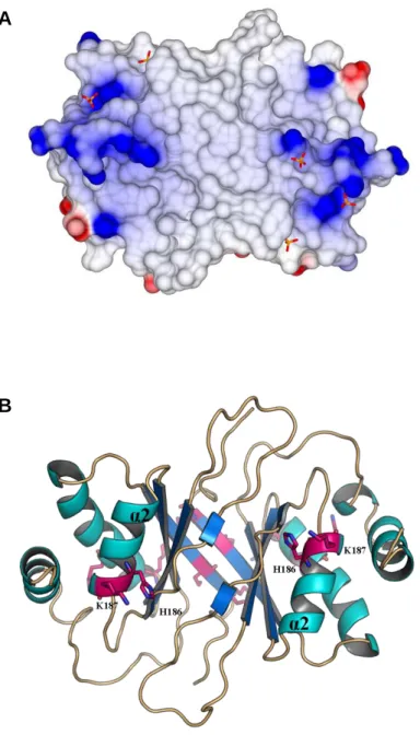
Figure 2.1. The ventral face of mLANA DBD ...34 Figure 2.2. vmLANAH186D/K187E recombinant virus displays normal lytic replication kinetics in vitro and in vivo ...37
Figure 2.3. mLANA binding to viral TR DNA is required for the establishment and maintenance
of MuHV-4 latency ...40
Figure 2.4. mLANA binding to viral TR DNA is essential for MuHV-4 latency expansion in GC B
cells ...42
Figure 2.5. Loss of mLANA-mediated TR DNA binding abolishes MuHV-4 colonization of
splenic follicles ...43 Figure 2.6. Phenotypic changes in vmLANAH186D/K187E recombinant virus are intrinsic to ORF73
locus and not the consequence of mutations elsewhere in MuHV-4 genome ...44 Figure 3.1. The dorsal positive patch of mLANA DBD ...50
Figure 3.2. Recombinant viruses containing mutations on mLANA dorsal positive patch display
normal lytic replication kinetics in vitro and in vivo ...53 Figure 3.3. The positive patch periphery of mLANA dorsal face is required for efficient expansion of MuHV-4 latency ...55 Figure 3.4. The positive patch periphery of mLANA dorsal face is required for efficient MuHV-4 colonization of GC B cells ...57 Figure 3.5. The positive patch periphery of mLANA dorsal face is required for efficient MuHV-4
latency amplification in splenic follicles...58 Figure 3.6. Phenotypic changes in dorsal recombinant viruses are intrinsic to ORF73 locus and
not the consequence of mutations elsewhere in MuHV-4 genome ...60 Figure 4.1. β2-β3 SOCS-box loop of mLANA ...66 Figure 4.2. mLANAV199A, mLANAV199A/L202A and mLANAP203A/P206A exhibit impaired ability to
inhibit NF-κB and to activate Myc transcriptional activities ...68 Figure 4.3. mLANAV199A, mLANAV199A/L202A and mLANAP203A/P206A exhibit diminished capability
to promote p65 and Myc poly-ubiquitination ...70 Figure 4.4. mLANAV199A, mLANAV199A/L202A and mLANAP203A/P206A weakly associate with
ElonginC and Cullin5 ...72 Figure 4.5. Impairment of mLANA E3 ubiquitin-ligase activity impacts on MuHV-4 latency amplification ...75
Figure 5.1. Schematic diagram of MuHV-4 ORF50-deficient virus (MuHV-4 M3-Luc/ORF50
-eGFP) ... 82 Figure 5.2. Experimental strategy to establish cell lines latently infected with MuHV-4
ORF50-deficient recombinant viruses ... 83 Figure 5.3. Schematic diagram of the left end of BAC-cloned MuHV-4 genome... 84 Figure 5.4. mLANAV199A/L202A efficiently mediates MuHV-4 episome persistence ... 85
Figure 6.1. Schematic diagram of mLANA constructs for expression in insect cells ... 90 Figure 6.2. Protocol for gene expression test in insect cells using MultiBac system ... 92
Figure 6.3. Yellow fluorescent protein (YFP) expression from recombinant EMBacY baculoviral
DNA ... 93 Figure 6.4. Full-length mLANA is expressed in insect cells using baculovirus MultiBac system
INDEX OF TABLES
Table 1.1. EBV-associated diseases ... 6 Table 1.2. Patterns of EBV latent gene expression in normal B cells and tumours ... 8 Table 2.1. Frequency of MuHV-4 latent infection in total splenocytes ...41 Table 2.2. Frequency of MuHV-4 latent infection in GC B cells ...42 Table 3.1. Frequency of MuHV-4 latent infection in total splenocytes ...56 Table 3.2. Frequency of MuHV-4 latent infection in GC B cells ...57 Table 4.1. Frequency of MuHV-4 latent infection in total splenocytes ...76 Table 5.1. Number of resistant clones obtained from infection of MEF cells with
ORF50-deficient viruses ...85 Table 8.1. Primary antibodies (commercially available) used in this study ...108 Table 8.2. Primers used to generate pCMV-Myc-mLANA mutants ...122 Table 8.3. Primers used to amplify ORF73 gene ...123 Table 8.4. Primers and probe specific for M9 gene used to detect MuHV-4 DNA ...135
CHAPTER 1
Introduction
Gammaherpesviruses, such as the human viruses Epstein-Barr virus and Kaposi´s sarcoma-associated herpesvirus, establish lifelong latent infections in B lymphocytes and exert causative roles in several malignancies. During latency, the viral genome is maintained as a non-integrated circular episome within the host cell nucleus, and viral protein expression is highly restricted. To persist in proliferating cells, viral episomes must replicate in step with normal cell division and segregate to newly formed nuclei after mitosis. This process is mediated by gammaherpesvirus episome maintenance proteins. Gamma-2-herpesviruses, a genus of gammaherpesvirus, encode a latency-associated nuclear antigen (LANA), which mediates episome persistence. In addition, LANA proteins have been described as cellular transcription modulators. Since LANA is essential for latent infection, understanding the mechanisms underlying its functions offers an opportunity to control gammaherpesvirus infection and associated pathologies. This introductory chapter will mainly focus on the current knowledge on gammaherpesvirus latency and LANA structure and function.
1.1.
Herpesvirus
1.1.1. General properties
Herpesviridae is a family of enveloped viruses containing large linear double-stranded DNA
genomes (>100 kb) with a vast coding capacity. Herpesviruses are widely disseminated in vertebrate species, with a large spectrum of animals harbouring at least one of these viruses. However, each herpesvirus exhibits a very narrow host range. Herpesviruses are remarkably well-adapted to their hosts, probably due to a long co-evolutionary history. Thus, herpesvirus infection of a natural immunocompetent host is rarely fatal, promoting virus transmission (Barton et al., 2011; Davison, 2002; Wu et al., 2010).
The hallmark of herpesviruses is the establishment of latent infections which persist for the life of the host. During latency, herpesviruses persist as a non-infectious form, from which they periodically reactivate to disseminate to new hosts. Most herpesvirus infections are asymptomatic or cause a mild illness, but effects can be devastating in immunocompromised hosts (Barton et al., 2011; Wu et al., 2010).
Herpesviruses have biphasic life cycles consisting of a lytic and a latent phase. Upon transmission to a naïve host, the virus establishes a productive acute infection at the site of infection, usually the epithelium of a mucosal surface. During this lytic phase, there is a sequential order of viral gene expression and the viral genome is replicated several times, leading to the
production of infectious virion progeny and the death of the infected cell. This primary infection is rapidly resolved by the host immune system and results in effective immunity against reinfection (Stoopler, 2005; Wu et al., 2010).
The lytic infection is followed by a lifelong latent infection in a specific type of cells. The targeted cell type is herpesvirus specific and is usually different from the cell type harbouring the productive infection. During latency, limited gene expression occurs. No virions are produced and viral genome is maintained as a non-integrated circular episome in the nucleus of the infected cell, being replicated by host DNA polymerase and equally distributed to daughter cells, when the latently infected cell divides during the course of cellular growth. The expression of viral proteins is severely reduced to those required for maintenance of viral genome, manipulation of host cell function and evasion of the host immune system. By expressing few proteins during latency, the virus limits the amount of antigen produced, thereby reducing the chance of being detected by the host immune system. A critical aspect of latency is the ability of the virus to sporadically reactivate in response to specific signals, ensuring the transmission of infectious virions to new hosts, as well as reinfection and establishment of latent infection in more cells of the same individual, thereby establishing a reservoir of infection for life (Barton et al., 2011; Wu et al., 2010).
Herpesviridae family is divided into three subfamilies: Alphaherpesvirinae, Betaherpesvirinae
and Gammaherpesvirinae. Alphaherpesviruses establish latency mainly in neurons, betaherpesviruses in myeloid cells, and gammaherpesviruses in lymphocytes. Humans are the natural hosts of eight herpesviruses: herpes simplex virus (HSV)-1, HSV-2, and varicella-zoster virus (VZV) (Alphaherpesvirinae); human cytomegalovirus (HCMV), human herpesvirus (HHV)-6, and HHV-7 (Betaherpesvirinae); Epstein-Barr virus (EBV), and Kaposi’s sarcoma-associated herpesvirus (KSHV), also known as HHV-8 (Gammaherpesvirinae) (Davison, 2002; Stevenson et
al., 2009; Wu et al., 2010). Since the work presented in this thesis is focused on the pathogenesis
of gammaherpesvirus latent infection, only this subfamily will be described in detail in the following sections.
1.1.2. The subfamily Gammaherpesvirinae
Members of the subfamily Gammaherpesvirinae are lymphotropic viruses, with the majority establishing latency in B lymphocytes. Gammaherpesviruses drive the proliferation of latently infected lymphocytes, in order to establish and maintain a large reservoir of latent viral genomes, and thus efficiently colonize the host. However, virus-driven lymphoproliferation is associated with the development of lymphoproliferative diseases, as well as several lymphoid and non-lymphoid cancers, since several gammaherpesviruses are able to induce neoplasia in natural or experimental hosts (Barton et al., 2011; Damania, 2004; Stevenson, 2004).
Gammaherpesvirinae subfamily is further subdivided into four genera, based on DNA
homology and genomic organization, being Lymphocryptovirus and Rhadinovirus the two main genera. Lymphocryptoviruses (or gamma-1-herpesviruses) have only been identified in primates, whereas Rhadinoviruses (or gamma-2-herpesviruses) infect a wide range of mammalian species, including primates (Barton et al., 2011; Davison et al., 2009; Simas and Efstathiou, 1998).
Gammaherpesviruses exhibit a very narrow host tropism, limiting the study of the human gammaherpesviruses EBV and KSHV. For this reason, there has been considerable interest in developing animal models to investigate the pathogenesis of human gammaherpesviruses. In particular, infection of laboratory mice with murid herpesvirus-4 (MuHV-4) has proved to be a good model system (Barton et al., 2011; Nash et al., 2001; Simas and Efstathiou, 1998). The following sections of this thesis will focus on the two known human gammaherpesviruses and then on MuHV-4 as an experimental model for the study of gammaherpesvirus pathogenesis.
1.2.
Epstein-Barr virus (EBV)
Epstein-Barr virus (EBV), a gamma-1-herpesvirus, was discovered more than 50 years ago in cultured tumour cells derived from Burkitt’s lymphoma tissue (Epstein et al., 1964). This was the first human tumour virus ever described (Thorley-Lawson and Allday, 2008). EBV prevalence in the normal population is extremely high, reaching over 95% of the adult population worldwide (Kutok and Wang, 2006; Lieberman, 2014). Primary EBV infection generally occurs in the first decade of life and is usually asymptomatic. However, if the infection is acquired during adolescence or later, it can result in infectious mononucleosis, a self-limiting lymphoproliferative disease (Henle et al., 1968; Kutok and Wang, 2006). After primary infection, which is rapidly controlled by both cellular and humoral immune mechanisms, EBV persists in a quiescent state in resting memory B lymphocytes that circulate in the peripheral blood for the life of healthy carriers (Babcock et al., 1998; Hislop et al., 2007; Kutok and Wang, 2006). Persistent infection is characterized by stable low levels of latently infected memory B cells, as a consequence of the dynamic equilibrium between virus-driven B cell proliferation and host immune control. A low level of active viral replication continues asymptomatically in EBV carriers, leading to virus secretion into the saliva and transmission from one human to another through the oral route (Hislop et al., 2007; Kutok and Wang, 2006).
Although EBV infection is benign and uneventful in the majority of humans, EBV has been associated with a number of diseases, particularly autoimmunity and cancer. Immunocompromised individuals are at risk for EBV lymphomas that are aggressive and often fatal (Table 1.1). However, EBV-associated malignancies can also develop in immunocompetent individuals (Table 1.1) (Kutok and Wang, 2006; Thorley-Lawson and Gross, 2004).
Table 1.1. EBV-associated diseases (adapted from Kutok and Wang, 2006).
Non-malignant disease
Infectious mononucleosis Chronic active infection Oral hairy leukoplakia
Malignant disease
Immunocompromised host
Acquired immunodeficiency syndrome (AIDS)-associated B cell lymphomas
Post-transplant lymphoproliferative disorder
Severe combined immunodeficiency-associated B cell lymphomas X-linked lymphoproliferative disorder-associated B cell
lymphomas Immunocompetent
host
Burkitt’s lymphoma
Classical Hodgkin’s lymphoma T cell lymphomas
Lymphoepithelioma-like carcinoma (salivary, thymus, lungs, stomach)
1.2.1. Germinal centre (GC) model of EBV infection
EBV is the best studied gammaherpesvirus and a biological model of EBV infection has been proposed, the germinal centre (GC) model. This model, described below, reveals strategies of infection which are believed to be common to all gammaherpesviruses that persist in B lymphocytes.
The GC model (Figure 1.1), proposed by Thorley-Lawson and co-workers, defends that EBV persists by exploiting normal B cell biology (Thorley-Lawson, 2001). This implies that new latently infected B cells pass through a series of differentiation stages, each employing a discrete viral gene transcription programme. Following oral transmission, EBV establishes a lytic infection in permissive cells of the oropharynx, leading to high levels of virus shedding in saliva. It is thought that both squamous epithelial cells and locally infiltrating B cells support this lytic infection. Thereafter, the virus colonises the general B cell system via growth-transforming latent infection of B cells in local lymphoid tissue such as the tonsil. According to the GC model, EBV directly infects naïve B cells, activating them into proliferating latently infected blasts. At this stage, EBV expresses its latency III or growth transcription programme (Table 1.2), which is characterized by the expression of the full repertoire of viral latent proteins: EBV nuclear antigens (EBNAs) 1, 2, 3A, 3B, 3C and LP, and latent membrane proteins (LMPs) 1, 2A and 2B. These proteins have all the necessary activities to push B cells to become activated blasts without external signalling (Babcock
et al., 2000; Joseph et al., 2000; Thorley-Lawson, 2005). In culture, EBV immortalises human
resting B cells by expressing the growth programme, driving the establishment of continuously proliferating lymphoblastoid cell lines (growth transformation or immortalisation) (Thorley-Lawson and Mann, 1985).
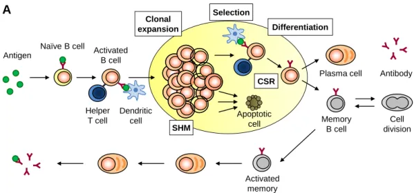
Figure 1.1. Normal B cell response and the parallel with the GC model of EBV persistence. (A) Normal B cell response. The epithelium of the tonsil continuously samples antigens entering the mouth. Underneath the epithelium, naïve B cells that encounter cognate antigen become activated and migrate into a follicle where they receive further activation signals from dendritic cells and helper T cells, and establish a GC reaction. In the GC, the blast undergoes repeated rounds of cell division, proliferating rapidly, in association with somatic hypermutation (SHM) and class switch recombination (CSR), followed by interaction with follicular dendritic cells and T cells to select those blasts that have the highest affinity for the antigen. The later receive further survival and differentiation signals, while the other cells die by apoptosis. The surviving ones differentiate into either a plasma cell or a memory B cell and leave the GC. Upon re-challenge with the antigen, memory B cells are quickly activated and differentiate into antibody producing plasma cells. (B) GC model of EBV persistence. EBV traverses the epithelium and infects a naïve B cell, activating it into a proliferating latently infected blast, as though the cell was responding to antigen, through expression of all nine known latent proteins, the growth transcription programme. These cells migrate to the follicle where the viral transcription programme changes to the more restricted default programme and a GC reaction is established. Subsequently the latently infected cells differentiate into memory B cells that leave the follicle. In the periphery, all viral protein expression is shut down, the latency programme, and the latently infected cells are maintained as normal memory B cells. These
Clonal expansion Selection Differentiation CSR SHM Apoptotic cell Antigen
Naïve B cell Activated B cell
Helper T cell
Dendritic cell
Plasma cell Antibody
Memory B cell Cell division Activated memory
A
Growth programme Default programme Latency programme GC B cells EBV Viral episome Memory B cellB
EBNA1 only Lytic programmememory B cells can occasionally divide to maintain the pool of latently infected cells and so express the genome tethering protein EBNA1, the EBNA1 only programme. At any time a small subset of latently infected memory B cells initiates lytic replication in association with terminal differentiation signals. Reactivation leads to the production of infectious virus and cell death. These virions can either replicate at a secondary tissue, where they are amplified and shed into saliva for transmission to new hosts, or infect new naïve B cells, thus restarting the cycle of infection (adapted from De Silva and Klein, 2015, Lawson, 2005 and Thorley-Lawson et al., 2013).
Table 1.2. Patterns of EBV latent gene expression in normal B cells and tumours (adapted from Thorley-Lawson, 2005).
Transcription
programme Genes expressed
a Infected normal B
cell type Function
Infected tumour type Growth (Latency III) EBNA1, 2, 3A, 3B, 3C and LP LMP1, 2A and 2B
Naïve B cell activation Immunoblastic
lymphoma
Default (Latency II)
EBNA1, LMP1 and
LMP2A Germinal centre
Differentiation of activated B cell into memory
Hodgkin’s disease
EBNA1
(Latency I) EBNA1 Dividing memory
Cellular division of latently infected memory B cells
Burkitt’s lymphoma Latency
(Latency 0) None Resting memory
Allows lifetime persistence
Lytic All lytic genes Plasma cell Viral replication in
plasma cell aDoes not include the non-coding EBER and BART RNAs.
Proliferating latently infected blasts then migrate into a follicle to participate in the GC reaction. Here, EBV expresses the latency II or default transcription programme (Table 1.2), consisting of a more restricted pattern of latent proteins: EBNA1, LMP1 and LMP2A. EBNA1 is required for replication and maintenance of the viral episome, mediating the association of viral genomes with host mitotic chromosomes, whereas LMP1 and LMP2A proteins provide the necessary surrogate signals to drive the differentiation of latently infected B cells into memory B cells that leave the GC and enter the peripheral circulation (Babcock et al., 2000; Caldwell et al., 1998; Casola et al., 2004; Gires et al., 1997; He et al., 2003; Marechal et al., 1999; Panagopoulos et al., 2004; Roughan and Thorley-Lawson, 2009; Sears et al., 2003; Thorley-Lawson, 2001; Yates et al., 1985).
In latently infected memory B cells, EBV expresses either EBNA1 transcription programme (latency I) or latency transcription programme (latency 0) (Table 1.2). During EBNA1 transcription programme, EBV only expresses the viral genome tethering protein EBNA1, so that viral episomal DNA is maintained and segregated when B cells divide. In contrast, in the latency transcription programme no viral proteins are expressed at all. The memory compartment has been considered
the site of long-term persistence, since the virus is quiescent and hence invisible to the immune response (Babcock et al., 2000; Hochberg et al., 2004; Thorley-Lawson, 2001).
In addition to latent proteins, EBV expresses several non-coding RNAs and microRNAs (miRNAs) during latency (Kang and Kieff, 2015). EBV-encoded RNA (EBER) 1 and 2 are small non-coding RNAs abundantly expressed in latently infected cells (Lerner et al., 1981). EBV-encoded miRNAs are expressed from two regions of EBV genome: BART and BHRF1. The EBV genome transcribes at least 25 pre-miRNAs and more than 40 mature miRNAs (Kang and Kieff, 2015; Lopes et al., 2013). Similarly to latent proteins, EBV-encoded miRNAs exhibit distinct patterns of expression according to the EBV latency programme (Qiu et al., 2011).
At any time, a small subset of latently infected memory B cells can reactivate and initiate lytic replication, which is thought to occur in response to the normal physiologic signals that drive terminal differentiation of memory B cells into plasma cells. Reactivation of EBV is subdivided into three phases: immediate early, when the transcription factors initiating viral replication are expressed, early, when the proteins involved in viral DNA replication are produced, and late, when viral DNA and structural proteins are assembled into virions. A crucial role for epithelial cells in amplifying the amount of infectious virus before shedding has been proposed. Released infectious virus can then be shed into saliva for viral spread to new hosts, or infect new naïve B cells, thus completing the cycle (Hadinoto et al., 2009; Laichalk and Thorley-Lawson, 2005; Thorley-Lawson
et al., 2013).
Each stage of the cycle of EBV infection has been demonstrated experimentally (Babcock et
al., 1998; Babcock et al., 2000; Laichalk and Thorley-Lawson, 2005) and, with the exception of the
memory compartment, is potentially regulated by the host immune response (Hislop et al., 2007). The GC model of EBV infection remains the only experimentally validated model that accounts for all the latent and lytic stages of the virus, providing an explanation for the origin and pathogenesis of EBV-associated lymphomas (Thorley-Lawson et al., 2013). However, a quantitative analysis of viral persistence and an understanding of the dynamic interactions between the different components of the GC model and how their regulation by the immune system produces the EBV pattern of persistence has been lacking. Recently, a mathematical description of the GC model has been developed that successfully recapitulates persistent EBV infection, correctly predicting the observed patterns of cytotoxic T lymphocytes (CTLs) regulation and the size of the infected GC and memory B cell populations (Delgado-Eckert and Shapiro, 2011; Hawkins et al., 2013). Importantly, this mathematical model predicts that it is the cycle of infection that explains persistence and provides the stability that allows EBV to persist at extremely low levels, rather than viral quiescence in the memory B cell compartment. This moves the focus away from a single infected stage, the memory B cell compartment, to the entire cycle of infection.
1.3.
Kaposi’s sarcoma-associated herpesvirus (KSHV)
Kaposi’s sarcoma-associated herpesvirus (KSHV) was first discovered in 1994 by Chang and colleagues, from patients with Kaposi’s sarcoma (KS) (Chang et al., 1994). KSHV, also known as human herpesvirus-8 (HHV-8), is a gamma-2-herpesvirus with an etiologic role in KS, the most prevalent tumour among human immunodeficiency virus (HIV)/acquired immunodeficiency syndrome (AIDS) patients worldwide (Achenbach et al., 2011). KSHV is also associated with other two human malignancies: primary effusion lymphoma (PEL) and multicentric Castleman’s disease (MCD) (Cesarman et al., 1995; Soulier et al., 1995). KS tumours are comprised of KSHV-infected cells of endothelial origin, whereas PEL and MCD are of B cell origin. Furthermore, KSHV was recently associated with an inflammatory cytokine syndrome (Uldrick et al., 2010).
Unlike EBV virus, KSHV is not ubiquitous. Among the general population, the seroprevalence of KSHV infection in northern Europe, Asia and America is less than 10%, but it reaches 30% in Mediterranean regions and over 50% in most of sub-Saharan Africa. The association between the incidence of KSHV-associated malignancies and KSHV seroprevalence is high. However, the presence of KSHV DNA alone in healthy individuals is not sufficient to cause disease, and the existence of cofactors as HIV infection or drug-induced immunosuppression are important for KSHV-associated disease progression. The transmission mode of KSHV has not been completely clarified yet. It is currently thought that KSHV is mainly transmitted through saliva, although transmission through organ transplants, blood and sexual fluids have also been documented (Cai
et al., 2010; Edelman, 2005; Mesri et al., 2010; Uldrick and Whitby, 2011; Uppal et al., 2014).
Being a herpesvirus, KSHV exhibits two distinct phases of infection: lytic replication and latency. Latency is characterized by limited gene expression without virion production, and it represents the main strategy used by the virus to escape host immune system, while maintaining its genome in infected cells. Periodically, the latent virus reactivates to enter lytic replication, during which the viral genes are fully expressed in a cascade manner (immediate early, early and late genes), leading to the production of infectious particles and transmission to new hosts. Little is known about KSHV primary infection, but there is evidence that it is benign in immunocompetent individuals, and it results in lifelong latency (Wang et al., 2001; Wu et al., 2012).
1.3.1. KSHV latency
During latency, KSHV genome is maintained as a circular episome within the host cell nucleus with highly restricted protein expression, in order to maintain the genome in the dividing cells and to limit host immune responses, while enhancing cell survival and virus persistence (Uppal et al., 2014). Only a small portion of the viral genome is actively transcribed during latency. The major latency locus includes four viral genes, encoding KSHV latency-associated nuclear antigen
(kLANA)/ORF73, vCyclin/ORF72, viral Fas-associated death domain-like interleukin-1β-converting enzyme-inhibitory protein (vFLIP)/ORF71, and K12/Kaposin family (Kaposin A, B and C), as well as twelve viral pre-miRNAs (Cai et al., 2005; Dittmer et al., 1998). kLANA is the viral episome maintenance protein and, in addition, it exerts transcriptional regulatory activities and affects cell growth, through interaction with several cell proteins (Ballestas and Kaye, 2011). vCyclin, the viral homolog of cellular cyclin D, regulates cell cycle and proliferation, whereas vFLIP, the viral homolog of cellular FLIP, has a role in inhibition of apoptosis (Uppal et al., 2014). The kLANA promoter controls the expression of kLANA, vCyclin and vFLIP, while the Kaposin promoter drives the expression of the three Kaposin transcripts, a bicistronic transcript for vCyclin and vFLIP, and the twelve viral pre-miRNAs which can be processed to yield mature miRNAs (Dittmer et al., 1998; Pearce et al., 2005). The major latency locus is abundantly and consistently transcribed in all latently infected cells (Speck and Ganem, 2010). Additionally, PEL cells, but not KS cells, express a second latency locus encoding viral interferon regulatory factor-3 (vIRF-3), also known as LANA-2, indicating that some latency genes may be lymphoid specific (Rivas et al., 2001; Speck and Ganem, 2010). More recently, a third latency locus encoding K1 protein has been identified (Chandriani and Ganem, 2010). Other viral genes expressed during latency, although at low levels, include viral interleukin-6 (vIL-6) and K15 (Giffin and Damania, 2014).
In vivo, KSHV has been detected in endothelial cells, epithelial cells, B cells and monocytes
(Ambroziak et al., 1995; Blasig et al., 1997; Dupin et al., 1999; Pauk et al., 2000). However, the main targets of KSHV long-term latent infection are B cells, particularly the subset expressing the lambda (λ) light chain of B cell receptor (Chadburn et al., 2008; Hassman et al., 2011). KSHV infection drives proliferation of primary B cells (Hassman et al., 2011) and individual KSHV latent genes, specifically the ones encoding kLANA and vFLIP, induce B cell proliferative phenotypes in transgenic mice (Ballon et al., 2011; Sin et al., 2010). B cell specific expression of multiple KSHV latent proteins, including kLANA, vFLIP, vCyclin and Kaposin, as well as the viral miRNAs, leads to sustained hyperplasia, lymphoma and hyper-responsiveness to antigen stimulation in transgenic mice (Sin and Dittmer, 2013).
The mechanisms by which KSHV establishes and maintains latency are not as well understood as for EBV. However, the overall strategy is believed to be the same: subversion of normal B cell developmental pathways to induce cell proliferation and achieve long-term persistence. A putative model of KSHV B cell pathogenesis has been proposed (Dittmer and Damania, 2013). According to this model, the primary infection event drives the infected cell into an activated state. In non-permissive cell subtypes the virus is rapidly lost or the cell dies. In contrast, in cells permissive to the establishment of latency, the virus persists and confers a survival advantage to the infected cell. The molecular nature of this advantage and the exact complement of viral genes which confer it are not well defined. This model suggests that the establishment of KSHV latent infection is analogous to that of EBV, which starts with expressing a more extensive set of genes and then contracts to latency I or latency 0 transcription programmes.