NEDDylation Is Essential for Kaposi
’
s
Sarcoma-Associated Herpesvirus Latency and
Lytic Reactivation and Represents a Novel
Anti-KSHV Target
David J. Hughes1¤a
*, Jennifer J. Wood1, Brian R. Jackson1,2, Belinda Baquero-Pérez1, Adrian Whitehouse1,2*
1School of Molecular and Cellular Biology, Faculty of Biological Sciences, University of Leeds, Leeds, United Kingdom,2Astbury Centre for Structural Molecular Biology, Faculty of Biological Sciences, University of Leeds, Leeds, United Kingdom
¤a Current address: Biomedical Sciences Research Complex, University of St. Andrews, St. Andrews, Fife, United Kingdom
*djh25@st-andrews.ac.uk(DJH); a.whitehouse@leeds.ac.uk(AW)
Abstract
Kaposi’s sarcoma-associated herpesvirus (KSHV) is the causative agent of Kaposi's sarco-ma (KS) and prisarco-mary effusion lymphosarco-ma (PEL), which are aggressive sarco-malignancies associ-ated with immunocompromised patients. For many non-viral malignancies, therapeutically targeting the ubiquitin proteasome system (UPS) has been successful. Likewise, laboratory studies have demonstrated that inhibition of the UPS might provide a promising avenue for the treatment of KSHV-associated diseases. The largest class of E3 ubiquitin ligases are the cullin-RING ligases (CRLs) that are activated by an additional ubiquitin-like protein, NEDD8. We show that pharmacological inhibition of NEDDylation (using the small molecule inhibitor MLN4924) is cytotoxic to PEL cells by inhibiting NF-κB. We also show that CRL4B is a novel regulator of latency as its inhibition reactivated lytic gene expression. Further-more, we uncovered a requirement for NEDDylation during the reactivation of the KSHV lytic cycle. Intriguingly, inhibition prevented viral DNA replication but not lytic cycle-associated gene expression, highlighting a novel mechanism that uncouples these two features of KSHV biology. Mechanistically, we show that MLN4924 treatment precluded the recruitment of the viral pre-replication complex to the origin of lytic DNA replication (OriLyt). These new findings have revealed novel mechanisms that regulate KSHV latency and reac-tivation. Moreover, they demonstrate that inhibition of NEDDylation represents a novel ap-proach for the treatment of KSHV-associated malignancies.
Author Summary
Kaposi’s sarcoma-associated herpesvirus (KSHV) causes Kaposi’s sarcoma (KS) and pri-mary effusion lymphoma (PEL), often fatal malignancies afflicting HIV-infected patients. a11111
OPEN ACCESS
Citation:Hughes DJ, Wood JJ, Jackson BR, Baquero-Pérez B, Whitehouse A (2015) NEDDylation Is Essential for Kaposi’s Sarcoma-Associated Herpesvirus Latency and Lytic Reactivation and Represents a Novel Anti-KSHV Target. PLoS Pathog 11(3): e1004771. doi:10.1371/journal.ppat.1004771
Editor:Klaus Früh, Oregon Health and Science University, UNITED STATES
Received:July 3, 2014
Accepted:February 28, 2015
Published:March 20, 2015
Copyright:© 2015 Hughes et al. This is an open access article distributed under the terms of the Creative Commons Attribution License, which permits unrestricted use, distribution, and reproduction in any medium, provided the original author and source are credited.
Data Availability Statement:All relevant data are within the paper and its Supporting Information files.
Funding:This work was funded in parts by The Wellcome Trust (http://www.wellcome.ac.uk), Yorkshire Cancer Research (http://
Previous research has shown that blockade of the ubiquitin proteasome system (UPS, a normal quality control pathway that degrades cellular proteins) is able to kill KSHV-infected lymphoma cells. A large component of the UPS is made up by the protein family known as the cullin-RING ubiquitin ligases (CRLs), which are activated by NEDD8 (a pro-cess known as NEDDylation). Recently, an inhibitor of NEDDylation (MLN4924) was de-veloped and is currently in clinical trials as an anti-cancer drug. As NEDDylation has not been investigated for many viruses, we used this to compound examine its importance in KSHV biology. Firstly we show that NEDDylation is essential for the viability of KSHV-infected lymphoma cells, and MLN4924 treatment killed these cells by blocking NF-κB activity (required for KSHV latency gene expression and KSHV-associated cancer). Fur-thermore, we show that NEDDylation is required for KSHV to replicate its genome, a criti-cal step in the production of new virus particles. Therefore, this research has identified a novel molecular mechanism that governs KSHV replication. Furthermore, it demonstrates that NEDDylation is a viable target for the treatment of KSHV-associated malignancies.
Introduction
The ubiquitin-proteasome system (UPS) and associated pathways are rapidly becoming accept-ed as major therapeutic targets for the treatment of malignancy [1], which potentially include those associated with oncogenic viruses. Additionally, small molecule inhibitors have been suc-cessfully used for dissecting the biological roles of these intriguing pathways, which is critical for our understanding of their mechanisms of cytotoxicity. Indeed, inhibition of the UPS is cy-totoxic to Kaposi’s sarcoma-associated herpesvirus (KSHV, also referred to as human herpesvi-rus 8 [HHV8]) infected cells [2–5]. Infection with KSHV is commonly associated with fatal malignancies, is the causative agent of primary effusion lymphoma (PEL) and Kaposi’s sarco-ma (KS) and is frequently associated with multicentric Castlesarco-man’s disease (MCD) [6,7]. Like all herpesviruses, KSHV infection is lifelong and has two distinct phases to its lifecycle; latent and lytic. During latency, viral gene expression is highly restricted and, in the tumor setting, in-volves the expression of the latency associated nuclear antigen (LANA), the viral FLICE inhibi-tory protein (vFLIP), viral cyclin, kaposin and various virally encoded miRNAs. Together these promote tumorigenesis in all known KSHV-associated malignancies. Nevertheless, at least for KS, the lytic phase of KSHV, which results in the expression of the complete viral genome and the production of infectious virions, is necessary for sarcomagenesis. For this reason, the mo-lecular mechanisms governing the switch from latency to lytic reactivation have received much attention as they may provide excellent targets for therapeutic intervention.
Current treatments of KSHV-associated malignancies have limited efficacy. PEL is treated using a combination of cyclophosphamide, doxorubicin, vincristine and prednisone (similar to CHOP therapy) and/or highly active retroviral therapy (HAART) [8,9]. For AIDS-related KS, HAART is also favored, and due to the requirement of KSHV lytic infection for the pathogene-sis of KS, anti-herpesviral drugs have also been used [10]. More recently, preclinical models have demonstrated that inhibition of the UPS using bortezamib [2–5], or bortezamib in combi-nation with a histone deacetylase (HDAC) inhibitor (vorinostat) may provide a promising new avenue [11]. Given the success of bortezamib (marketed as Velcade) for the treatment of multi-ple myeloma and mantle cell lymphoma, there are now various additional small molecule inhibitors of the UPS at various stages of development [1], as well as drugs that target ubiqui-tin-like pathways such as the NEDDylation cascade [12].
The attachment of ubiquitin-like (Ubl) proteins to substrates represents one of the most vital posttranslational modifications in the cell and, so far, twelve Ubls have been identified in-cluding ubiquitin (Ub), NEDD8 and SUMO. The covalent attachment of Ubls to substrates re-quires an enzyme cascade involving an E1 activating enzyme, an E2 conjugating enzyme and an E3 ligase that provides substrate specificity, all of which are potential‘drugable’targets. Ubi-quitylation is the principal mechanism by which proteins are targeted for proteasomal degrada-tion, and the largest class of E3 ligases are the Cullin-RING ligases (CRLs) [13]. These modular proteins are centered on one of several Cullin scaffolds (Cul1, 2, 3, 4A, 4B, 5 and 7) and consist of a RING-binding protein (RBX1 or RBX2) that interacts with the Ub-loaded E2 and a strate adapter protein that brings the target into close proximity with the Ub-E2. Many sub-strate adapters have been identified, and the various combinations of Cul-adapter complexes means there are several hundred possible CRLs, with several-fold more substrates [14]. Intrigu-ingly, the activity of CRLs is dependent on themselves being modified by the Ubl NEDD8, which requires the activity of the NEDD8 activating enzyme, NAE1 [15]. Recently, a specific NAE1 inhibitor has been developed (MLN4924) and is currently in clinical trials for various malignancies. MLN4924-mediated inhibition of NEDDylation blocks CRL activity resulting in the global stabilization of CRL targets [12].
While the ubiquitin and SUMO pathways have been extensively studied in infectious dis-eases [16,17], much less attention has been given to the role of NEDD8. Recent work has shown that the large tegument protein of gammaherpesviruses has deNEDDylase activity and this was important for EBV infection [18]. Another study demonstrated that inhibition of NEDDylation blocked the ability of HIV to degrade the cellular restriction factor APOBEC3G via Vif-mediated hijack of Cul5 [19]. There are indications in the literature that NEDDylation is likely to play a central role in viral pathogenesis. For example, HIV is known to modulate the activity of at least three cullins [20,21], the KSHV LANA protein forms a CRL complex with Cul5 and targets p53 [22], and it was recently shown that human cytomegalovirus (HCMV) utilizes the nucleotide excision repair (NER) pathway, which is regulated predominantly by Cul4A/B [23], to repair its genomes during viral replication [24].
Here we asked if NEDDylation is important for the KSHV lifecycle, and to begin to dissect the functional consequences of its inhibition. Treatment of cancer cells with MLN4924 leads to dramatic cytotoxicity, and some of the best characterized mechanisms include the induction of DNA re-replication by blocking the degradation of Cdt-1 [25,26], or by inhibiting NF-κB sig-naling via stabilization of the inhibitor of NF-κB protein IκBα[27], both of which eventually lead to apoptosis. Given the essential role of NF-κB for the maintenance of viral latency and the survival of PEL [28], we reasoned that these cells would be highly sensitive to MLN4924. We found that, indeed, MLN4924 led to significant PEL cytotoxicity and this was mediated via inhibition of NF-κB signaling. Intriguingly, we also showed that NEDDylation was essential for amplification of the KSHV genome during reactivation of the lytic cycle and that treatment with MLN4924 prevented the recruitment of RTA to the origin of lytic replication (OriLyt). This work has highlighted the essential role of NEDD8 and CRL-mediated ubiquitylation dur-ing the life cycle of KSHV and suggests that NEDDylation may provide a novel therapeutic tar-get for the treatment of KSHV-associated malignancies.
Results
Inhibition of NEDDylation is cytotoxic to PEL cells and leads to lytic
cycle-associated gene expression
followed by immunoprecipitation of NEDD8 and immunoblot analysis confirmed that Cullin proteins were modified in infected cells (S1 Fig). As MLN4924 has demonstrated anti-prolifer-ative activity in various cancer cell lines, we assessed its cytotoxic effects in PEL cells. We treat-ed PEL cells (BCBL-1, TREx-BCBL-1-RTA and BC-3) with varying concentrations of
MLN4924 and determined cell viability after 96 h using a luminescence ATP detection assay. These data demonstrated that MLN4924 was indeed cytotoxic to latently infected PEL cells with approximate EC50values of 1.10μM and 0.15μM, for TREx-BCBL-1-RTA and BC-3
re-spectively (Fig. 1A). However, it was interesting to note that a low level of ORF57 expression was observed in TREx-BCBL-1-RTA (see 10μM lane) and low levels of endogenous RTA in
BC-3 cells treated with MLN4924, suggesting that drug treatment alone induced lytic gene ex-pression (Fig. 1B & C). We also noted that MLN4924 led to apoptosis in BC-3 cells as shown by the cleavage of PARP (Fig. 1C). To investigate MLN4924-induced cytotoxicity, cell cycle analysis of TREx-BCBL-1-RTA cells was performed 24 h after treatment with 1μM MLN4924.
Compared to untreated TREX-BCBL-1-RTA cells (Fig. 1D(i)), MLN4924 treatment led to a reduction in S-phase and an accumulation of cells in G2/M (Fig. 1D(ii)). Clearly, inhibition of NEDDylation was toxic to PEL cells, and as shown for BC-3 this led to apoptosis. To further in-vestigate this, we tested the activation of caspases in TREx-BCBL-1-RTA cells. Similar to BC-3, treatment of TREx-BCBL1-RTA led to the cleavage of PARP, indicative of Caspase 3/7 activa-tion, as did the reactivation of the KSHV lytic cycle after the addition of Dox. (Fig. 1E). It has been shown previously that induction of the lytic cycle activates initiator Caspase 8, but not Caspase 9 in order to activate effector caspases such as Caspase 3 and 7 [29]. In agreement with this, we also showed that lytic reactivation did not activate Caspase 9; nevertheless, MLN4924 did, resulting in the activation of Caspase 3 (Fig. 1E). Importantly however, caspase inhibition (using the pan-caspase inhibitor, z-VAD-FMK) did not prevent PEL cell death after treatment with MLN4924 (Fig. 1F). This result showed that PEL cytotoxicity was not solely a result of ap-optosis and suggests other factors, such as the inhibition latency-associated gene expression may contribute to cell death.
Therefore, we next measured the effect of treatment on viral gene expression. Latently in-fected TREx-BCBL-1-RTA were treated with 1μM MLN4924 and harvested 24 h later. As
shown inFig. 2A, the expression of all lytic-cycle associated genes tested were induced after in-hibition of NEDDylation. In contrast, the expression of latency-associated transcriptsORF73 (latency associated nuclear antigen [LANA]) andORF71(vFLIP) were reduced in the presence of drug. Interestingly, the expression ofK12followed that of latency-associated genes. While K12 is dramatically induced upon activation of the KSHV lytic cycle [30], its expression during latency is initiated from the LANA promoter [31]. This suggested that MLN4924 treatment modulated latency-associated viral gene expression, rather than inducing the full KSHV lytic cycle. To further investigate the potential of MLN4924 to reactivate lytic cycle gene expression, rKSHV.219 cells were cultured for 36 h in the presence of 0.1μM MLN4924, a concentration
that was tolerated while in culture. These HEK293-based cells are latently infected with recom-binant KSHV that contains a red fluorescent protein (RFP) reporter gene under the control of an RTA-responsive promoter (thePANpromoter); hence expression of RTA (and thus reacti-vation of the lytic cycle) leads to RFP expression. As shown inFig. 2B, RFP expression was ob-served in numerous cells demonstrating that MLN4924 was indeed able to induce the
MLN4924-induced cytotoxicity is due to the inhibition of NF-
κ
B
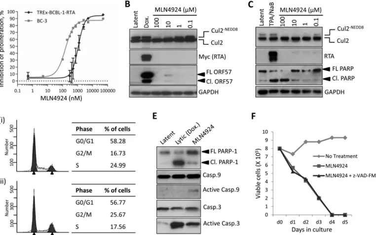
The reported mechanisms of MLN4924-induced cytotoxicity is the induction of DNA re-replication(by blocking the CRL-mediated degradation of Cdt-1) [26], or by inhibiting NF-κB signaling as CRL-mediated degradation of IκBαis prevented, thus precluding NF-κB’s tran-scriptional activity [27]. It is well established that PEL is dependent on NF-κB signaling, and that KSHV vFLIP drives the constitutive activation of NF-κB in order to maintain viability. NF-κB activity also inhibitsRTA-mediated transactivation of lytic genes, and thus it is impor-tant for maintaining latency. As the viral gene expression profiles following MLN4924 treat-ment (Fig. 2A) are consistent with an inhibition of NF-κB, we tested whether it was inhibited after MLN4924 treatment. The NF-κB transcription factor is tightly regulated via its interac-tion with IκBαwhich maintains it in the cytoplasm and away from its transcriptional targets. Upon stimulation, the Iκκcomplex phosphorylates IκBαat ser32 and 36, which signals for theFig 1. Inhibition of NEDDylation is cytotoxic in PEL cells.(A) TREx-BCBL-1-RTA and BC-3 cells were plated into 96-well plates and treated at varying concentrations of MLN4924. Cells viability was determined 96 h later using an ATPlite assay, and shown is a representative trace of several experiments, and EC50s were calculated (TREx-BCBL-1-RTA = 1.10μM; BC-3 = 0.15μM). (B) Immunoblot analysis of TREx-BCBL-1-RTA cells showing that MLN4924 effectively inhibits NEDDylation in PEL cells as shown by reductions in NEDDylated Cul2 after 24 h treatment. Also, treatment appeared to reactivate KSHV lytic gene expression (see ORF57 expression in 10μM lane). (C) Similar to (B), but with a second PEL cell line, BC-3. As for TREx-BCBL-1-RTA, treatment led to the activation of lytic gene expression as shown by endogenous RTA expression. Cleavage of PARP also indicated that MLN4924 activates apoptosis in this cell line. (D) Cell cycle analysis of TREx-BCBL-1-RTA cells treated for 24 h with 1μM MLN4924 showing a reduction in S-phase and an accumulation of cells in G2/M. (E) TREx-BCBL-1-RTA cells treated with 10μM MLN4924 for 24 h activated caspase 3/7 as indicated by PARP-1 cleavage. In contrast to lytic reactivation by Dox., MLN4924 led to the activation of initiator Caspase 9 which presumably led to Caspase 3 cleavage (activation). (F) Stationary cultures of BCBL-1 cells were treated with MLN4924 and cell viability was monitored by trypan blue exclusion over 5 days. Cells were pre-treated with z-VAD-FMK just prior to treatment to inhibit caspase-mediated apoptosis; however, this did not prevent cytotoxicity. FL PARP-1 denotes full length PARP-1; Cl. PARP-1 denotes cleaved PARP-1. FL ORF57 denotes full length ORF57 and Cl. ORF57 denotes the caspase 7-cleaved ORF57 [29].
Fig 2. MLN4924 induced lytic cycle—associated expression.(A) Latently infected TREx-BCBL-1-RTA cells were treated with 1μM MLN4924 and total
RNA was isolated from samples harvested 24 h later. The normalized expression (usingGAPDHexpression) of lytic cycle genes and latency associated
(ORF73,ORF71 and K12) genes were analyzed by qRT-PCR and compared to the levels found in DMSO-treated control samples. Three biological replicates
were independently analyzed and the error bars represent the standard deviation from the mean. (B) The inhibition of NEDDylation leads to reactivation of lytic cycle protein expression; rKSHV.219 cells were treated with 0.1μM MLN4924 (a concentration that was tolerated for the duration of the experiment) for 36 h. Red fluorescent protein (RFP) expression was observed in numerous MLN4924-treated cells, indicative of RTA protein expression. RFP expression was observed in a minority DMSO-only treated cells (as expected due to low level spontaneous reactivation). (C) ORF57 expression was also detected by immunoblot analysis of MLN4924-treated rKSHV.219 cells. (D) Inhibition of NEDDylation enhances reactivation. MLN4924-treated rKSHV.219 cells were incubated with or without TPA (able to reactivate KSHV) for 36 h.
recruitment of the Cul1-containingβTrCP E3 ligase leading to the subsequent degradation of IκBα. This releases NF-κB allowing to translocate to the nucleus and drive transcription [32]. Therefore, MLN4924 inhibition of CRL1 function should stabilize phosphorylated IκBα
(pIκBα) leading to an inhibition in NF-κB function.
To investigate the most likely mechanism of MLN4924-induced cytotoxicity we performed a timecourse experiment (Fig. 3A). Here we show that as little as 1 h treatment of
TREX-BCBL-1-RTA cells with MLN4924 led to an inhibition of NEDDylation (as shown by the lack of NEDDylated Cul2 compared to DMSO-treated cells). We also observed the stabili-zation of pIκBαas early as 1 h post-treatment. By contrast, the accumulation of Cdt1 did not occur until 4 h with significant levels not observed until 6–8 h later. Furthermore, cell cycle analysis of TREx-BCBL-1-RTA cells revealed an accumulation of cells in G2/M after 24 h, in-dicative of a block at the DNA damage check point. Concomitant with this, we observed the phosphorylation of p53 at 24 h post treatment, but not at earlier time points. Likewise, we did not observe significant alterations in the levels of phosphorylated H2Ax (γH2Ax)—a marker of DNA strand breaks—althoughγH2Ax is known to be present in latently infected cells due to
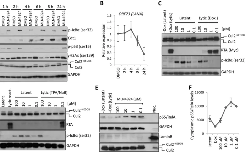
Fig 3. PEL cytotoxicity is due to NF-κB inhibition.(A) TREx-BCBL-1-RTA cells were treated with 5μM MLN4924 for the indicated times. Cell lysates were
analyzed by immunoblot using the indicated antibodies to determine the mode of cytotoxicity. (B) Transcription of NF-κB-regulated latency-associated gene LANAwas determined at 2, 4, 6 and 8 h post treatment with 5μM MLN4924 to investigate the point at which the inhibition of NEDDylation had an effect. (C) MLN4924 treatment led to the stabilization of phosphorylated IκBα(pIκBα[ser32]) in TREx-BCBL-1-RTA cells, IκBαexpression is reduced in lytic cells reactivated by the addition of Dox. (D) Similar to (C), pIκBαis stabilized in MLN4924-treated BC-3 cells. (E) Cytoplasmic fractions were enriched (as shown by the lack of nuclear Lamin B and enrichment of GAPDH—compare to nuclear fraction [Nuc.]) from TREx-BCBL-1-RTA cells treated with varying
concentrations of MLN4924 which shows the stabilization of the p65/RelA subunit of NF-κB in the cytoplasm. (F) Densitometry analysis using ImageJ of the immunoblot analysis shown in (E).
its association with LANA expression [33]. Importantly, the MLN4924-associated reduction of ORF73mRNA expression occurs prior to any indication of DNA damage (Fig. 3B). Along with cell cycle analysis (Fig. 1D) showing that treatment of TREx-BCBL-1-RTA cells led to a reduc-tion of cells in S-Phase (which is inconsistent with Cdt1-associated DNA-rereplicareduc-tion), and the rapid stabilization of pIκBαsuggests that inhibition of NF-κB signaling was responsible for cytotoxicity. Additionally, pIκBαlevels increased in an MLN4924-dose-dependent manner in latently-infected TREx-BCBL-1-RTA (Fig. 3C) and BC-3 cells (Fig. 3D) treated for 24 h, show-ing that, as expected, treatment did stabilize pIκBα. In accordance with this, the cytoplasmic levels of the p65/RelA subunit of NF-κB were increased in a dose-dependent manner (Fig. 3E-F—enrichment of the cytoplasmic compartment was verified by the lack of nuclear Lamin B—compared to the nuclear fraction control which maintained Lamin B but lacked GAPDH). Previous reports have shown that upon lytic reactivation,IκBαis
posttranscription-ally downregulated [34] and consistent with this, cytoplasmic p65/RelA decreased in Dox-treated (reactivated) cells further highlighting the involvement of IκBαfor MLN4924-induced inhibition of NF-κB.
Additionally, we did not observe pIκBαexpression in reactivated cells (those with RTA ex-pression) regardless of MLN4924 treatment (Fig. 3C-D). This was also highlighted in our con-focal analysis of p65/RelA localization showing it had translocated to the nucleus in reactivated cells even when treated with MLN4924 (S2 Fig). We therefore surmised that inhibition of
NF-κB, via stabilization of IκBαwas responsible for cytotoxicity and demonstrated that inhibition of NEDDylation may provide a therapeutic option for the treatment of NF-κB-dependent PEL.
Identification of Cul4B as a novel regulator of KSHV latency
expression of various lytic cycle-associated genes (Fig. 4D). Interestingly, expression of the la-tency-associated geneLANAdid not alter suggesting that Cul4B specifically regulated the ex-pression of lytic genes. Here, we also investigated the exex-pression ofK12, which in contrast to the reduced expression we observed in the presence of MLN4924, was increased by Cul4B knockdown (Fig. 4D). Although a thorough investigation of individual CRLs is required (for ex-ample, using RNAi experiments to inhibit the various CRL components), this data suggests that MLN4924’s effect on KSHV latency is through its inhibition of CRL activity. In addition, we have identified CRL4B as a novel regulator of KSHV latency.
Fig 4. The role of individual CRLs for the regulation of KSHV latency.(A) Dominant-negative versions of each Cullin (FLAG-DNCul that inhibits CRL activity [35]) was expressed in latently infected rKSHV.219 cells and ORF57 expression (as a marker of lytic reactivation) was tested by immunoblot analysis 36 h later. Treatment of cells with MLN4924 and TPA/NaB (sodium butyrate) served as positive controls for reactivation.*denotes non-specific bands and error bars represent the standard deviation of the mean of two independent transfection experiments. (B) Densitometry analysis of (A). (C) qRT-PCR analysis ofCul4BmRNA from TREx-BCBL-1-RTA cells transfected with either a scramble control shRNA expression vector (Scr.) or four independent shRNA expression vectors targeted atCul4B(shCul4B). RNA was harvested four days post transfection. (D) qRT-PCR analysis of viral gene expression in Cul4B-knockdown cells normalized againstGAPDHexpression. Error bars represent the standard deviation from three independent transfections per condition. OLT denotes OriLyt transcript.
NEDDylation is essential for replication of the KSHV genome
The KSHV lytic cycle is intimately linked to the pathogenesis of KSHV malignancies and the role of NEDDylation or the importance of CRL-mediated ubiquitylation during this process is currently unknown. To investigate if ubiquitylation is indeed a feature of KSHV reactivation, we stained reactivated cells with anti-Ub FK2 (which only recognizes ubiquitylated proteins and not free ubiquitin) and to increase the stringency of this assay, we removed soluble nuclear material prior to fixation (seeMaterials and Methods). As shown inFig. 5A, reactivation is as-sociated with significant levels of ubiquitylation which appeared to localize to the edges of the replication compartments (Fig. 5Ainset). However, in the presence of low concentration MLN4924 (1μM), the degree of replication compartment ubiquitylation was reduced. There
are two potential reasons that explain this observation; that CRLs are the predominant E3 li-gases that ubiquitylate replication compartment factors, or that NEDDylation is required with-in replication compartments, and this is important for recruitwith-ing ubiquitwith-in E3 ligases (as recently demonstrated during the DNA double-strand break repair mechanism [39]). As NED-Dylation-dependent ubiquitylation occurred around replication compartments, we asked whether this modification was required for KSHV lytic reactivation. For these experiments, we employed TREx-BCBL-1-RTA cells, where the lytic cycle is induced by the addition of doxycy-cline (Dox.) by virtue of Dox-inducible expression of exogenous RTA-Myc. The advantage of using TREx-BCBL-1-RTA cells is that the lytic cycle is robustly induced (more so than with TPA/NaB) and any effect is a result of viral gene expression and not due to the general effects of TPA/NaB. Cells were treated with varying concentrations of MLN4924, the lytic cycle was induced and 24 h later cells were harvested and viral protein expression was assessed by immu-noblotting. As shown inFig. 5B, MLN4924 was able to inhibit the expression of RTA-Myc and its downstream target ORF57 in a dose-responsive manner, corresponding to the levels of Cul2 NEDDylation. As RTA-Myc represents exogenous protein expression (i.e. not from the KSHV genome) in TREx-BCBL-1-RTA cells, these results showed that MLN4924 inhibited all tran-scription in these cells. Using qPCR analysis of viral genomes, we also confirmed that
MLN4924 was able to inhibit KSHV DNA replication (Fig. 5C). However, KSHV genome rep-lication was inhibited at lower concentrations than was required to inhibit viral protein expres-sion (compare protein expresexpres-sion and genome replication levels in cells treated with 1μM)
highlighting a potentially novel mechanism, mediated by NEDDylation, that uncouples these two features of KSHV biology. These data show for the first time that NEDDylation (or NED-Dylation-dependent ubiquitylation) plays a significant role during the KSHV lytic cycle.
MLN4924-mediated inhibition of KSHV genome replication is not due to
caspase activation
and investigated viral genome copy number by qPCR, viral DNA replication was still inhibited (Fig. 6B).
An additional measure of viral genome replication involves the incorporation of EdU into DNA specifically marking replicating viral genomes in replication compartments, where
Fig 5. NEDDylation is required for lytic reactivation.(A) To test if NEDDylation-associated ubiquitylation is a feature of KSHV lytic reactivation, immunofluorescence analysis was used. TREx-BCBL-1-RTA cells were left untreated, or treated with MLN4924 and reactivated by the addition of Dox. Soluble nuclear proteins (i.e. those not associated with chromatin) were removed prior to fixation (seeMaterials and Methods). Ubiquitylated proteins (using antibodies specific for ubiquitylated proteins and not free ubiquitin) were seen associated with the edges of KSHV replication compartments (inset; RC denotes replication compartment; Ub denotes ubiquitin-modified proteins), as indicated by RTA staining in untreated, but dox. reactivated cells and the level of ubiquitylation was reduced in the presence of 1μM MLN4924. (B) Cell lysates were prepared from TREx-BCBL-1-RTA cells that were treated with MLN4924 and reactivated with Dox for 24 h. Immunoblot analysis of viral proteins shows that lytic-associated protein expression is inhibited in a dose-dependent manner (C) TREx-BCBL-1-RTA cells were treated at varying concentrations of MLN4924, reactivated with doxycycline and total DNA was isolated 72 h later. Normalized (to cellular GAPDH) viral genome replication was measured using qPCR analysis of three biological replicates; error bars represent standard deviation from the mean.
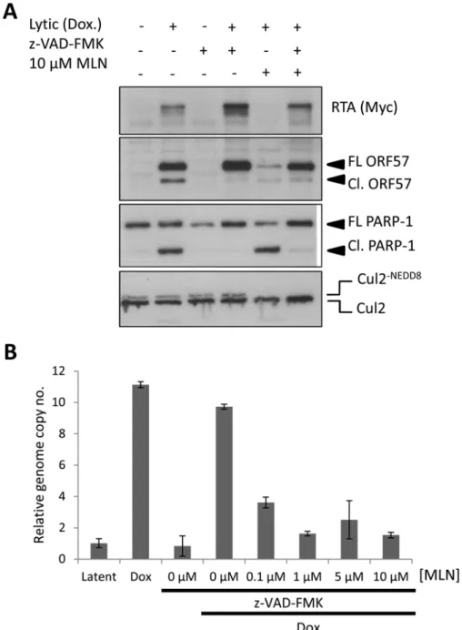
proteins such as RTA are also localized (Fig. 7A). However, even in cells clearly expressing RTA, MLN4924 treatment prevented virus replication, as shown by the lack of EdU-positive replication compartments. Inhibition of caspases restored viral protein expression in MLN4924-treated cells; however, viral DNA replication was still inhibited (Fig. 6B). We
Fig 6. Inhibiting MLN4924-induced apoptosis does not restore lytic reactivation.(A) Inhibition of caspase activity (using the pan-caspase inhibitor z-VAD-FMK) restored lytic cycle protein expression in reactivated cells treated with MLN4924. Successful inhibition of caspase activity was monitored by immunoblot analysis of PARP-1 and ORF57 cleavage. FL ORF57 denotes full length ORF57; Cl. ORF57 denotes cleaved ORF57. (B) Despite restoration of protein expression upon caspase inhibition, viral genome replication was still blocked. TREx-BCBL-1-RTA cells were treated as indicated and cells were harvested 72 h later. Total DNA was harvested and qPCR was used to determine KSHV genome copies relative to latent cells (as inFig. 5C).
therefore repeated our EdU analysis of drug-treated cells in the presence of z-VAD-FMK. Inhi-bition of caspases permitted the formation of replication centers and amplification of KSHV DNA in non-treated cells. Nevertheless, z-VAD-FMK was still unable to restore viral genome replication in MLN4924-treated cells (Fig. 7B). These data confirm that MLN4924-mediated activation of caspases was not responsible for the inhibition of KSHV genome replication upon MLN4924 treatment.
Fig 7. MLN4924 prevents the proper organization of replication compartments and the recruitment of RTA to OriLyt.(A) Replication compartments were observed in TREx-BCBL-1-RTA cells reactivated by Dox. for 16 h by imaging for the incorporation of EdU into replicating KSHV DNA, and its colocalization with RTA (arrows, top panel). Treatment with MLN4924 inhibited the formation of replication compartments (as observed by pan-nuclear staining of RTA, bottom panels) and blocked the replication of viral DNA (as indicated by the absence of EdU in RTA-positive cells). (B) Reactivated cells treated with caspase inhibitor z-VAD-FMK still formed replication compartments, yet z-VAD-FMK was unable to restore KSHV DNA replication in MLN4924 treated cells. (C) Latent TREx-BCBL-1-RTA cells were treated with 1μM MLN4924 for 18 h and ChIP analysis was performed using antibodies specific for IgG (isotype control), RNA Pol II and Myc-tag (for RTA), and primers specific for the RTA responsive element (RRE) of OriLyt. Treatment induced RNA Pol II recruitment (with a slight increase in RTA) corroborating our transcriptional analyses (seeFig. 2). (D) The same analysis was performed but in cells
reactivated with Dox. This analysis showed that MLN4924 treatment blocked the recruitment of RTA onto OriLyt during lytic reactivation. Error bars represent the standard deviation of the mean from two independent ChIP experiments.
NEDDylation is required for the proper formation of KSHV replication
compartments and the recruitment of RTA to OriLyt
Throughout our studies, we noticed that reactivated cells treated with MLN4924 displayed an unusual RTA expression pattern; as shown in Figs.5A&7A, MLN4924 treatment is associated with pan-RTA localization, rather than in discrete, EdU-positive foci. We also observed the same phenotype when we costained for RTA and a second replication compartment-associated factor, RNA Pol II (S3 Fig). We therefore hypothesized that this would have implications for RTA’s ability to mediate KSHV genome replication, and therefore provide a rationale for the MLN4924-induced block in replication. OriLyt is thecis-acting loci where proteins required for KSHV genome replication assemble. The mechanisms of this have been well characterized and RTA is central to this process. For example, it has been shown that the pre-replication complex (which involves RTA, K8, the core replication proteins and various cellular proteins) is formed prior to its loading onto OriLyt, and that RTA, through its direct DNA binding activ-ity with RRE, recruits these factors to OriLyt [40–42]. Therefore, as MLN4924 inhibited viral genome replication and prevented the correct localization of RTA, we asked whether treatment precluded the loading of the replication complex. To do this, TREx-BCBL-1-RTA cells were treated with 1μM MLN4924 (a concentration that permits viral protein expression but inhibits
replication) and 18 h later, ChIP analysis was performed using antibodies specific for IgG (iso-type control), RNA Pol II and Myc-tag (for RTA), and primers specific for the RTA responsive element (RRE) of OriLyt. Firstly, we performed this analysis on latently infected cells (Fig. 7C), where we observed an enrichment of RNA Pol II (6.45-fold over IgG), whereas RTA levels were lower than those observed for IgG control. Interestingly, when cells were treated, RNA Pol II occupancy at OriLyt approximately doubled, and RTA levels increased 1.6-fold over IgG. These results confirm that MLN4924 was able to induce transcription in latently infected cells; however, they show that only low levels of RTA are required. During lytic replication, both RNA Pol II (63-fold over IgG) and RTA (44-fold over IgG) were increased at OriLyt (Fig. 7D). However, after MLN4924 treatment, RNA Pol II occupancy reduced ca. 3-fold (to 21-fold over IgG) and RTA occupancy decreased ca. 4-fold (to 10-fold over IgG) confirming that the inhibi-tion in KSHV genome replicainhibi-tion was due to a block in the recruitment of the viral pre-replica-tion complex. Consequently, by using MLN4924 to investigate the role of NEDDylapre-replica-tion in KSHV biology, we have uncovered a novel mechanism that regulates lytic reactivation.
Discussion
we asked whether MLN4924 was able to kill these cells. Furthermore, as the lytic cycle plays a significant role during the pathogenesis of KS, we also asked whether the NEDDylation cascade was necessary for virus reactivation, and if so, what role it plays.
The activation of NF-κB is central to KSHV infection (by modulating viral gene expression) and for the pathogenesis of KSHV-associated malignancies (via induction of inflammatory me-diators and the expression of antiapoptotic genes). Inhibiting NEDDylation was clearly cyto-toxic to PEL cells, and mechanistically, it appears that this was due to the inhibition of NF-κB. For example, we showed that MLN4924 led to: i) accumulation of pIκBα, ii) a KSHV gene ex-pression profile consistent with inhibition of NF-κB and iii) the induction of apoptosis (Fig. 8). This corroborates recent work demonstrating that MLN4924 killed NF-κB-dependent
ABC-DLBCL cells in a comparable fashion [27]. In fact, ABC-DLBCL cells were more sensitive than GCB-DLBCLs (that are not reliant on NF-κB) [27] suggesting that this drug may be more efficacious for cancers such as PEL and KS, where NF-κB activation is a requirement for malig-nancy. NF-κB is also required for transcription of the latency control locus which expresses an alternatively spliced transcript that producesORF71(vFLIP),ORF72(vCyclin) andORF73 (LANA). As LANA is essential for maintenance of the KSHV genome during mitosis, NF-κB signaling is clearly required for sustained infection. Interestingly, MLN4924 did not appear to overtly affect LANA expression (despite reducing its mRNA level) which is in line with previ-ous reports showing that its half-life exceeds the time course of our experiments [43] and signi-fying that cytotoxicity was not a result of KSHV genome loss. Moreover, the histological signature of KS involves elongated, spindle-shaped endothelial cells and vFLIP expression alone is sufficient to bring about this morphological change in endothelial cell cultures [44,45]. Importantly, this phenotypic alteration has been shown to be NF-κB-dependent; therefore, it will be of particular interest to investigate if MLN4924 can inhibit this characteristic feature of KS.
In contrast to its role in latency-associated transcription, NF-κB is a negative regulator of numerous lytic cycle-associated genes, including those analyzed in this study (RTA,ORF57 andORF47) [46]. The KSHV protein RTA is both necessary and sufficient for the induction of the lytic cycle, resulting in the expression of the entire KSHV genome and virion production. Many RTA-responsive genes contain RBP-Jκbinding sites, and the translocation of NF-κB into the nucleus allows it to bind RBP-Jκand prevent its association with RTA. Hence, the in-duction of lytic cycle-associated gene expression in PEL and rKSHV.219 cells is very likely due to MLN4924-induced inhibition of NF-κB. The Cul1-containingβTrCP is the principal E3 li-gase responsible for IκBαdegradation [37]. In agreement the above hypothesis, we found that individual expression of dominant-negative versions of Cul1 (DNCul) was able to activate lytic cycle gene expression. Importantly, these data also suggest that MLN4924’s effect on viral gene expression was due to its inhibition of the CRL activity as opposed to a direct role of NEDDyla-tion in KSHV gene expression or the over-arching effects a global block in NEDDylaNEDDyla-tion would have on cellular function. However, an in-depth investigation of the various CRL com-ponents is required to fully elucidate the question of specificity.
Fig 8. Summary.(A) NF-κB signaling is essential for the survival of PEL as it drives the expression of KSHV latency genes, suppresses lytic cycle-associated genes and promotes antiapoptotoic gene expression [28]. Normally, IκBαinhibits NF-κB by preventing its nuclear translocation. Upon stimulation, or modulation by viral proteins such as vFLIP, IκBαis phosphorylated which signals for the recruitment of the Cul1-containing
βTrCP ubiquitin ligase, leading to its polyubiquitylation and degradation [32]. This releases NF-κB allowing it to translocate to the nucleus to activate transcription. This study has shown that MLN4924 blocks the degradation of IκBαthus preventing the nuclear translocation of NF-κB and subsequent PEL cell death. (B) The lytic cycle of KSHV is required for the pathogenesis of KS [7], and therefore the molecular mechanisms governing KSHV reactivation need to be understood. This study has shown that inhibiting NEDDylation prevents lytic reactivation by blocking the recruitment of the viral replication complex and the RNA
are centered on chromatin regulation, such as chromosome condensation, heterochromatin formation and DNA replication and repair processes. Therefore, although Cul4’s potential role in the maintenance of KSHV latency is novel, it might not be surprising. The two Cul4 proteins are virtually identical apart from an extended N-terminal portion found in Cul4B that encodes a nuclear localization signal. Therefore, Cul4B predominantly resides in the nucleus, whereas Cul4A is recruited to the nucleus in response to genotoxic stress. This provides further cre-dence to the hypothesis that Cul4B is important for the regulation of KSHV gene expression. The DNA damage response is a recurring theme in virus biology as it either aids or is a conse-quence of infection [24,47–49]. Cul4A and 4B play a pivotal role in the nucleotide excision re-pair (NER) process, and it was recently reported that human cytomegalovirus (HCMV) is dependent on NER during replication of its genome. Therefore, the role of Cul4 during KSHV infection merits further investigation.
A striking observation however, was that MLN4924 treatment blocked viral genome replica-tion despite appreciable levels of viral gene expression. At 1μM MLN4924, ORF57 expression
was not significantly reduced, but genome replication was reduced by ca. 80%. We considered the possibility that the induction of apoptosis may be responsible for this, and so we repeated the experiments in the presence of a pan-caspase inhibitor (z-VAD-FMK). Even though this led to a recovery in KSHV protein expression even when up to 10μM MLN4924 was used,
ge-nome replication was still not observed. Over the course of our studies, we did notice aberrant RTA localization in BCBL-1 cells treated with MLN4924, followed by reactivation (e.g.Fig. 7). Given that RTA’s function is to recruit the viral pre-replication complex, along with various cellular factors required for KSHV genome replication, its localization is intimately linked with sites of viral genome replication occurs. These sites are termed replication compartments (or replication centers) and can be observed as discrete foci at the nuclear periphery that co-stain with RTA and newly replicated viral DNA (this can be observed using BrdU or EdU that spe-cifically marks viral DNA due to KSHV’s ability to block cellular DNA replication during reac-tivation). We therefore hypothesized that NEDDylation, or CRL-mediated ubiquitylation was required for KSHV genome replication (but not viral gene expression).
Firstly, we showed that reactivation of KSHV is associated with ubiquitylation within repli-cation compartments, and that MLN4924 treatment inhibited this. We next investigated whether NEDDylation was required for RTA’s recruitment to the origins of lytic replication (OriLyt). Using ChIP analysis, we observed that treatment of latent cells led to an increase in Pol II (ca. 2-fold) and RTA (ca. 1.6-fold) occupancy, which agrees with our data showing that treatment led to activation of viral gene expression. Treatment followed by reactivation with Dox. however showed us that NEDDylation was required for RTA recruitment to OriLyt. Here we showed that 1μM MLN4924 (a concentration that still permits viral gene expression)
blocked RTA recruitment to OriLyt. Likewise, Pol II occupancy was reduced to similar levels. This confirmed to us that NEDDylation was required for the proper recruitment of the RTA (and therefore the pre-replication complex) to OriLyt, and offers and explanation for the aber-rant localization of RTA and the inhibition of genome replication.
There are various hypotheses that may explain the importance of NEDDylation for the re-cruitment of the pre-replication complex to OriLyt (Fig. 8). It might be possible that proteins within the pre-replication complex are themselves targets for NEDD8 modification. We have
K48-linked ubiquitin (thus targeting them for degradation) or K63-linked ubiquitin suggesting a CRL-mediated signaling requirement during lytic reactivation. Modification (either Ub or NEDD8) may also be required for the proper processing of proteins that are essential for reactivation. Nevertheless, this study has highlighted that NEDDylation may be a novel target for the treatment of various KSHV-associated malignancies.
addressed this for RTA and showed that RTA is not NEDDylated (S4 Fig); given that RTA is the protein responsible for recruitment, this result suggested that this hypothesis is unlikely [40]. It might also be possible that NEDDylation or CRL-mediated ubiquitylation of OriLyt-associated chromatin might be important for the recruitment of the pre-replication complex, as has recently been reported for the recruitment of repair enzymes during a DNA damage re-sponse [50]. Although it is still possible that these modifications play a role in cells, RTA binds to DNA in a sequence-specific manner, and it can do thisin vitroin the absence of chromatin suggesting that this may not be the role of NEDDylation during reactivation [51]. A further hy-pothesis is that modification of a factor that resides at OriLyt during latency (in order to main-tain latency) may be required during reactivation. This may include CRL-mediated
ubiquitylation (via K48) that targets a latency-associated protein for degradation thus allowing access to OriLyt by the pre-replication complex. It may also involve Ub-modification via K63 linkage that might enhance bindingin vivo. Various cellular proteins have been reported to in-teract with OriLyt during reactivation, but in that study, proteins that bound during latency were not investigated [42]. All of the above hypotheses merit further investigation but are un-fortunately beyond the scope of this study.
In summary, we have demonstrated that NEDDylation is essential for various aspects of KSHV infection. Inhibition of NEDDylation using MLN4924 proved cytotoxic to PEL cells due to their dependence on NF-κB. Remarkably, we also show that a functioning NEDDylation cascade is essential for KSHV genome replication as it was required for the recruitment of the RTA-mediated pre-replication complex to OriLyt. These new findings have opened up new av-enues of investigation regarding the regulation of herpesvirus latency and reactivation. More-over, they demonstrate that inhibition of NEDDylation represents a novel approach for the treatment of KSHV-associated malignancies, including KS that is dependent on both lytic rep-lication and the latency-associated activation of NF-κB.
Materials and Methods
Cell lines and drug treatments
BCBL-1 and BC-3 are primary effusion lymphoma (PEL) B cell lines latently infected with KSHV. TREx-BCBL-1-RTA cells (a kind gift of Dr. Jae Jung, University of Southern California) are a BCBL-1-based cell line that has been engineered to inducibly express exogenous Myc-tagged RTA by the addition of doxycycline, leading to a robust reactivation of the full KSHV lytic cycle [52]. The rKSHV.219 cell line maintains KSHV as a latent infection and was generat-ed by infecting HEK293T cells with a recombinant KSHV that contains a constitutively active puromycin resistance and GFP gene, and an RFP gene that is fused to an RTA-responsive lytic cycle (PAN) promoter; hence, expression of RFP can be used as a reporter of RTA activity [53]. HEK293T cells were also used for co-immunoprecipitation experiments. All cells were main-tained at 37°C in a humidified incubator with 5% CO2. The PEL cell lines were maintained in RPMI-1640 (Lonza) supplemented with 10% FBS (Life Technologies). TREx-BCBL-1-RTA media also contained 200μg/ml hygromycin B (Life Technologies). The rKSHV.219 and
HEK293T cell lines were maintained in DMEM (Lonza) supplemented with 10% FBS (Life Technologies). The rKSHV.219 cultures also contained 1μg/ml puromycin. KSHV reactivation
was induced in BCBL-1 and rKSHV.219 by the addition of 20 ng/ml 12-O-tetradecanoylphor-bol 13-acetate (TPA) and 1.5 mM sodium butyrate (NaB) or 1μg/ml doxycycline hyclate
(Sigma) in TREx-BCBL-1-RTA cells.
enzymes was achieved by the addition of 50μM z-VAD-FMK or FMK-negative control (MBL
International) 30 min prior to MLN4924 and/or reactivation treatment. Inhibitors remained on the cells for the duration of the experiments.
Viability assays
Cell viability was determined using the standard trypan blue exclusion method and using the ATPlite Luminescence ATP Detection Assay (Perkin Elmer). For the ATPlite assay, 10,000 cells were seeded into white 96-well plates and allowed to settle for 16 h at 37°C. Varying con-centrations of MLN4924 were added to the cells followed by 96 h incubation at 37°C. Cellular proliferation was determined according to the manufacturer’s instructions.
Cell cycle analysis
Cells (106) were treated with 1μM MLN4924 (or left untreated) for 24 h, washed in cold PBS
and fixed for 24 h in cold 70% ethanol at -20°C. Prior to analysis, cells were washed in PBS and treated with 1 ml PBS, 10μg/ml propidium iodide (Sigma), 0.5 mg RNase A for 3 h at 37°C.
Cells were then pelleted, resuspended in 1 ml PBS and analysed using a Becton Dickinson BD-LSRFortessa flow cytometer. Data were fitted using ModFit software.
Expression vectors and transfection
Most expression vectors were obtained from Addgene: pcDNA3-DN-hCul1-FLAG (plasmid 15818), pcDNA3-DN-hCul2-FLAG (plasmid 15819), pcDNA3-DN-hCul3-FLAG (plasmid 15820), pcDNA3-DN-hCul4A-FLAG (plasmid 15821), pcDNA3-DN-hCul4B-FLAG (plasmid 15822), pcDNA3-DN-hCul5-FLAG (plasmid 15823) [35], pcDNA3-HA-Cullin4A (plasmid 19907), pcDNA3-Myc-NEDD8 (plasmid 19943). pFLAG-CMV-4-NEDD8 was a kind gift from Dr Eric Stebbins (Rockefeller University) [54]. RTA expression vectors (pRTA and pRTAH145L) were gifts of Dr Gary Hayward (Johns Hopkins University). Cells were plated into 6-well plates and transfections routinely used 1μg plasmid DNA and Lipofectamine 2000 (Life
Technologies) following the manufacturer’s instructions.
Knockdown of gene expression
Knockdown of Cul4B expression was accomplished using four individual SureSilencing shRNA expression vectors (Qiagen). TREx-BCBL-1-RTA cells (3 x 10⁶per transfection) were transfected with 20μg scramble shRNA (control) or 5μg of each of four shCul4B vectors by
nucleofection (Lonza; solution V and nucleofector program T-01). Four days post-transfection, RNA was extracted using Trizol (Life Technologies) and used to generate cDNA for qRT-PCR analysis (see below).
Immunoblot analysis
Cells were washed in PBS and proteins extracted in lysis buffer containing 50 mM Tris (pH 7.4), 150 mM NaCl, 1% NP-40 and 1x protease inhibitor cocktail (Roche) for 15 min on ice and clarified by centrifugation at 12,000 xgfor 10 min, 4°C. Cells used in Nuclear/cytoplasmic enrichments assays (ca. 106) were washed in PBS, lysed in 50μl PBS, 1% Triton X-100 and 1x
Sigma), mouse mAb anti-HA-tag (1:5000; Life Technologies), mouse mAb anti-ORF57 (1:1000; Santa Cruz), rabbit mAb Cullin 2 (1:1000; Life Technology), mouse mAb anti-PARP-1 (1:1000; Cell Signaling Technology), mouse mAb anti-GAPDH (1:5000; Sigma), mouse mAb anti-Lamin B (1:1000; Santa Cruz), rabbit mAb anti-Phospho-IκBα(ser32) (1:1000; Cell Signaling Technology), mouse mAb anti-p65/RelA (1:1000; Santa Cruz), rabbit mAb anti-Caspase 3 (8G10) (1:1000; Cell Signaling Technology), rabbit mAb anti-active Cas-pase 3 (Abcam; 1:250), mouse mAb anti-CasCas-pase 9 (Cell Signaling Technology), mouse mAb anti-Cdt-1 (1:1000; Santa Cruz), mouse mAb anti-γH2Ax (ser139) (1:1000, Santa Cruz) rabbit mAb anti-Phospho-p53 (ser15) (1:1000; Cell Signaling Technology). Signals were detected using chemiluminescence and densitometry (ImageJ) was used to semi-quantify
expression levels.
Quantitative RT-PCR (qRT-PCR)
As previously reported [56], total cellular RNA was extracted from cells using Trizol (Life Technologies) according to the manufacturer’s instructions and contaminating DNA was re-moved using the DNA-freekit (Ambion). Complimentary DNA (cDNA) was generated from 1μg RNA in 20μl reaction volumes using M-MuLV reverse transcriptase (RT; NEB) according
to the manufacturer’s recommendations with 5 ng oligo(dT). In parallel, negative control reac-tions were performed for each RNA by omitting RT in order to confirm that quantification represented cDNA and not contaminating DNA. Quantitative PCR reaction mixes (20μl)
in-cluded 1x SensiMix SYBR green master mix (Bioline), 0.5μM each primer and 1μl cDNA
reac-tion mix. Cycling was performed in a RotorGene Q machine (Eppendorf) and included an initial 10 min denaturation step at 94°C, followed by 40 cycles of 30 s at 94°C, 30 s at 60°C and 30 s at 72°C. Melting curve analysis was performed between 65 and 95°C (with 0.2°C incre-ments) to verify amplicon specificity. Quantification ofGAPDHmRNA was used to normalize between samples, and the average cycle threshold (CT) was determined from three independent
RNA samples from independent cultures. Relative expression compared to non-treated or DMSO-treated control cells was calculated using theΔΔCTmethod.
Indirect immunofluorescence microscopy
anti-ubiquitinylated proteins clone FK2 mouse mAb (1:250; Millipore), mouse mAb anti RNA Pol II (Millipore).
Virus reactivation assays
Virus reactivation was determined by two complimentary assays—identification of viral repli-cation compartments by imaging the incorporation of EdU into replicating viral DNA and by quantitative PCR analysis of viral genome amplification. Incorporation of EdU was performed using a Click-iT EdU Imaging Kit (Life Technologies). Briefly, 1 x 10⁶TREx-BCBL-1-RTA cells were drug-treated as appropriate, virus reactivation was induced by the addition of doxy-cycline and the cells were plated onto poly-L-lysine-coated coverslips. After 16 h, cells were pulsed for 45 min with 10μM EdU, washed, fixed and permeablized according to the
manufac-turer’s recommendations. After detection of EdU (according to the manufacturer’s protocol), the cells were washed and further incubated with RTA antisera (1:100 in PBS, 2% BSA) for 1 h at 37°C, washed again and incubated with Alexa Fluor 488 goat anti-rabbit antibody (Life Technologies; 1:500 in PBS, 2% BSA) for 1 h at 37°C. Finally, DNA was stained using Hoechst 33342 (1:2000 in PBS) and mounted in VECTORSHIELD (Vectorlabs). Images were captured using an LSM700 laser scanning microscope (Carl Zeiss) using 639 nm, 488 nm and 405 nm la-sers for the detection of EdU, RTA and DNA respectively and processed using ZEN imaging software (Carl Zeiss).
For quantitative PCR analysis 1 x 10⁶TREx-BCBL-1-RTA cells were drug-treated as appropriate and virus reactivation was induced by the addition of doxycycline. At 24 h post-reactivation, total DNA was extracted using a DNA Minikit (Qiagen) and quantified by UV spectrophotometry. Viral DNA was quantified using 3.4 ng DNA, in 20μl reaction volumes as
described above for qRT-PCR, using primers specific for the ORF57 gene. Quantification of GAPDH was used to normalize between samples, and the average cycle threshold (CT) was
de-termined from three independent samples from independent cultures. Relative levels of viral DNA was calculated using theΔΔCTmethod.
Immunoprecipitation (NEDDylation assay)
HEK293T cells were plated into 6-well dishes and co-transfected with the indicated plasmids with pcDNA-Myc-NEDD8 or pFLAG-CMV-4-NEDD8 [54] for 24 h. Cells were washed in PBS and proteins extracted in 1 ml lysis buffer (see above) and incubated with anti-c-Myc aga-rose (Sigma) or goat anti-DDDDK conjugated agaaga-rose (for detection of FLAG-tagged proteins; Abcam) following the manufacturer’s recommendations. Immunoprecipitated (NEDDylated) proteins were eluted in Laemmli buffer and subject to immunoblot analysis (see above).
Chromatin immunoprecipitation (ChIP)
Chromatin immunoprecipitation (ChIP) assays were carried out as previously stated [59] using the EZ-ChIP chromatin immunoprecipitation kit (Millipore). Per condition, 107cells were treated with 1% formaldehyde for 10 min prior to three washes with ice-cold PBS. Cells were then resuspended in 3 ml SDS lysis buffer (1% SDS, 10 mM EDTA, 50 mM Tris, [pH 8.1]) containing 1x protease inhibitor cocktail II and sonicated 15 time with 20 s pulses at 4°C. Insoluble material was removed by centrifugation for 10 min at 12,000 xg, 4°C. For each immu-noprecipitation (IP), 100μl of prepared chromatin was added to 900μl of dilution buffer
1 h at 4°C. The Protein G agarose-antibody/chromatin complexes were then washed sequen-tially in 1 ml of the provided buffers; low salt immune complex wash buffer (0.1% SDS, 1% Tri-ton X-100, 2 mM EDTA, 20 mM Tris-HCl, [pH 8.1], 150 mM NaCl), high salt immune complex wash buffer (0.1% SDS, 1% Triton X-100, 2 mM EDTA, 20 mM Tris-HCl, [pH 8.1], 500 mM NaCl), LiCl immune complex wash buffer (0.25 M LiCl, 1% IGEPAL CA630, 1% deoxycholic acid sodium salt, 1 mM EDTA, 10 mM Tris, [pH 8.1]) and TE buffer (10 mM Tris-HCl, [pH 8.0], 1 mM EDTA). The DNA/protein complexes were eluted in 200μl elution
buffer (1% SDS, 0.1 M NaHCO3) before reversal of crosslinks with 5 M NaCl for 4 h at 65°C. Samples were then treated with RNase A at 37°C for 30 min followed by incubation with Pro-teinase K for 2 h at 45°C. The resulting DNA was purified over the provided spin filter col-umns, with elution in 50μl of elution buffer C. The purified DNA was then subject to qPCR
amplification using primers directed to the OriLyt (Forward primer: 5’- ACG GGC CTG GAA TCT CGC CTC TGG-3’and Reverse primer: 5’- ATG GGC GTA ACC GTA GGA CAA GCT G-3’). Each qPCR reaction performed in triplicate containing 5μl purified DNA and was
quan-tified as described above.
Oligonucleotides
Oligonucleotide primer sequences are available upon request.
Supporting Information
S1 Fig. Cullin proteins are NEDDylated in KSHV-infected cells.NEDDylation assays were
carried out in latently-infected rKSHV.219 cells. Cells were transfected with FLAG-NEDD8 along with the indicated Myc-tagged protein. FLAG-NEDDylated proteins were immunopre-cipitated and modified proteins were detected by immunoblot analysis usingαMyc antibodies. Myc-Hey1, a protein not expected to be NEDDylated was used as a negative control.
(TIF)
S2 Fig. MLN4924-induced inhibition of NF-κB.Immunofluorescence (confocal) analysis
NF-κB in untreated TREx-BCBL-1-RTA cells maintaining a latent infection (top left) showed low levels of NF-κB subunit RelA (p65) in the nucleus—a requirement for latency-associated gene expression and PEL viability. Upon lytic reactivation (dox-treated), RelA translocated to the nucleus (top right) due to the lytic cycle-associated downregulation of IκBα[34]. In 10μM
MLN4924-treated latent cells (bottom left), the nucleus was devoid of RelA in most cells sup-porting the hypothesis that MLN4924 stabilized IκBα, thus preventing the nuclear transloca-tion of NF-κB (seeFig. 3); however, in MLN4924-treated cells (10μM) where the lytic cycle
was induced (bottom right), RelA still readily translocated to the nucleus, further highlighting the role of IκBαstabilization for the MLN4924-associated inhibition of NF-κB signaling. (TIF)
S3 Fig. MLN4924 prevents the proper organization of replication compartments.Upon
dox-induced reactivation of the KSHV lytic cycle in TREx-BCBL-1-RTA cells, the proteins re-quired for KSHV gene expression and genome replication are recruited to discrete foci known as replication compartments (arrows, top panels). These include various viral proteins (such as RTA—red) and cellular factors (e.g. RNA Pol II—green). Treatment of cells with 1μM
MLN4924 (here for 16 h) prevents the proper organisation of KSHV replication compartments (bottom panels). NT denotes no treatment.
(TIF)
S4 Fig. RTA is not NEDDylated.NEDDylation assays were carried out in transfected
agarose was used to immunoprecipitate NEDDylated proteins. HA-Cul4A and HA-PP2A served as positive and negative controls, respectively. Expression of RTA or the RTAH145L mu-tant was detected using RTA antisera.
(TIF)
Acknowledgments
We are grateful to members of the Whitehouse laboratory for helpful discussions. The authors would like to thank Dr Sally Boxall (University of Leeds) for assistance with cell cycle analysis, Dr. Jae Jung (University of Southern California) for the TREx-BCBL-1-RTA cells, Dr. Jeffery Vieira for rKSHV.219–293T cells, Dr Eric Stebbins (Rockefeller University) for pFLAG-CMV-4-NEDD8 and Dr. Gary Hayward (Johns Hopkins University) for RTA expression vectors. We are indebted to Millennium Pharmaceuticals (Cambridge, MA) for their provision of
MLN4924.
Author Contributions
Conceived and designed the experiments: DJH AW. Performed the experiments: DJH JJW BRJ BBP AW. Analyzed the data: DJH JJW AW. Wrote the paper: DJH AW.
References
1. Kisselev AF, van der Linden WA, Overkleeft HS. Proteasome inhibitors: an expanding army attacking a unique target. Chem Biol. 2012; 19: 99–115. doi:10.1016/j.chembiol.2012.01.003PMID:22284358
2. Boulanger E, Meignin V, Oksenhendler E. Bortezomib (PS-341) in patients with human herpesvirus 8-associated primary effusion lymphoma. Br J Haematol. 2008; 141: 559–561. doi:10.1111/j.1365-2141. 2008.07057.xPMID:18341641
3. Brown HJ, McBride WH, Zack JA, Sun R. Prostratin and bortezomib are novel inducers of latent Kapo-si's sarcoma-associated herpesvirus. Antivir Ther. 2005; 10: 745–751. PMID:16218174
4. Matta H, Chaudhary PM. The proteasome inhibitor bortezomib (PS-341) inhibits growth and induces apoptosis in primary effusion lymphoma cells. Cancer Biol Ther. 2005; 4: 77–82. PMID:15662128
5. Sarosiek KA, Cavallin LE, Bhatt S, Toomey NL, Natkunam Y, et al. Efficacy of bortezomib in a direct xenograft model of primary effusion lymphoma. Proc Natl Acad Sci U S A. 2010; 107: 13069–13074. doi:10.1073/pnas.1002985107PMID:20615981
6. Dittmer DP, Damania B. Kaposi sarcoma associated herpesvirus pathogenesis (KSHV)—an update. Curr Opin Virol. 2013; 3: 238–244. doi:10.1016/j.coviro.2013.05.012PMID:23769237
7. Mesri EA, Cesarman E, Boshoff C. Kaposi's sarcoma and its associated herpesvirus. Nat Rev Cancer. 2010; 10: 707–719. doi:10.1038/nrc2888PMID:20865011
8. La Ferla L, Pinzone MR, Nunnari G, Martellotta F, Lleshi A, et al. Kaposi' s sarcoma in HIV-positive pa-tients: the state of art in the HAART-era. Eur Rev Med Pharmacol Sci. 2013; 17: 2354–2365. PMID:
24065230
9. Simonelli C, Spina M, Cinelli R, Talamini R, Tedeschi R, et al. Clinical features and outcome of primary effusion lymphoma in HIV-infected patients: a single-institution study. J Clin Oncol. 2003; 21: 3948–3954. PMID:14581418
10. Gantt S, Casper C. Human herpesvirus 8-associated neoplasms: the roles of viral replication and antivi-ral treatment. Curr Opin Infect Dis. 2011; 24: 295–301. doi:10.1097/QCO.0b013e3283486d04PMID:
21666458
11. Bhatt S, Ashlock BM, Toomey NL, Diaz LA, Mesri EA, et al. Efficacious proteasome/HDAC inhibitor combination therapy for primary effusion lymphoma. J Clin Invest. 2013; 123: 2616–2628. doi:10.1172/ JCI64503PMID:23635777
12. Soucy TA, Smith PG, Milhollen MA, Berger AJ, Gavin JM, et al. An inhibitor of NEDD8-activating en-zyme as a new approach to treat cancer. Nature. 2009; 458: 732–736. doi:10.1038/nature07884
PMID:19360080
14. Bennett EJ, Rush J, Gygi SP, Harper JW. Dynamics of cullin-RING ubiquitin ligase network revealed by systematic quantitative proteomics. Cell. 2010; 143: 951–965. doi:10.1016/j.cell.2010.11.017PMID:
21145461
15. Rabut G, Peter M. Function and regulation of protein neddylation. 'Protein modifications: beyond the usual suspects' review series. EMBO Rep. 2008; 9: 969–976. doi:10.1038/embor.2008.183PMID:
18802447
16. Wimmer P, Schreiner S, Dobner T. Human pathogens and the host cell SUMOylation system. J Virol. 2012; 86: 642–654. doi:10.1128/JVI.06227-11PMID:22072786
17. Everett RD, Boutell C, Hale BG. Interplay between viruses and host sumoylation pathways. Nat Rev Microbiol. 2013; 11: 400–411. doi:10.1038/nrmicro3015PMID:23624814
18. Gastaldello S, Hildebrand S, Faridani O, Callegari S, Palmkvist M, et al. A deneddylase encoded by Ep-stein-Barr virus promotes viral DNA replication by regulating the activity of cullin-RING ligases. Nat Cell Biol. 2010; 12: 351–361. doi:10.1038/ncb2035PMID:20190741
19. Stanley DJ, Bartholomeeusen K, Crosby DC, Kim DY, Kwon E, et al. Inhibition of a NEDD8 Cascade Restores Restriction of HIV by APOBEC3G. PLoS Pathog. 2012; 8: e1003085. doi:10.1371/journal. ppat.1003085PMID:23300442
20. Romani B, Engelbrecht S, Glashoff RH. Antiviral roles of APOBEC proteins against HIV-1 and suppres-sion by Vif. Arch Virol. 2009; 154: 1579–1588. doi:10.1007/s00705-009-0481-yPMID:19669862
21. Yu X, Yu Y, Liu B, Luo K, Kong W, et al. Induction of APOBEC3G ubiquitination and degradation by an HIV-1 Vif-Cul5-SCF complex. Science. 2003; 302: 1056–1060. PMID:14564014
22. Cai QL, Knight JS, Verma SC, Zald P, Robertson ES. EC5S ubiquitin complex is recruited by KSHV la-tent antigen LANA for degradation of the VHL and p53 tumor suppressors. PLoS Pathog. 2006; 2: e116. PMID:17069461
23. Hannah J, Zhou P. Regulation of DNA damage response pathways by the cullin-RING ubiquitin ligases. DNA Repair (Amst). 2009; 8: 536–543. doi:10.1016/j.dnarep.2009.01.011PMID:19231300
24. O'Dowd JM, Zavala AG, Brown CJ, Mori T, Fortunato EA. HCMV-infected cells maintain efficient nucle-otide excision repair of the viral genome while abrogating repair of the host genome. PLoS Pathog. 2012; 8: e1003038. doi:10.1371/journal.ppat.1003038PMID:23209410
25. Soucy TA, Dick LR, Smith PG, Milhollen MA, Brownell JE. The NEDD8 Conjugation Pathway and Its Relevance in Cancer Biology and Therapy. Genes Cancer. 2010; 1: 708–716. doi:10.1177/ 1947601910382898PMID:21779466
26. Milhollen MA, Narayanan U, Soucy TA, Veiby PO, Smith PG, et al. Inhibition of NEDD8-activating en-zyme induces rereplication and apoptosis in human tumor cells consistent with deregulating CDT1 turn-over. Cancer Res. 2011; 71: 3042–3051. doi:10.1158/0008-5472.CAN-10-2122PMID:21487042
27. Milhollen MA, Traore T, Adams-Duffy J, Thomas MP, Berger AJ, et al. MLN4924, a NEDD8-activating enzyme inhibitor, is active in diffuse large B-cell lymphoma models: rationale for treatment of NF-{kappa}B-dependent lymphoma. Blood. 2010; 116: 1515–1523. doi:10.1182/blood-2010-03-272567
PMID:20525923
28. de Oliveira DE, Ballon G, Cesarman E. NF-kappaB signaling modulation by EBV and KSHV. Trends Microbiol. 2010; 18: 248–257. doi:10.1016/j.tim.2010.04.001PMID:20452220
29. Majerciak V, Kruhlak M, Dagur PK, McCoy JP, Zheng ZM Jr.. Caspase-7 cleavage of Kaposi sarcoma-associated herpesvirus ORF57 confers a cellular function against viral lytic gene expression. J Biol Chem. 2010; 285: 11297–11307. doi:10.1074/jbc.M109.068221PMID:20159985
30. Sun R, Lin SF, Staskus K, Gradoville L, Grogan E, et al. Kinetics of Kaposi's sarcoma-associated her-pesvirus gene expression. J Virol. 1999; 73: 2232–2242. PMID:9971806
31. Pearce M, Matsumura S, Wilson AC. Transcripts Encoding K12, v-FLIP, v-Cyclin, and the MicroRNA Cluster of Kaposi's Sarcoma-Associated Herpesvirus Originate from a Common Promoter. J Virol. 2005; 79: 14457–14464. PMID:16254382
32. Karin M, Ben-Neriah Y. Phosphorylation meets ubiquitination: the control of NF-[kappa]B activity. Annu Rev Immunol. 2000; 18: 621–663. PMID:10837071
33. Jha HC, Upadhyay SK, M AJP, Lu J, Cai Q, et al. H2AX phosphorylation is important for LANA-mediat-ed Kaposi's sarcoma-associatLANA-mediat-ed herpesvirus episome persistence. J Virol. 2013; 87: 5255–5269. doi:
10.1128/JVI.03575-12PMID:23449797
34. Lei X, Bai Z, Ye F, Xie J, Kim CG, et al. Regulation of NF-kappaB inhibitor IkappaBalpha and viral repli-cation by a KSHV microRNA. Nat Cell Biol. 2010; 12: 193–199. doi:10.1038/ncb2019PMID:20081837
36. Lee DF, Kuo HP, Liu M, Chou CK, Xia W, et al. KEAP1 E3 ligase-mediated downregulation of NF-kappaB signaling by targeting IKKbeta. Mol Cell. 2009; 36: 131–140. doi:10.1016/j.molcel.2009.07. 025PMID:19818716
37. Yaron A, Hatzubai A, Davis M, Lavon I, Amit S, et al. Identification of the receptor component of the IkappaBalpha-ubiquitin ligase. Nature. 1998; 396: 590–594. PMID:9859996
38. Nakagawa T, Xiong Y. X-linked mental retardation gene CUL4B targets ubiquitylation of H3K4 methyl-transferase component WDR5 and regulates neuronal gene expression. Mol Cell. 2011; 43: 381–391. doi:10.1016/j.molcel.2011.05.033PMID:21816345
39. Ma T, Chen Y, Zhang F, Yang CY, Wang S, et al. RNF111-dependent neddylation activates DNA dam-age-induced ubiquitination. Mol Cell. 2013; 49: 897–907. doi:10.1016/j.molcel.2013.01.006PMID:
23394999
40. Rossetto CC, Susilarini NK, Pari GS. nteraction of Kaposi's sarcoma-associated herpesvirus ORF59 with oriLyt is dependent on binding with K-Rta. J Virol. 2011; 85: 3833–3841. doi: 10.1128/JVI.02361-10PMID:21289111
41. Wang Y, Li H, Chan MY, Zhu FX, Lukac DM, et al. Kaposi's sarcoma-associated herpesvirus ori-Lyt-dependent DNA replication: cis-acting requirements for replication and ori-Lyt-associated RNA tran-scription. J Virol. 2004; 78: 8615–8629. PMID:15280471
42. Wang Y, Li H, Tang Q, Maul GG, Yuan Y. Kaposi's sarcoma-associated herpesvirus ori-Lyt-dependent DNA replication: involvement of host cellular factors. J Virol. 2008; 82: 2867–2882. doi:10.1128/JVI. 01319-07PMID:18199640
43. Chen W, Hilton IB, Staudt MR, Burd CE, Dittmer DP. Distinct p53, p53:LANA, and LANA complexes in Kaposi's Sarcoma—associated Herpesvirus Lymphomas. J Virol. 2010; 84: 3898–3908. doi:10.1128/ JVI.01321-09PMID:20130056
44. Alkharsah KR, Singh VV, Bosco R, Santag S, Grundhoff A, et al. Deletion of Kaposi's sarcoma-associ-ated herpesvirus FLICE inhibitory protein, vFLIP, from the viral genome compromises the activation of STAT1-responsive cellular genes and spindle cell formation in endothelial cells. J Virol. 2011; 85: 10375–10388. doi:10.1128/JVI.00226-11PMID:21795355
45. Grossmann C, Podgrabinska S, Skobe M, Ganem D. Activation of NF-kappaB by the latent vFLIP gene of Kaposi's sarcoma-associated herpesvirus is required for the spindle shape of virus-infected endothe-lial cells and contributes to their proinflammatory phenotype. J Virol. 2006; 80: 7179–7185. PMID:
16809323
46. Izumiya Y, Izumiya C, Hsia D, Ellison TJ, Luciw PA, et al. NF-kappaB serves as a cellular sensor of Ka-posi's sarcoma-associated herpesvirus latency and negatively regulates K-Rta by antagonizing the RBP-Jkappa coactivator. J Virol. 2009; 83: 4435–4446. doi:10.1128/JVI.01999-08PMID:19244329
47. Jackson BR, Noerenberg M, Whitehouse A. A novel mechanism inducing genome instability in Kapo-si's sarcoma-associated herpesvirus infected cells. PLoS Pathog. 2014; 10: e1004098. doi:10.1371/ journal.ppat.1004098PMID:24788796
48. Karttunen H, Savas JN, McKinney C, Chen YH, Yates JR 3rd, et al. Co-opting the Fanconi Anemia Ge-nomic Stability Pathway Enables Herpesvirus DNA Synthesis and Productive Growth. Mol Cell. 2014; 55: 111–22. doi:10.1016/j.molcel.2014.05.020PMID:24954902
49. Turnell AS, Grand RJ. DNA viruses and the cellular DNA-damage response. J Gen Virol. 2012; 93: 2076–2097. doi:10.1099/vir.0.044412-0PMID:22855786
50. Jackson SP, Durocher D. Regulation of DNA damage responses by ubiquitin and SUMO. Mol Cell. 2013; 49: 795–807. doi:10.1016/j.molcel.2013.01.017PMID:23416108
51. Bu W, Carroll KD, Palmeri D, Lukac DM. Kaposi's sarcoma-associated herpesvirus/human herpesvirus 8 ORF50/Rta lytic switch protein functions as a tetramer. J Virol. 2007; 81: 5788–5806. PMID:
17392367
52. Nakamura H, Lu M, Gwack Y, Souvlis J, Zeichner SL, et al. Global changes in Kaposi's sarcoma-associated virus gene expression patterns following expression of a tetracycline-inducible Rta transac-tivator. J Virol. 2003; 77: 4205–4220. PMID:12634378
53. Vieira J O'Hearn PM. Use of the red fluorescent protein as a marker of Kaposi's sarcoma-associated herpesvirus lytic gene expression. Virology. 2004; 325: 225–240. PMID:15246263
54. Jubelin G, Taieb F, Duda DM, Hsu Y, Samba-Louaka A, et al. Pathogenic bacteria target NEDD8-conjugated cullins to hijack host-cell signaling pathways. PLoS Pathog. 2010; 6: e1001128. doi:
10.1371/journal.ppat.1001128PMID:20941356
56. Jackson BR, Boyne JR, Noerenberg M, Taylor A, Hautbergue GM, et al. An interaction between KSHV ORF57 and UIF provides mRNA-adaptor redundancy in herpesvirus intronless mRNA export. PLoS Pathog. 2011; 7: e1002138. doi:10.1371/journal.ppat.1002138PMID:21814512
57. Boyne JR, Jackson BR, Taylor A, Macnab SA, Whitehouse A. Kaposi's sarcoma-associated herpesvi-rus ORF57 protein interacts with PYM to enhance translation of viral intronless mRNAs. EMBO J. 2010; 29: 1851–1864. doi:10.1038/emboj.2010.77PMID:20436455
58. Goodwin DJ, Hall KT, Giles MS, Calderwood MA, Markham AF, et al. The carboxy terminus of the her-pesvirus saimiri ORF 57 gene contains domains that are required for transactivation and transrepres-sion. J Gen Virol. 2000; 81: 2253–2265. PMID:10950983

![Fig 4. The role of individual CRLs for the regulation of KSHV latency. (A) Dominant-negative versions of each Cullin (FLAG-DNCul that inhibits CRL activity [35]) was expressed in latently infected rKSHV.219 cells and ORF57 expression (as a marker of lytic](https://thumb-eu.123doks.com/thumbv2/123dok_br/18367997.354951/9.918.62.804.117.664/individual-regulation-dominant-inhibits-activity-expressed-infected-expression.webp)


![Fig 8. Summary. (A) NF-κB signaling is essential for the survival of PEL as it drives the expression of KSHV latency genes, suppresses lytic cycle-associated genes and promotes antiapoptotoic gene expression [28].](https://thumb-eu.123doks.com/thumbv2/123dok_br/18367997.354951/16.918.298.783.111.871/summary-signaling-essential-expression-suppresses-associated-antiapoptotoic-expression.webp)
