Biopolymers and Cell. 2015. Vol. 31. N 1. P. 5–14 doi: http://dx.doi.org/10.7124/bc.0008C7
UDC 576.322 577.22
Characteristic of mTOR signaling and its involvement in the regulation
of cell movements through remodeling the cytoskeleton architecture
V. R. Kosach, O. V. Cherednyk, A. I. Khoruzhenko
Institute of Molecular Biology and Genetics, NAS of Ukraine, 150, Zabolotnoho Str., Kyiv, Ukraine, 03680
mTOR kinase is one of the basic links at the crossroad of several signal transduction pathways. De re gulated mTOR kinase signaling accompanies the progress of cancer, diabetes, neurodegenerative disorders and ag-ing. Implication of mTOR inhibitor rapamycin decreases migration and invasion of malignant cells, and metastasis formation. However, a precise mechanism of the regulation of cellular locomotion by mTOR kinase is not fully understood. This article focuses on the recent fi ndings that demonstrated a possible role of mTOR kinase in the regulation of cytoskeleton remodeling and cell migration properties. Detailed studies on this non-canonical mTOR function will extend our knowledge about cell migration and metastasis for-mation and might improve anti-cancer therapeutic approaches.
K e y w o r d s: mTOR signaling, rapamycin, cytoskeleton remodeling, intermediate fi laments, microtu-bules, cancer metastasis.
Introduction
The mTOR (mammalian target of rapamycin) kinase is a central link of several signaling pathways that integrates the signals from growth factors, hor mones, stress, energy status, and amino acids to con trol the organism growth, homeostasis and aging. mTOR acts through two functionally and struc turally distinct complexes, named mTORC1 and mTORC2 (mTOR complex 1 and 2).
Taken together, active mTOR complexes stimu-late the cellular growth and proliferation by positive regulation of transcription, translation, riboso me bio-genesis, cell survival, inhibition of autopha gy and apoptosis [1]. Overactivation of the mTOR kinase was found in a range of human diseases, such as dif-ferent types of cancer, type 2 diabetes, obesity, and neurodegenerative disorders. The re fore, mTOR is con-sidered as a perspective target of anti-cancer and an-ti-aging therapies [2].
One of the most dangerous stages of oncogenesis is the metastasis formation. At this stage the tumor is considered malignant. It is known, that the primary tumor causes death only in 10 % of the patients, whe-reas 90 % of deaths are caused by metastases [3]. The metastasis formation is directly dependent on the cell motility and invasion, which allow the cells to change a position within the tissues. It was shown, that known mTOR inhibitor rapamycin and its syn-thetic analogs can demonstrate not only cytostatic effects, but a decrease in the motility of cancer cells as well [4, 5]. However, the mechanism of the regu-lation of cell migration and invasion by mTOR ki-nase is not fully understood.
The cytoskeleton, a cytoplasmic system of fi bers, is critical to sustain cell motility. Intermediate fi la-ments, actin-containing microfi laments and microtu-bules are the three main cytoskeletal systems of ver-tebrate and many inverver-tebrate cells. The rearrange-ments of the cytoskeleton architecture are the main
reason of the cell locomotion [6]. Early studies re-vealed that mTORC2 regulates the actin cytoskele-ton polarization in yeasts. Moreover, further research showed that inactivation of both mTOR complexes impairs the movement of normal and cancer cells [7]. This article focuses on the recent studies reveal-ing the role of mTOR kinase in the cytoskeleton re-modeling and cell locomotion.
Structure and functions
of mTOR kinase complexes
TOR (Target of Rapamycin) is a serine/threonine protein kinase, the activity of which is inhibited specifi cally by macrolide rapamycin (the other na-me sirolimus). Rapamycin, produced by fi lamentous bacterium Streptomyces hygroscopicus, was ini-tially found in the soil sample of the Easter Island (Rapa Nui) in 1970s and subsequently discovered to have antifungal, immunosuppressive and cytostatic effects. Biochemical studies and genetic scree ning in the yeast mutants, resistant to the growth-inhibi-tory properties of rapamycin, led to the identifi ca-tion of TOR kinase [8]. Interestingly, rapamycin does not directly inhibit TOR, but it forms a com-plex with cytosolic protein FKBP12 (FK506 bind-ing protein 12 kDa, the other name FKBP1A). The rapamycin-FKBP12 complex binds to the C termi-nal part of TOR molecule, termed FRB (FKBP12-rapamycin binding domain), thereby in hibiting TOR kinase activity and functions [9]. Further studies re-vealed the homologues of yeast TOR in the fl ies (Drosophila me lanogaster), worms (Caenorhabditis elegans), fun gus Cryptococcus neoformans, plants (Arabi dop sis tha liana) and mammals. That indi-cates a high evolutionary conservatism of the ki-nase, and hence its im por tant role in the regulation of intracellular processes.
It should be noted that although mTOR originally stood for ‘mammalian TOR’, it is now also used
of-fi cially as an abbreviation for ‘mechanistic TOR’. Un-fortunately, sometimes the expression ‘me cha nistic TOR’ is used to indicate TOR kinase from any spe-cies that brings some confusion in the fi eld [10]. To prevent further confusion we will use the term mTOR when discussing kinase in mammalian organisms.
In mammalian cells mTOR is a catalytic compo und of two different complexes mTORC1 and mTORC2, which coordinates anabolic and catabolic processes in response to growth factors and nutrients. The mTOR-containing complexes have different sensitivities to rapamycin as well as upstream regulators and down-stream targets.
Components and substrates of mTOR complex 1
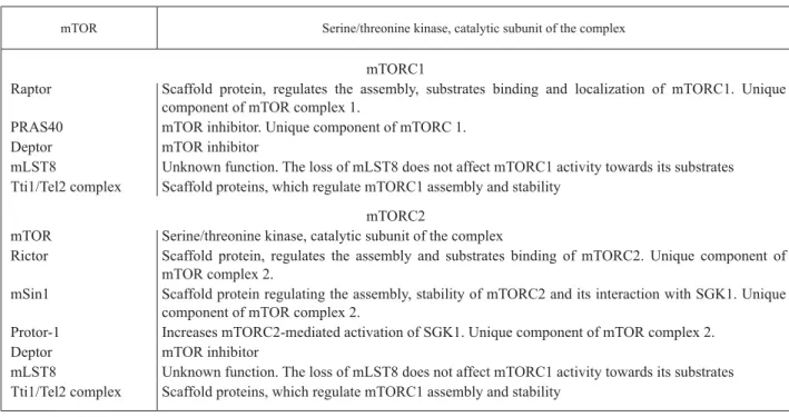
The most studied complex is mTORC1. It consists of mTOR, Raptor (regulatory-associated protein of mTOR), mLST8 (mammalian lethal Sec13 protein 8), PRAS40 (proline-rich kt substrate 40 kDa), Dep-tor (DEP-domain-containing mTOR-interacting pro-tein), and the Tti1/Tel2 complex. Raptor and PRAS40 are unique components of mTORC1. The known fun-ctions of mTOR partner proteins are listed in the Tab-le 1 [11]. Cryo-eTab-lectron studies reveaTab-led that mTORC1 is an obligate dimer with an overall rhomboid shape and a central cavity. It was shown that the dimeric interfaces were formed by interlocking interactions between the mTOR and Raptor subunits. It was also proposed that some mTORC1 substrates with multi-ple phosphorylation sites could shuttle between the two mTOR active sites within the dimer [12].
mTORC1 acts as a signal integrator for four major regulatory inputs: nutrients, growth factors, energy and stress. Growth factors and hormones regulate mTORC1 through several different signaling path-ways, such as PI3K/Akt network, Ras–Raf–MAPK/ Erk signaling and Wnt pathway. The implication of multiple growth factor-initiated pathways in mTORC1 regulation is likely to allow mTOR to participate in many developmental and physiological processes [1].
phosphorylation and activation. In turn, activated Akt phosphorylates TSC2 (also known as tuberin), a large protein that, together with TSC1 (also known as hamartin), forms the TSC1–TSC2 complex. TSC1– TSC2 acts as a GTPase activating protein (GAP) for RHEB and promotes its loading with GDP. Akt-mediated phosphorylation of TSC2 inhibits GAP ac-tivity of the TSC1–TSC2 complex and induces RHEB to bind GTP. The GTP-bound form of Rheb directly interacts with mTORC1 and strongly stimu-lates its kinase activity [13, 14].
Nutrients activate mTORC1 through amino acids availability. Import of the amino acids causes small Rag GTPases to switch to the active conformation. The active Rag heterodimer physically interacts with Raptor, causing mTORC1 to cluster onto the surface of late endosomes and lysosomes, where the Rag GTPases reside. This relocalization enables mTORC1 to interact with its activator RHEB [15, 16].
In mammalian cells the most extensively studied substrates of mTORC1 are S6Ks (ribosomal protein S6 kinases) and 4E-BP1 (eIF4E- binding protein 1). The main function of these proteins is the regulation of mRNA translation initiation and progression, thus
controlling the rate of protein synthesis. Previous studies have shown that both proteins contain TOS (TOR signaling) motifs. Mu tations in the amino acid sequence of the TOS motif signifi cantly reduces the level of phosphorylation of S6K and 4E-BP1 under
in vitro conditions, due to the impaired ability of these proteins to interact with Raptor. Besides the canonical function of the mentioned mTOR targets, they are involved in the regulation of cell viability, migration, cytoskeleton remodeling etc. [17–19].
Components and substrates
of mTOR complex 2
Com pared to mTORC1, less is known about mTORC2. It is insensitive to amino acids, but responds to growth factors through a poorly defi ned mechanism.
mTORC2 is formed by mTOR, Rictor (rapa my-cin-insensitive companion of mTOR), mLST8, Dep-tor, mSIN1 (mammalian stress-activated pro tein ki-nase interacting protein) and Protor-1 (pro tein ob-served with Rictor-1, also known as PRR5). Ric tor, mSin1 and Protor-1 are unique components of mTORC2. The known functions of mTORC2 pro-teins are listed in the Table 1. In yeasts, TORC2 is
Table 1. The known functions of mTORC1 and mTORC2 proteins
mTOR Serine/threonine kinase, catalytic subunit of the complex
mTORC1
Raptor Scaffold protein, regulates the assembly, substrates binding and localization of mTORC1. Unique component of mTOR complex 1.
PRAS40 mTOR inhibitor. Unique component of mTORC 1.
Deptor mTOR inhibitor
mLST8 Unknown function. The loss of mLST8 does not affect mTORC1 activity towards its substrates Tti1/Tel2 complex Scaffold proteins, which regulate mTORC1 assembly and stability
mTORC2
mTOR Serine/threonine kinase, catalytic subunit of the complex
Rictor Scaffold protein, regulates the assembly and substrates binding of mTORC2. Unique component of mTOR complex 2.
mSin1 Scaffold protein regulating the assembly, stability of mTORC2 and its interaction with SGK1. Unique component of mTOR complex 2.
Protor-1 Increases mTORC2-mediated activation of SGK1. Unique component of mTOR complex 2.
Deptor mTOR inhibitor
oligomeric and forms homodimers, but whether mTORC2 can form dimers/multimers in ma m ma lian cells is unknown. mTORC2 is insensitive to acute rapamycin treatment. However, chro nic treat ment with rapamycin inhibits mTORC2 functions in many cell lines, possibly, by sequestration of all mTOR molecules and, therefore, prevention of the de novo
mTORC2 assembly [20].
It was revealed, that mTORC2 substrates are the AGC kinase family members, such as: Akt, cPKCs (conventional protein kinases C) and SGK (serum- and glucocorticoid-regulated kinase). Thro ugh these kinases mTORC2 takes part in the regulation of cell survival, cell cycle progression and anabolism [1, 20].
Participation of mTOR kinase in the regulation
of cytoskeleton reorganization
The initial characterization of mTORC2 led to the discovery of the participation of mTOR signaling in actin cytoskeleton polarization and cell movements [21]. Recent studies revealed that both complexes: mTORC1 and mTORC2 play a crucial role in the processes of cell motility and invasion through regu-lation of cytoskeleton remodeling [22].
Interplay between mTOR kinase
and intermediate fi laments
Intermediate fi laments (IFs) form an extensive cy-toskeletal network within the cell (Fig. 1, A).
The subunits composing intermediate fi laments constitute a superfamily of α-helical proteins that are found in the cytoplasm of different tissues and on the nuclear membrane. In humans, there are at least 67 genes that encode IF proteins, which ma kes this gene family one of the largest in the human genome. The various members of the intermediate fi lament pro-tein family are expressed differentially in complex patterns during embryonic development and in the terminally differentiated tissues. So, this superfamily has been divided into fi ve distinct types on the basis of similarities in sequence and their patterns of ex-pression in cells (Table 2) [6, 23, 24].
Phosphorylation rate plays an important role in the assembly and disassembly of the intermediate
fi laments. Hyperphosphorylation of multiple sites of the IFs during mitosis causes rapid disassembly of the fi laments and their separation to the daughter cells. Recent studies also showed high level of fl ex-ibility of the IFs even in stationary interphase cells. These fi ndings suggest that dynamic of IF cytoskel-eton remodeling is under the control of kinases and phosphatases [23, 25].
For a long time, IFs have been considered as com-ponents of the cell that maintain the cellular shape and provide resistance to mechanical stress. However, a lot of recent studies revealed novel non-canonical functions of intermediate fi lament proteins. For ex-ample, it was shown that keratins mediate localiza-tion of the hemidesmosomes and desmosomes in the human keratinocytes. Dep le tion of all keratins by genome engineering caused altered distribution of the hemidesmosomal proteins, which resulted in a faster adhesion and migration of keratin-free cells [26]. Moreover, analyses of vimentin −/− mice have revealed that loss of vimentin leads to impaired wound healing due to defects in the capacity of fi -broblasts to migrate [27]. These fi ndings support a hypothesis that intermediate fi laments play impor-tant role in cell motility and that altered regulation of
Table 2. Classifi cation of the intermediate fi laments
Type
of IFs IFprotein Tissue distribution
Type I Acidic keratins All types of the epithe-lia
Type II Basic keratins
Type III Vimentin Desmin
Glial fi brillary acidic protein
Peripherin
Mesenchymal cells Muscle
Glial cells, astrocytes
Neurons Type IV NF(Neurofi laments)-L
NF-M NF-H Nestin
Internexin Syncolin
Neurons Neurons Neurons
Neuroepithelial stem cells
Neurons Muscle cells Type V Lamins A, C, B1, B2 Nuclear lamina of all
types of the cells Beaded
fi laments
Phakinin Filensin
IFs assembly could be involved in cancer cell spread-ing. Also, intermediate fi laments take part in the apoptosis regulation and cell signaling.
Studies on the keratin 17 (K17)-null mouse skin keratinocytes revealed that K17 regulates cell growth and size through mTOR signaling. Keratin 17 is an intermediate filament protein rapidly induced in wounded stratified epithelia that alters cellular vis-coelastic properties and optimizes tissue repair. Mo-use skin keratinocytes lacking K17 show dep ressed protein translation and are of smaller size, correlat-ing with decreased Akt/mTOR signalcorrelat-ing activity. It was discovered that K17 regulates mTOR activation through binding to the adaptor protein 14-3-3σ. Two amino acid residues located in the amino-terminal head domain of keratin 17 are required for the se-rum-dependent relocalization of 14-3-3σ from the nucleus to the cytoplasm, and for the sti mulation of mTOR activity and cell growth [28].
Another evidence of the cooperation between IFs and mTOR kinase comes from the research of the transgenic mice lacking the entire keratin multipro-tein family. All keratin-null embryos die from severe growth retardation at embryonic day 9.5. Em bryonic epithelia suffer no cytolysis but display mislocalized desmo- somes and glucose transporters GLUT1 and GLUT3. An altered localization of glucose trans-porters subsequently activates the energy sensor ad-enosine monophosphate kinase (AMPK). AMPK is a negative mTORC1 regulator, it inactivates mTOR signaling, thereby represses protein biosynthesis in keratin-null embryos [29].
Treatment of a human HaCaT keratinocyte cell line with mTOR inhibitors (rapamycin, temsirolimus or everolimus) resulted in selective keratin 6a (K6a) repression. Furthermore, treatment of the HaCaT cell line with the siRNAs targeting components of the mTOR pathway altered the levels of K6a expres-sion. Oral rapamycin administration also improves the symptoms in pachyonychia congenita patients, suggesting mTOR inhibitors may be a therapeutic option for people with mutations that disrupt the in-termediate fi laments formation. The se results show a possible bidirectional interplay between mTOR ki-nase and intermediate fi lament proteins [30].
It is known that the site-specifi c phosphorylation of IF proteins induces the disassembly of the fi la-ment structures. During mitosis, the hyperphos pho-rylation of intermediate fi laments by Cdk1 (Cyc lin-dependent kinase 1), Plk1 (Polo-like kinase 1), Rho- and Aurora-B kinases is essential for the effi cient segregation of IF networks into daughter cells [31]. However, it was revealed that IF network is also a highly mobile structure in the interphase cells and its remodeling is under the control of protein kinases and phosphatases, such as protein kinase C (PKC) and protein kinase A (PKA) [25].
Regulation of actin cytoskeleton
reorganization by mTOR kinase
A globular protein actin forms microfi laments, which are 7 nm in diameter polar fi brils that organize an extensive network in the cytoplasm of all eukaryotic cells (Fig. 1, B). Actin can be present as either a free monomer G-actin (globular) or a part of a linear polymer microfi lament called F-actin (fi lamentous). All actin subunits in the microfi lament point toward the same end of the fi lament. Actin fi lament exhibits polarity: the end that possesses an actin subunit that has its ATP binding site exposed is called the «(-) end», whereas the opposite end where the cleft is di-rected at a different adjacent monomer is called the «(+) end». The assembly of G-actin into F-actin is accompanied by the hydrolysis of ATP. Actin par-ticipates in many important cellular processes, in-cluding muscle contraction, cell motility, cell divi-sion, vesicle and organelle traffi c, and the establish-ment and maintenance of cell junctions [6, 32].
The actin cytoskeletal rearrangements are regu-lated by intracellular signaling pathways directed by Rac (Ras-related C3 botulinum toxin substrate), Rho (Ras homolog gene family), and Cdc42 (Cell divi-sion control protein 42 homolog), all Ras-like mol-ecules belonging to the GTPase superfamily of switch proteins [6]. So, it was interesting whether mTOR could infl uence actin cytoskeleton architec-ture through these proteins. Indeed, further research showed that in yeasts TOR2 activated Rho1 and Rho2 via their exchange factor ROM2 (Rho1 gua-nine nucleotide exchange factor 1). However, the actual mechanism by which TORC2 regulates the Rho1 GTPase pathway is not well studied [35, 36].
Depletion of mTOR and Rictor, but not Raptor, impairs actin polymerization, leading edge estab-lishment, and directional migration in neutrophils
stimulated with chemoattractants. It was shown that depletion of Rictor inhibits Rac and Cdc42 activi-ties, supposing that they are the target of mTORC2. Interestingly, depletion of mSin1, an integral com-ponent of mTORC2, caused no detectable changes in neutrophil polarity and chemotaxis [37, 38].
Several recent studies pointed to the mTORC2 in-volvement in the formation of long-term memory by regulating and stabilizing the actin cytoskeleton in the dendritic spines of neurons. Rictor-defi cient mice showed a reduction in the ratio of fi brilar actin (F-actin) to actin monomers, as well as a reduction in the expression of a number of upstream positive reg-ulators of actin polymerization. These data suggested that mTORC2 is required for the long-term memory formation by increasing the F-actin important for dendritic spine growth and remodeling [39, 40].
Fig. 1. The architecture of different types of the cytoskeleton fi bers in the human breast adenocarcinoma MCF-7 cell line: intermedi-ate fi laments (A), microfi laments (B), microtubules (C)
Fig. 2. Immunofl uorescent analysis of the phospho-mTOR Ser2481 (B) colocalization with tubulin β (A) during cytokinesis in the human breast adenocarcinoma MCF-7 cell line. Nuclei were counterstained with Hoechst 33342 (C)
A B C
10 m
5 m 5 m 5 m 5 m
Merge
10 m 20 m
Current research revealed that mTORC1 also could be implicated in the actin cytoskeleton reassembly in different cells. It was shown that rapamycin treat-ment induced S6K inactivation, inhibited actin stress
fi ber formation and cell migration in a wide range of mammalian cell lines. Further studies discovered that S6K, Akt, PDK1, and activated mTOR were localized to the actin arc of the Swiss 3T3 fi broblasts [10]. Rapamycin treatment blocked the epidermal growth factor (EGF)-induced actin arc formation in these cells, supporting a hypothesis, that mTORC1/S6K axis is also important for the cytoskeleton regulation [41]. It was observed that rapamycin inhibited IGF-I-induced F-actin reorganization and phosphorylation of the focal adhesion proteins, such as FAK (Focal adhesion ki-nase), paxillin and p130Cas, by inhibition of the S6K1 activity [7, 33]. Knockdown of mTORC1 and mTORC2 induced a mesenchymal-epithelial transition in the col-orectal cancer cells, due to increased cell-cell contacts as well as decreased actin cytoskeletal remodeling and decreased activation of the small GTPases, RhoA and Rac1 [36]. It supports the idea that mTOR could regu-late cytoskeleton rearrangement through phosphoryla-tion of the actin-remodulating proteins.
The present study showed that activated PI3K-Akt-mTOR signaling pathway promotes invasion and me-tastasis in hepatocellular carcinoma though up-regu-lation of MMP-9 (Matrix metalloproteinase 9), tho-ugh, indicating that mTORC1 could infl uence cellular locomotion by several distinct directions [42, 43].
The crosstalk between mTOR kinase
and microtubules
A microtubule is a polymer of globular tubulin sub-units, which are arranged in a cylindrical tube mea-suring 25 nm in diameter – the thickest fi brils of the cytoskeleton (Fig. 1, C). Similar to F-actin a micro-tubule is polarized and has (+) end and (-) end. Po ly-merization of the tubulin subunits requires the hy-drolysis of the GTP molecules. In addition to regula-tion of the cell motility, microtubules play a major role in organization of the cell polarity through a spe-cial structure called the microtubule-organizing cen-ter (MTOC). Loca ted near the nucleus, the MTOC directs the assembly and orientation of microtubules,
the ro u te of vesicle tra ffi cking, and the orientation of organelles. Mic ro tubules play a crucial role during mitosis by the formation of mitotic spindle, which is used to separate eukaryotic chromoso mes. A large number of proteins infl uences the assembly and sta-bility of microtubules and their association with oth-er cell structures. These proteins are collectively cal-led microtubule-associated prote ins (MAPs) [6, 32]. Involvement of TOR1 and TOR2 in the control of various aspects of microtubule dynamics was report-ed in yeasts [44]. However, the role of TORs in the regulation of microtubule dynamics has not been fully elucidated yet.
In mammalian cells mTOR was found to bind di-rectly to and phosphorylate cytoplasmic linker pro-tein of 170 kDa (CLIP-170), which is a MAP that binds to the (+) end of the microtubule and stabilizes it [45]. However, the exact function of this phospho-rylation is not fully understood. It was revealed that TSC2 knockout resulted in a greater abundance of stabilized microtubules underneath the cellular cor-tex. Time-lapse imaging of dynamic microtubules al so revealed disorganized movements of the grow-ing microtubule plus-ends in the cellular cortex re-gion, including growth in a direction that is parallel to the cortex. The authors suggested that the func-tional mTOR-CLIP-170 interaction helps microtu-bules grow to the cellular cortex [46].
It was shown that mTOR kinase is also involved in the regulation of intracellular transport associated with microtubules. Inhibition of the expression of TSC2, which is involved in the activation of mTOR, leads to disruption of the caveolin (scaffold protein, involved in the endocytosis) transport to the plasma membrane. Instead, it was detected in the vesicles, randomly located in the cytoplasm. Incubation of rat
fi broblasts in the media that contain high concentra-tions of mTOR inhibitor rapamycin led to the same result, and chaotic arrangement of microtubules in the cortical zone was observed in the cells [47].
coim-munoprecipitation. Inhibition of dynein fun ction us-ing RNAi hinders the mTORC1 activity in the hu-man fi broblasts and the human glioblastoma–astro-cytoma cell line U373-MG [48].
Moreover, the phosphorylated form of mTOR ki-nase (phospho-mTOR Ser2481) was observed to lo-calize at the cleavage furrow of different cell lines during cytokinesis. Inhibition of the polymerization of microtubules by nocodazole leads to the loss of phospho- mTOR (Ser2481) ability to target the spin-dle midzone and the cleavage furrow during cytoki-nesis. At these conditions phospho-mTOR was ran-domly dispersed across the entire mitotic cytoplasm, indicating that mitotic traveling of phospho-mTOR (Ser2481) requires dynamic microtubules [49].
Using anti-phospho-mTOR (Ser2481) antibodies (Merck Millipore) we revealed the colocalization of phospho-mTOR (Ser2481) and tubulin β at the cleavage furrow that has not been demonstrated earlier (Fig. 2). Immunofl uorescent analysis was performed as de-scribed [50]. Our fi ndings support a hypothesis that mTOR phosphorilated at Ser 2481 interacts with micro-tubules during cytokinesis. However, further studies are needed to understand the mechanism of this process.
Conclusion
Novel fi ndings in the mTOR signaling fi eld shed light on the non-canonical functions of the mTOR kinase. The bidirectional crosstalk bet ween mTOR and all three types of the cytoskeleton points to the important role of mTOR signaling pathway in the normal cell lo-comotion during embryonic development, wound heal-ing, and che mo taxis as well as in the cancer cells spreading. Further detailed investigation of this new as-pect of the mTOR activity might lead to the optimiza-tion of the current anti-cancer therapeutic approaches.
REFERENCES
1. Zoncu R, Efeyan A, Sabatini DM. mTOR: from growth sig-nal integration to cancer, diabetes and ageing. Nat Rev Mol Cell Biol. 2011;12(1):21–35.
2. Lamming DW, Ye L, Sabatini DM, Baur JA. Rapalogs and mTOR inhibitors as anti-aging therapeutics. J Clin Invest. 2013;123(3):980–9.
3. Chambers AF, Groom AC, MacDonald IC. Dissemination and growth of cancer cells in metastatic sites. Nat Rev Can-cer. 2002;2(8):563–72.
4. Hong SM, Park CW, Cha HJ, et al. Rapamycin inhibits both mo-tility through down-regulation of p-STAT3 (S727) by dis-rupting the mTORC2 assembly and peritoneal dissemina-tion in sarcomatoid cholangiocarcinoma. Clin Exp Me tas ta-sis. 2013;30(2):177–87.
5. Feng W, Jia S. Rapamycin inhibits the invasive ability of thyroid cancer cells by down-regulating the expression of VEGF-C in vitro. Cell Biochem Funct. 2012;30(6):487–91. 6. Lodish H, Arnold B, Kaiser Chris, Monty K. Molecular Cell
Biology. New York: W. H. Freeman, 2012; 973 p.
7. Zhou H, Huang S. Role of mTOR signaling in tumor cell mo-tility, invasion and metastasis. Curr Protein Pept Sci. 2011;
12(1):30–42.
8. Laplante M, Sabatini DM. mTOR Signaling. Cold Spring Harb Perspect Biol. 2012;4(2): a011593.
9. Yang H, Rudge DG, Koos JD, Vaidialingam B, Yang HJ, Pavletich NP. mTOR kinase structure, mechanism and reg-ulation. Nature. 2013;497(7448):217–23.
10. Hall MN. Talks about TORCs: recent advancesin target of rapamycin signalling. On mTOR nomenclature. Biochem Soc Trans. 2013;41(4):887–8.
11. Laplante M, Sabatini DM. mTOR signaling in growth con-trol and disease. Cell. 2012;149(2):274–93.
12. Yip CK, Murata K, Walz T, Sabatini DM, Kang SA. Structure of the human mTOR complex I and its implications for ra-pamycin inhibition. Mol Cell. 2010;38(5):768–74. 13. Manfredi GI, Dicitore A, Gaudenzi G, Caraglia M,
Persa-ni L, Vitale G. PI3K/Akt/mTOR signaling in medullary thy-roid cancer: a promising molecular target for cancer therapy. Endocrine. 2015;48(2):363–70.
14. Follo MY, Manzoli L, Poli A, McCubrey JA, Cocco L. PLC and PI3K/Akt/mTOR signalling in disease and cancer. Adv Biol Regul. 2015;57:10–6.
15. Efeyan A, Zoncu R, Sabatini DM. Amino acids and mTORC1: from lysosomes to disease. Trends Mol Med. 2012;18(9): 524–33.
16. Bar-Peled L, Chantranupong L, Cherniack AD, et al. A tu-mor suppressor complex with GAP activity for the Rag GT-Pases that signal amino acid suffi ciency to mTORC1. Sci-ence. 2013;340(6136):1100–6.
17. Filonenko VV. PI3K/mTOR/S6K signaling pathway – new pla yers and new functional links. Biopolym Cell. 2013;29
(3):207–14.
18. Lyzogubov V, Khozhaenko Y, Usenko V, et al. Immunohis-tochemical analysis of Ki-67, PCNA and S6K1/2 expression in human breast cancer. Exp Oncol. 2005;27(2): 141–4.
19. Filonenko VV, Tytarenko R, Azatjan SK, et al. Immunohis-tochemical analysis of S6K1 and S6K2 localization in hu-man breast tumors. Exp Oncol. 2004;26(4): 294–9. 20. Oh WJ, Jacinto E. mTOR complex 2 signaling and
func-tions. Cell Cycle. 2011;10(14):2305–16.
22. Sen B, Xie Z, Case N, et al. mTORC2 regulates mechani-cally induced cytoskeletal reorganization and lineage selec-tion in marrow-derived mesenchymal stem cells. J Bo ne Miner Res. 2014;29(1):78–89.
23. Magin TM, Vijayaraj P, Leube RE. Structural and regu latory functions of keratins. Exp Cell Res. 2007;313(10): 2021–32. 24. Chang L, Goldman RD. Intermediate fi laments mediate
cyto-skeletal crosstalk. Nat Rev Mol Cell Biol. 2004;5(8): 601–13. 25. Sihag RK, Inagaki M, Yamaguchi T, Shea TB, Pant HC.
Ro-le of phosphorylation on the structural dynamics and func-tion of types III and IV intermediate fi laments. Exp Cell Res. 2007;313(10):2098–109.
26. Seltmann K, Roth W, Kröger C, et al. Keratins mediate lo-calization of hemidesmosomes and repress cell motility. J In vest Dermatol. 2013;133(1):181–90.
27. Ivaska J, Pallari HM, Nevo J, Eriksson JE. Novel functions of vimentin in cell adhesion, migration, and signaling. Exp Cell Res. 2007;313(10):2050–62.
28. Kim S, Wong P, Coulombe PA. A keratin cytoskeletal protein regulates protein synthesis and epithelial cell growth. Na-ture. 2006;441(7091):362–5.
29. Vijayaraj P, Kröger C, Reuter U, Windoffer R, Leube RE, Magin TM. Keratins regulate protein biosynthesis through localization of GLUT1 and -3 upstream of AMP kinase and Raptor. J Cell Biol. 2009;187(2):175–84.
30. Hickerson RP, Leake D, Pho LN, Leachman SA, Kaspar RL. Rapamycin selectively inhibits expression of an inducible keratin (K6a) in human keratinocytes and improves sym p-toms in pachyonychia congenita patients. J Dermatol Sci. 2009;56(2):82–8.
31. Izawa I, Inagaki M. Regulatory mechanisms and functions of intermediate fi laments: a study using site- and phosphory-lation state-specifi c antibodies. Cancer Sci. 2006; 97(3):167– 74.
32. Lewin B. Cells. MA:"Jones & Bartlett Learning" 2007. 863p. 33. Liu L, Chen L, Chung J, Huang S. Rapamycin inhibits
F-ac-tin reorganization and phosphorylation of focal adhesion proteins. Oncogene. 2008;27(37):4998–5010.
34. Goncharova EA, James ML, Kudryashova TV, Goncharov DA, Krymskaya VP. Tumor suppressors TSC1 and TSC2 diffe-rentially modulate actin cytoskeleton and motility of mou se embryonic fi broblasts. PLoS One. 2014;9(10): e111476. 35. Ohsawa M, Kobayashi T, Okura H, Igarashi T, Mizuguchi M,
Hino O. TSC1 controls distribution of actin fi bers through its effect on function of Rho family of small GTPases and regulates cell migration and polarity. PLoS One. 2013;8 (1): e54503. 36. Gulhati P, Bowen KA, Liu J, et al. mTORC1 and mTORC2
regulate EMT, motility, and metastasis of colorectal cancer via RhoA and Rac1 signaling pathways. Cancer Res. 2011;
71(9):3246–56.
37. Byles V, Covarrubias AJ, Ben-Sahra I, et al. The TSC-mTOR pathway regulates macrophage polarization. Nat Commun. 2013; 4:2834.
38. He Y, Li D, Cook SL, et al. Mammalian target of rapamycin and Rictor control neutrophil chemotaxis by regulating Rac/ Cdc42 activity and the actin cytoskeleton. Mol Biol Cell. 2013;24(21):3369–80.
39. Josselyn SA, Frankland PW. mTORC2: actin on your me-mory. Nat Neurosci. 2013;16(4):379–80.
40. Angliker N, Rüegg MA. In vivo evidence for mTORC2-me-diated actin cytoskeleton rearrangement in neurons. Bioar-chitecture. 2013;3(4):113–8.
41. Berven LA, Willard FS, Crouch MF. Role of the p70(S6K) pathway in regulating the actin cytoskeleton and cell migra-tion. Exp Cell Res. 2004;296(2):183–95.
42. Mendes Sdos S, Candi A, Vansteenbrugge M, et al. Mic ro ar-ray analyses of the effects of NF-kappaB or PI3K pathway inhibitors on the LPS-induced gene expression profi le in RAW264.7 cells: synergistic effects of rapamycin on LPS-induced MMP9-overexpression. Cell Signal. 2009; 21(7): 1109–22.
43. Zhou HY, Wong AS. Activation of p70S6K induces expres-sion of matrix metalloproteinase 9 associated with hepato-cyte growth factor-mediated invasion in human ovarian cancer cells. Endocrinology. 2006;147(5):2557–66. 44. Choi JH, Adames NR, Chan TF, Zeng C, Cooper JA, Zheng XF.
TOR signaling regulates microtubule structure and fun ction. Curr Biol. 2000;10(14):861–4.
45. Malik AR, Urbanska M, Macias M, Skalecka A, Jaworski J. Beyond control of protein translation: what we have lear ned about the non-canonical regulation and function of mam-malian target of rapamycin (mTOR). Biochim Bio phys Acta. 2013;1834(7):1434–48.
46. Zhou Q, Wong CH, Lau CP, et al. Enhanced antitumor activ-ity with combining effect of mTOR inhibition and microtu-bule stabilization in hepatocellular carcinoma. Int J Hepa-tol. 2013;2013:103830.
47. Barnes EA, Kenerson HL, Jiang X, Yeung RS. Tuberin regu lates E-cadherin localization: implications in epitheli al-me se nchymal transition. Am J Pathol. 2010;177(4):1765–78.
48. Clippinger AJ, Alwine JC. Dynein mediates the localizati on and activation of mTOR in normal and human cytome-galovirus-infected cells. Genes Dev. 2012;26(18):2015–26. 49. Vazquez-Martin A, Sauri-Nadal T, Menendez OJ, et al.
Ser2481-autophosphorylated mTOR colocalizes with chro mo-somal passenger proteins during mammalian cell cytokine-sis. Cell Cycle. 2012;11(22):4211–21.
В. . К , . В. Ч , А. І. Х Ха а а mTOR
а а а
а
mTOR a є є ,
-.
Д mTOR є
, ,
. mTOR
є
. ,
mTOR . Д
-,
mTOR
. Д є
mTOR
-.
К а: mTOR , ,
, , ,
-.
В. . К , . В. Ч , А. . Х Ха а а mTOR а
а а
mTOR a ,
-. Д mTOR
-, ,
.
mTOR
. ,
mTOR
. Э
-, mTOR
.
mTOR
-.
К а: mTOR , ,
-, ,
-, .