Molecular evolution and transcriptional profile of GH3 and GH20
b
-N-acetylglucosaminidases in the entomopathogenic fungus
Metarhizium
anisopliae
.
Eder Silva de Oliveira
1, Ângela Junges
1, Nicolau Sbaraini
1, Fábio Carrer Andreis
1, Claudia Elizabeth
Thompson
1, Charley Christian Staats
1and Augusto Schrank
11
Centro de Biotecnologia, Universidade Federal do Rio Grande do Sul, Porto Alegre, RS, Brazil.
Abstract
Cell walls are involved in manifold aspects of fungi maintenance. For several fungi, chitin synthesis, degradation and recycling are essential processes required for cell wall biogenesis; notably, the activity of b -N-acetylglucosa-minidases (NAGases) must be present for chitin utilization. For entomopathogenic fungi, such asMetarhizium anisopliae, chitin degradation is also used to breach the host cuticle during infection. In view of the putative role of NAGases as virulence factors, this study explored the transcriptional profile and evolution of putative GH20 NAGases (MaNAG1 and MaNAG2) and GH3 NAGases (MaNAG3 and MaNAG4) identified in M. anisopliae. While MaNAG2 orthologs are conserved in several ascomycetes, MaNAG1 clusters only with Aspergilllus sp. and entomopathogenic fungal species. By contrast, MaNAG3 and MaNAG4 were phylogenetically related with bacterial GH3 NAGases. The transcriptional profiles ofM. anisopliae NAGase genes were evaluated in seven culture condi-tions showing no common regulatory patterns, suggesting that these enzymes may have specific roles during the Metarhizium life cycle. Moreover, the expression of MaNAG3 and MaNAG4 regulated by chitinous substrates is the first evidence of the involvement of putative GH3 NAGases in physiological cell processes in entomopathogens, indi-cating their potential influence on cell differentiation during theM. anisopliae life cycle.
Keywords: NAGases, GH20 and GH3,Metarhizium, chitinolytic system, entomopathogenesis. Received: December 08, 2017; Accepted: February 23, 2018.
Introduction
Chitin is the second most abundant polymer on Earth and its recycling from carapaces, cuticles and fungal cell walls impacts on carbon and nitrogen cycles. The chitin polymer is composed of b-1,4-linked
N-acetyl-D-gluco-samine (GlcNAc) subunits (Beier and Bertilsson, 2013) and its degradation can be driven in two ways: i) chitin can be deacetylated to chitosan by action of chitin deacetylases (EC 3.5.1.41), which yields glucosamine monomers via the enzymatic hydrolysis by chitosanase (EC 3.2.1.132); or ii) by the chitinolytic degradation process generating GlcNAc monomers, which involves the initial hydrolysis of the
b-1,4 glycoside bonds by the action of a group of enzymes,
including chitinases (EC 3.2.1.14), lytic polysaccharide monooxygenases (LPMOs) (of the auxiliary activity 10 family - AA10; EC N/A) andb-N-acetylglucosaminidases
(NAGases; EC 3.2.1.52) (Beier and Bertilsson, 2013; Tho-ratet al., 2017). The enzymes evolved in the chitinolytic
degradation process act in a consecutive fashion to
com-pletely degrade chitin (Patilet al., 2000; Hartlet al., 2012;
Brzezinskaet al., 2014). LPMOs and endo-acting GH18
chitinases insert strand breaks at random positions within the chitin polymer, while exo-acting GH18 chitinases sub-sequently cleave chito-oligosaccharides (Chavan and Deshpande, 2013; Langner and Göhre, 2015). Finally, NAGases hydrolyzeb-1,4 linkages on
N-acetylglucosami-ne dimers (chitobiose), producing GlcNAc monosaccha-rides (Duo-Chuan, 2006).
NAGases are classified into three glycoside hydro-lase (GH) families, 3, 20, and 84, on the basis of their amino acid sequence similarities (Cantarelet al., 2009). GH3 and GH84 NAGases are distributed in several bacterial and metazoan cells, respectively, while members from the GH20 family are versatile enzymes abundant in fungi and insects. Although these three families encompass function-ally related enzymes, they possess no sequence homology, differing in their structure and catalytic mechanism (Slá-mováet al., 2010; Liuet al., 2012).
The genomes of ascomycetous filamentous fungi contain, on average, 15 to 25 chitinase-encoding genes, but only one or two genes encoding GH20 NAGases (Seidl, 2008; Jungeset al., 2014). Notably, as has been shown for DOI: http://dx.doi.org/10.1590/1678-4685-GMB-2017-0363
Send correspondence to Augusto Schrank. Centro de Biotecnolo-gia, Universidade Federal do Rio Grande do Sul, P. O. Box 15005, 91501-970 Porto Alegre, RS, Brazil. E-mail: [email protected]
the mycopathogenic fungusTrichoderma atroviride, chitin
could not be used as a nutrient source if NAGase activity is absent, despite the presence of approximately 30 chitinase genes, emphasizing the importance of these enzymes for the full degradation of the chitin polymer (López-Mondéjar
et al., 2009). In this way, the diversity of chitinase genes
contrasts with the relatively low number of NAGase genes and their fundamental importance on chitin metabolism.
Potential functions for NAGases in fungi include the use of exogenous chitin as a nutrient source and cell wall turnover during the fungal life cycle (Seidlet al., 2006).
These functions have already been described for GH20 NAGases inT. atroviride(Seidlet al., 2006;
López-Mon-déjaret al., 2009),Aspergillussp.(Kimet al., 2002), and Neurospora crassa (Tzelepis et al., 2012). In addition,
GH20 NAGases participate in processes related to fungal hyphal extension and branching (Rastet al., 1991), fungal
cell wall degradation during autolysis (Díezet al., 2005),
and have a putative role in insect pathogenesis (St. Legeret al., 1991).
In contrast, NAGases belonging to the GH3 family consist of a small group of bacterial enzymes that possess a broad range of functions depending on the organism. Simi-larly to GH20 NAGases, some GH3 NAGases participate in chitin catabolism, as in marine chitinolytic bacteria, such as Vibrio furnissii and Alteromonas sp. (Tsujibo et al.,
1994; Chitlaru and Roseman, 1996). Notably, only re-cently, the first fungal GH3 NAGase was described (Yang
et al., 2014). The RmNag enzyme from the zygomycete Rhizomucor miehei exhibited hydrolysis activity on
N-acetylchitooligosaccharide (GlcNAc)2-3substrates. This
report further supports the existence of GH3 NAGases in other fungal species, especially in ascomycetes, consider-ing their expansion of chitinolytic machinery genes (Seidl 2008; Jungeset al., 2014).
In recent years the chitin degradation machinery has attracted much attention, especially in entomopathogenic fungi, such asMetarhizium anisopliae(Hypocreales:
Cla-vicipitaceae). In these species, the chitinolytic system has, probably, two main biological functions: Firstly, as chitin is the major component of fungal cell walls, chitin-degrading enzymes act on the cell wall remodeling, which is neces-sary for hyphal vegetative growth (Seidl, 2008). Secondly, the infection of arthropod hosts requires a prior chitin hy-drolysis of the exoskeleton (St. Legeret al., 1991).
Further-more, M. anisopliae has the ability to differentiate into
specialized cell types during its infection cycle. The switch between conidia to hyphae and the formation of infection structures (i.e., appressorium and blastospore), are pro-cesses that require chitin degradation (Schrank and Vains-tein, 2010). Notably, the importance of someM. anisopliae
chitinase genes in infection process have been suggested and functionally verified using knockout constructions (da Silvaet al., 2005; Boldoet al., 2009; Staatset al., 2013).
Despite the knowledge gained by the study of chiti-nases inMetarhizium, the role of NAGases in the life cycle
and infection process of entomopathogens has not been fully elucidated. This study surveyed putative NAGase genes from GH3 and GH20 families inM. anisopliaeand
investigated their evolutionary relationships to those of other filamentous ascomycetes. To further characterize NAGase genes inM. anisopliae, their expression patterns
were evaluated in different cell types and various nutri-tional conditions. The results suggest new possibilities for studying NAGases participation inM. anisopliaebiology.
Material and Methods
NAGase gene mining of theM. anisopliaegenome
The survey of NAGase genes was performed in the
M. anisopliae E6 genome assembly (accession number
PRJNA245858) (Staatset al., 2014). In order to identify
putative GH20 NAGase genes, three well described NAGase sequences of filamentous fungi were used as the query in a tBLASTn search: NagA from A. nidulans
(XP_659106) (Kimet al., 2002), and Nag1 and Nag2 from T. atroviride(EHK40646 and EHK46127) (Brunneret al.,
2003; López-Mondéjaret al., 2009). Further screening was
performed using the conserved GH20 domain sequence found in GH20 hexosaminidases (InterProScan IPR015883) as the query. To identifyM. anisopliae
puta-tive NAGases of the GH3 family, the NagA protein se-quence from the bacteria Streptomyces thermoviolaceus
OPC-520 (BAA32403) was used as a query in the tBLASTn search (Tsujiboet al., 1998). Additionally, the
GH3 RmNag sequence from the zygomycete R. miehei
CAU-432 (AGC24356), the only fungal GH3 family mem-ber with NAGase activity to date (Yanget al., 2014), was
also used a query. Further screening was performed using the conserved GH3 domain sequence from GH3 hexosami-nidases (InterProScan IPR001764) as a query. All NAGase sequences were extracted from the BROAD Institute and NCBI databases.
Each identified NAGase sequence was applied to search for similarity onM. anisopliaecontigs employing
the tBLASTn algorithm in the BioEdit software (Hall, 1999). The positive NAGase containing contigs were screened for GH20 and GH3 family domains. The same screening methodology was applied using the conserved sequence motif from GH20 NAGases (H/N-x-G-A/C/G/M-D-E-A/I/L/V) (Slámová et al., 2010) and the
conserved motif from GH3 NAGases (K-H-F/I-P-G-H/L-G-x-x-x-x-D-S/T-H) (Mayeret al., 2006).
NAGase sequence analyses
To further confirm and analyze the specific GH20 and GH3 NAGases domains identified by thein silicosurvey,
dbCAN (Yinet al., 2012) and CDD (Conserved Domain
database) databases (Marchler-Baueret al., 2009).
Addi-tionally, BLASTx and manual inspection (search for ca-nonical 5’ and 3’ splice sites) was employed to predict and compare the number and position of introns betweenM. anisopliae putative NAGase gene sequences and public
NAGase sequences. Theoretical isoelectric points and mo-lecular mass values were obtained from Compute pI/Mw tool (Bjellqvist et al., 1993, 1994). Transmembrane
do-mains were investigated by TMHMM v.2.0 (Kroghet al.,
2001). Theoretical signal peptide cleavage sites were ana-lyzed by the SignalP 4.1 server (Petersen et al., 2011).
GPI-anchoring signals were predicted by the big-PI Fungal Predictor software (Eisenhaberet al., 2004). Non-classical
secretion pathway prediction was evaluated by the Secre-tomeP server 2.0 (Bendtsenet al., 2004) and the number of N-glycosylation sites was predicted by the GlycoEP Pre-dictor (Chauhanet al., 2013).
NAGase protein phylogeny
M. anisopliaeputative GH20 and GH3 NAGase
se-quences were employed to identify ortholog sese-quences in 15 filamentous fungi species (Table 1). RmNAG of the zygomyceteR. mieheiand 10 well described bacterial GH3
NAGases were added to the phylogenetic analysis of GH3 NAGases. Additionally, M. anisopliae b-glucosidases,
characterized fungalb-glucosidases and putativeb
-gluco-sidases from species described in Table 1, were used as outgroup for the phylogenetic analysis.
Only fungal sequences were used for the inference of the phylogenetic tree of GH20 NAGases, since alignment errors are more frequent when divergent sequences are in-cluded in the analysis. The amino acid alignments were built and trimmed with GUIDANCE2 (Selaet al., 2015)
us-ing PRANK (Löytynoja and Goldman, 2010) as an MSA algorithm with 100 bootstrap replicates and the additional default parameters. The cut-off score for filtering unreli-ably aligned amino acids was chosen to be 0.60, after the multiple alignments were manually checked. The best-fit evolutionary model was evaluated using ProtTest 3.4 (Dar-ribaet al., 2011). MrBayes 3.2.5 (Ronquistet al., 2012) and PhyML 3.1 (Guindonet al., 2010) were used to infer the GH3 and GH20 NAGase phylogenetic trees using Ba-yesian inference (BI) and maximum likelihood (ML), re-spectively. Four chains were run for 1,000,000 generations, sampled every 100 steps, with an average standard devia-tion of split frequencies < 0.01 as convergence criterion and 25% of genealogies discarded as burn-in in the BI analysis. In the ML analysis, a fast approximate likelihood ratio test (aLRT) was used for determining the branch support, which is a an appropriate alternative for the computa-tionally demanding bootstrap analysis (Anisimova and Gascuel, 2006; Anisimovaet al., 2011).
Fungal strain and culture conditions
Metarhizium anisopliaeE6 strain was isolated from
the insect Deois flavopicta in Brazil. Conidia were
col-lected from agar plate cultures and filtered with glass wool to remove the mycelium.M. anisopliae conidial
suspen-sions (1106conidia/mL) were cultured under seven
differ-ent growth conditions prior to RNA extraction: i) Cove’s Complete medium (MCc) containing (w/v) 1% glucose, 0.6% NaNO3, 0.15% casein hydrolisate, 0.05% yeast
ex-tract, 0.2% peptone, pH 7.0 plus 2% (v/v) salts solution [2.6% KCl, 2.6% MgSO4.7H2O and 7.6% KH2PO4(w/v)]
and 0.04% (v/v) Trace Elements Solution [0.04% Na2Ba4O7.7H2O, 0.4% CuSO4.5H2O, 0.01% FeSO4, 0.8%
Na2MNO4.7H2O, 0.8% MnSO4.7H2O and 0.8%
ZnSO4.7H2O (w/v)] (Pintoet al., 1997); ii) 0.25% GlcNAc
in minimum medium composed of 0.6% NaNO3(w/v) plus
0.25% GlcNAc) (w/v) as carbohydrate source, with salts and trace element solutions (Jungeset al., 2014); iii) 1%
Chitin in minimum medium composed of 0.6% NaNO3
(w/v) plus 1% crystalline chitin from crab shells as a carbo-hydrate source, with salts and trace element solutions (Jun-geset al., 2014).M. anisopliaecultures i, ii and iii were
maintained on a shaker (180 rpm) for 72 h at 28 °C, then washed with sterile distilled water and filtered through
Miraclothand frozen in liquid nitrogen for total RNA
ex-traction; iv) Autolysis: medium for mycelium autolysis induction (1% glucose (w/v) and 0.6% NaNO3(w/v),
sus-tained for 9 days) (Jungeset al., 2014; Kappelet al., 2016);
v) Sporulation: on MCc agar plates for conidia RNA ex-traction; vi) Blastospores: Inoculation of 5104conidia/mL
on ADAMEK medium for blastospore production [3% corn steep solids, 4% glucose and 3% yeast extract (w/v)], shaking for 64 h at 28 ºC (Adamek, 1965); vii) Appres-sorium induction medium: 5105conidia/mL was inoculated
in 0.004% yeast extract solution on 500 glass coverslips for 16 h at 28 ºC (Jungeset al., 2014). Blastospore and
appres-sorium induction were confirmed by microscopic observa-tion of randomly selected coverslips (Figure S1).
RNA sample preparation
Total RNA extraction fromM. anisopliaecells
Quantitative PCR (qPCR) experiments
Polymerase chain reactions were carried out on ABI-7500 Real-Time PCR System (Applied Biosystems, Foster City, CA, USA). Platinum SYBR Green qPCR SuperMix-UDG (Life Technologies, Grand Island, NY, USA) was used to monitor dsDNA synthesis. Each biological sample was analyzed in technical triplicates; template and no-reverse transcriptase controls were included.
Primers for qPCR assays were designed using VECTOR NTI software (Thermo Fisher Scientific, Wal-tham, MA, USA) (Table S1). Five housekeeping genes were evaluated: act (g-actin), gapdh (glyceraldehyde
3-phosphate dehydrogenase),tef1-a (translation elongation
factor 1-a),trp1(tryptophan biosynthesis enzyme), andtub
(a-tubulin). The efficiency of each reference gene across samples was analyzed usinggeNormversion 3.5 (Vande-sompeleet al., 2002) andNormFinder (Andersen et al., 2004). The best reference gene identified by both analyses for the samples tested wastef1-a, which was subsequently
used in all qPCR assays (Table S1).
Melting curves from each qPCR reaction were ana-lyzed to confirm specificity of the synthesized products and absence of primer dimers. Relative transcript expressions were analyzed by Cq (quantification cycle) values, apply-ing the 2-DDCt method (Livak and Schmittgen, 2001).
Re-sults were processed in GraphPad Prism (La Jolla, CA, USA) for graphics and statistical data acquisition. One-way
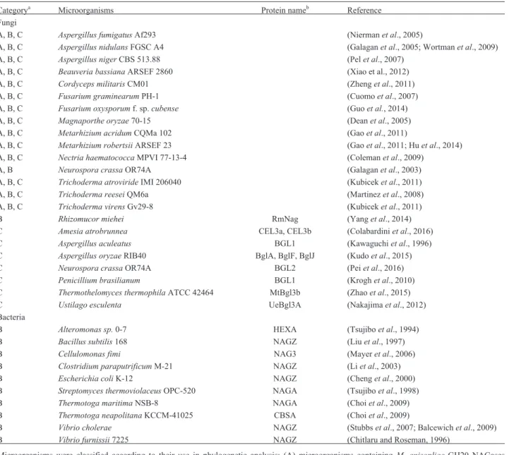
Table 1- List of microorganisms used in GH20 and GH3 NAGases phylogenetic analysis.
Categorya Microorganisms Protein nameb Reference
Fungi
A, B, C Aspergillus fumigatusAf293 (Niermanet al., 2005)
A, B, C Aspergillus nidulansFGSC A4 (Galaganet al., 2005; Wortmanet al., 2009)
A, B, C Aspergillus nigerCBS 513.88 (Pelet al., 2007)
A, B, C Beauveria bassianaARSEF 2860 (Xiao et al., 2012)
A, B, C Cordyceps militarisCM01 (Zhenget al., 2011)
A, B, C Fusarium graminearumPH-1 (Cuomoet al., 2007)
A, B, C Fusarium oxysporumf. sp.cubense (Guoet al., 2014)
A, B, C Magnaporthe oryzae70-15 (Deanet al., 2005)
A, B, C Metarhizium acridumCQMa 102 (Gaoet al., 2011)
A, B, C Metarhizium robertsiiARSEF 23 (Gaoet al., 2011; Huet al., 2014)
A, B, C Nectria haematococcaMPVI 77-13-4 (Colemanet al., 2009)
A, B Neurospora crassaOR74A (Galaganet al., 2003)
A, B, C Trichoderma atrovirideIMI 206040 (Kubiceket al., 2011)
A, B, C Trichoderma reeseiQM6a (Martinezet al., 2008)
A, B, C Trichoderma virensGv29-8 (Kubiceket al., 2011)
B Rhizomucor miehei RmNag (Yanget al., 2014) C Amesia atrobrunnea CEL3a, CEL3b (Colabardiniet al., 2016)
C Aspergillus aculeatus BGL1 (Kawaguchiet al., 1996) C Aspergillus oryzaeRIB40 BglA, BglF, BglJ (Kudoet al., 2015) C Neurospora crassaOR74A BGL2 (Peiet al., 2016) C Penicillium brasilianum BGL1 (Kroghet al., 2010) C Thermothelomyces thermophilaATCC 42464 MtBgl3b (Zhaoet al., 2015)
C Ustilago esculenta UeBgl3A (Nakajimaet al., 2012) Bacteria
B Alteromonas sp.0-7 HEXA (Tsujiboet al., 1994) B Bacillus subtilis168 NAGZ (Liuet al., 1997) B Cellulomonas fimi NAG3 (Mayeret al., 2006)
B Clostridium paraputrificumM-21 NAGZ (Liet al., 2003) B Escherichia coliK-12 NAGZ (Chenget al., 2000) B Streptomyces thermoviolaceusOPC-520 NAGA (Tsujiboet al., 1998) B Thermotoga maritimaNSB-8 NAGA (Choiet al., 2009) B Thermotoga neapolitanaKCCM-41025 CBSA (Choiet al., 2009)
B Vibrio cholerae NAGZ (Stubbset al., 2007; Balcewichet al., 2009) B Vibrio furnissii7225 NAGZ (Chitlaru and Roseman, 1996)
aMicroorganisms were classified according to their use in phylogenetic analysis: (A) microorganisms containingM. anisopliaeGH20 NAGases
orthologs; (B) microorganisms containingM. anisopliaeGH3 NAGases orthologs; and (C) microorganisms containingb-glucosidases included as an
outgroup in GH3 NAGase phylogenetic analysis.
analysis of variance (ANOVA), followed by Tukey’s mul-tiple comparisons test (p< 0.05) were performed to
deter-mine statistical differences among 2-DDCtvalues of the seven
experimental conditions.
Results
M. anisopliaeputative GH20 and GH3 NAGases
The survey of NAGase genes of theM. anisopliae
ge-nome, using NagA fromA. nidulansand NAG1 and NAG2
fromT. atrovirideas queries, resulted in the identification
of two putative GH20 NAGases, named MaNAG1 (MANI_010908; GenBank accession number KFG80340) and MaNAG2 (MANI_029504; GenBank accession num-ber KFG85702). All other fungal GH20 NAGase sequen-ces and GH20 conserved domain sequensequen-ces used as queries resulted in alignments with the same two previously de-tected contigs. Therefore, MaNAG1 and MaNAG2 are most probably the only M. anisopliae putative GH20
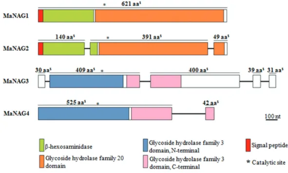
NAGases. The GH20 family domain (IPR015883) and the conserved motif of GH20 proteins (H/N-x-G-A/C/G/M-D-E-A/I/L/V) were found in both MaNAG1 and MaNAG2 sequences (Figure S2). Additionally, the putative GH20 NAGases also exhibited a chitobiase/beta-hexosaminidase N-terminal domain (IPR029018) (Figure 1).
The GH3 domain screening of theM. anisopliae
ge-nome allowed the identification of seven positive matches. However, phylogenetic analysis clearly revealed that only two sequences, named MaNAG3 (MANI_122030; GenBank accession number KFG78085) and MaNAG4
[MANI_128875; (Figure S3)] could be putative GH3 NAGases. Furthermore, these sequences exhibit higher similarity with bacterial GH3 NAGases and the RmNag GH3 (Figure S4). MaNAG3 and MaNAG4 share a con-served domain with GH3 family members (IPR001764) and exhibit the conserved sequence motif of GH3 proteins (K-H-F/I-P-G-H/L-G-x-x-x-x-D-S/T-H) (Figure S4). Fur-thermore, MaNAG3 and MaNAG4 sequences present a conserved GH3 C-terminal domain (IPR002772) (Figure 1). The other five putative GH3 proteins (KFG84234, KFG86760, KFG85258, KFG81708, and KFG84481) dis-play higher sequence conservation and are phylogenetic re-lated with fungalb-glucosidases (Figure 2 and Figure S4), raising the possibility of functional equivalence.
All properties of the proposedM. anisopliaeputative
NAGases are listed in Table 2. Putative GH20 NAGase genes have similar ORF sizes and exhibit no intron conser-vation between sequences. While MaNAG1 does not show any intron insertions, the MaNAG2 sequence has two in-tron insertions (Figure 1). The predicted molecular masses for MaNAG1 and MaNAG2 (66.98 kDa and 61.42 kDa, re-spectively) are similar to other fungal GH20 NAGases,A. nidulansNagA (68 kDa) (Kimet al., 2002),T. atroviride
Nag1 (73 kDa) (Brunneret al., 2003) andT. harzianumP1
Nag1 (72 kDa) (Peterbaueret al., 1996).
The theoretical pI ofM. anisopliaeGH20 NAGases
predicts that they are acidic enzymes, with MaNAG2 ex-hibiting a more acidic pI than MaNAG1, 4.85 and 6.07, re-spectively. Both putative GH20 NAGases have the same four N-glycosylation sites. Putative GH3 NAGase genes
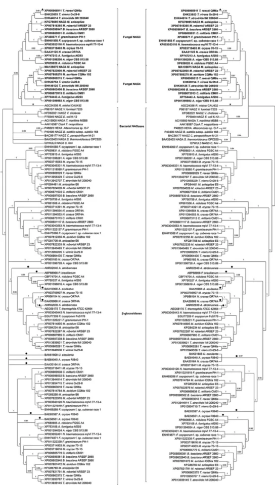
Figure 2- Phylogenetic relationships among GH3 NAGases from filamentous fungi, bacteria and zygomycetes. Putative and characterized fungal GH3
b-glucosidases were included as an outgroup. The phylogenies were obtained using MrBayes 3.2.5 (left side) and PhyML 3.1 (right side).Ø: NAGase
exhibit different physicochemical properties. MaNAG3 is the largest gene (3,223 bp), containing the highest expected number of introns (4) and theoretical molecular mass (98.71 kDa), with N-glycosylation translational modifica-tion signals on six sites. In contrast, MaNAG4 ORF size is 2,057 bp, the theoretical molecular mass is 60.67 kDa and the pI of predicted mature protein is 5.6. The predicted mo-lecular mass of MaNAG4 is similar to most known bacte-rial GH3 NAGases, asS. thermoviolaceusNagA (60 kDa)
(Tsujiboet al., 1998). None of the putative NAGase protein
sequences contain GPI-anchoring sites or non-classical se-cretion pathway prediction signals. Interestingly, both MaNAG1 and MaNAG2 have predicted secretion signal peptides, from which extracellular functions can be in-ferred. In contrast, putative GH3 NAGases are apparently cytoplasmic enzymes as they do not present any predicted secretion signals.
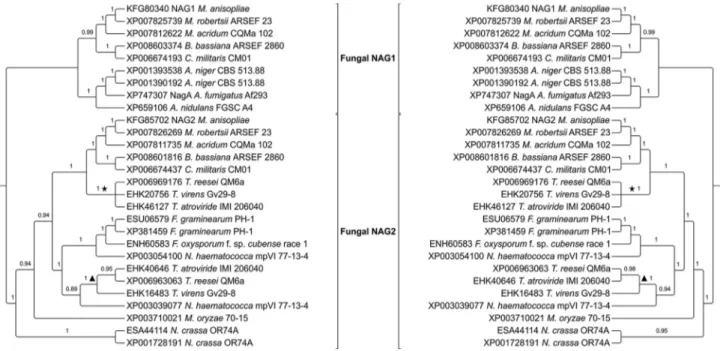
Phylogeny of putative GH20 NAGases
Twenty-six MaNAG1 and MaNAG2 orthologs were identified in 15 filamentous fungi genomes. Most of them are single copy of each putative GH20 NAGase of M. anisopliae. The conserved motif of GH20 proteins and the
highly conserved catalytic residues, aspartic and glutamic acids (D-E), were recognized in all of GH20 orthologs (Fig-ure S1).
The best-fit evolutionary model for GH20 NAGases was LG+I+G, which was used for the phylogenetic infer-ence. Phylogenetic analyses of GH20 NAGases fromM. anisopliaeand the other fifteen ascomycetes revealed an
early duplication event in GH20 NAGases, resulting in two distinct main clades (Figure 3). MaNAG1 formed a mono-phyletic group with other entomopathogenic fungi NAGase sequences (Metarhizium robertsii,Metarhizium acridum, Cordyceps militarisandBeauveria bassiana). This cluster
also formed a statistically supported clade with species from theAspergillusgenus. In contrast, MaNAG2 exhibits
a more diverse evolutionary history, with orthologs present in Trichoderma sp., Fusarium sp, Neurospora sp., and Magnaporthe sp. Interestingly, the present evolutionary
analysis revealed that both NAG1 and NAG2 from the mycoparasiteT. atroviride, used in theM. anisopliae
ge-nome screening, are evolutionarily more related to MaNAG2 (Figure 3).
For the majority of the 15 fungi analyzed, only one ortholog to MaNAG1 and one ortholog to MaNAG2 were detected in each species. Duplication events on a specific lineage resulting in paralogous proteins was only observed forAspergillus niger, which has two MaNAG1 orthologs,
and for Nectria haematococca, N. crassa and Fusarium graminearum, with two MaNAG2 orthologs.
Phylogeny of putative GH3 NAGases
Twenty-three MaNAG3 and MaNAG4 orthologs were identified on the filamentous fungi genomes
ined. Conserved sequence motifs of GH3 proteins (K-H-F / I-P-G-H / L-G-x-x-x-x-D-S / T-H) were found in all of them, however, few amino acid residues substitutions were observed (Figure S2). All 15 filamentous fungi have MaNAG3 orthologs. However, the M. acridum gene
ortholog was not included in the phylogenetic analysis, since it was not properly annotated in theM. acridum
ge-nome. In turn, onlyTrichodermasp.,Aspergillussp., and
the entomopathogens C. militaris and B. bassiana have
MaNAG4 orthologs.
To better understand GH3 NAGases evolutionary re-lationships, 10 well described bacterial GH3 NAGases and the characterized GH3 NAGase from the zygomyceteR. Miehei(Yanget al., 2014) were added to the phylogenetic
analysis (Table 1). Since several GH3 family fungal mem-bers withb-glucosidase activity have also been described (Kawaguchiet al., 1996; Kroghet al., 2010; Nakajimaet al., 2012; Kudoet al., 2015; Zhaoet al., 2015; Colabardini
et al., 2016; Peiet al., 2016), the phylogenetic relationships among the fungal, bacterial, andR. mieheiGH3 NAGases were inferred including as outgroup putative b
-glucosi-dases fromM. anisopliaeE6, characterized fungalb
-glu-cosidases and putative b-glucosidases from species described in Table 1. The best-fit evolutionary model for GH3 NAGases was LG+I+G. The evolutionary relation-ship of all GH3 proteins showed two distinct clades sepa-rating fungal and bacterial NAGases fromb-glucosidases
(Figure 2).
The phylogenetic tree revealed that MaNAG3 and MaNAG4 orthologs formed two distinct clusters (Figure 2). Both MaNAG3 and MaNAG4 grouped to other
Metarhizium species, but in contrast with the GH20
NAGases phylogeny, putative GH3 NAGases from
Metarhiziumsp. are evolutionarily more distant from
puta-tive GH3 NAGases of other entomopathogenic fungi (B. bassianaandC. militaris). Additionally, gene duplication
of MaNAG3 and MaNAG4 orthologs was not observed. Bacterial sequences did not form a monophyletic group, but they are basal in relation to fungal NAG3 and NAG4 (Figure 2). The difference between bacterial NAGases apparently is not related to gram-positive or gram-negative structural classification. It was also obser-ved that even bacterial NAGases with high chitinolytic sub-strate specificity (S. thermoviolaceus NagA, Clostridium paraputrificum NagZ, Alteronomonas sp. HexA, V. furnissii NagZ, Thermotoga maritma NagA and T. neapolitanaCbsA) grouped into distinct clades from fun-gal NAGases. This is probably due to the fact that some bacterial NAGases do not necessarily have GlcNAc hydro-lysis specificity over chitooligosaccharides. For example,
E. coliNagZ cleaves GlcNAc from muropeptides present in the bacterial cell wall (Chenget al., 2000).C. fimiNag3 is also an unusual GH3 NAGase, because it is ab
-N-acetyl-hexosaminidase with a wide range of substrates, hydrolyz-ing both b-N-acetylglucosaminedes and b-glucosides
(Mayeret al., 2006).
Patterns of transcript relative expression of putative NAGases
The expression profile of M. anisopliae putative
NAGases was investigated in different cell types under dif-ferent culture conditions: mycelium grown on glucose 1%, GlcNAc 0.25%, chitin 1% or autolysis conditions; and in-duced conidia, blastospore and appressorium. The four
pu-Figure 3- Phylogenetic relationships among GH20 NAGases from filamentous fungi. The phylogenies were obtained using MrBayes 3.2.5 (left side) and
tative NAGase gene transcripts were detected in all M. anisopliaecell types and culture conditions, validating the
annotation of the proposed genes.
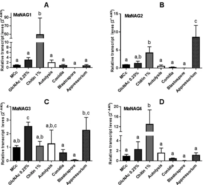
To gain information on the regulation of the putative NAGases by substrate, the transcript level of genes fromM. anisopliaecultured in MCc medium was established as a reference condition (Figure 4). Interestingly, the expression of MaNAG1, MaNAG2 and MaNAG4 were induced by 1% chitin, albeit at different levels (Figure 4). Notably, MaNAG1 showed the most pronounced expression induc-tion on this carbon source (Figure 4A). Addiinduc-tionally, MaNAG3 was the only MaNAGase induced in cultures with added 0.25% GlcNAc (Figure 4C). When different cellular types were taken into account, MaNAG3 exhibited detectable transcripts in cells forming appressorium, while
MaNAG2 was strongly induced in this cell type (Figure 4B). The expression of the four putative NAGases gene showed only basal levels in conidia and blastospores(
Fig-ure 4). These results indicate the minor participation of
pu-tative GH3 and GH20 in conidia and blastospores.
Discussion
Virulence determinants are the main focus in the study of entomopathogenic fungi (Schrank and Vainstein, 2010). As chitin is present in the exoskeleton of several ar-thropods, enzymes involved in chitin degradation and assimilation are predicted to play essential roles in host-entomopathogen interactions (Schrank and Vainstein, 2010). While chitinases are widely explored in entomo-pathogens and several fungal species with diverse
patho-Figure 4- Relative expression of GH20 and GH3 NAGase genes inM. anisopliae, considering MCc as the reference condition. Transcriptional profiles of GH20 NAGase genes (MaNAG1 and MaNAG2) and GH3 NAGase genes (MaNAG3 and MaNAG4) in seven different conditions (mycelium growth
on different carbon source media, autolysis, and different cell types), usingtef1aas a reference gene and applying the 2-DDCtmethod. A)nag1; B)nag2; C)
genic traits, the role of NAGases in the fungal life cycle and their importance in infection has not been explored. Here, four putative NAGase genes belonging to the GH20 family (MaNAG1andMaNAG2) and GH3 family (MaNAG3and MaNAG4) ofM. anisopliaegenome were analyzed.
St. Legeret al.(1991) purified a secreted NAGase
fromM. anisopliaeby gel-filtration, with a pI of 6.4 and
molecular mass of 110-120 kDa. We hypothesize that this
M. anisopliaepurified enzyme could be the MaNAG1
pre-sented here, based on the predicted pI (6.07) and molecular mass (66.98 kDa) of MaNAG1, likely forming a homo-dimer. In fact, some fungal GH20 NAGases (Kogaet al.,
1991; Rylavá et al., 2011) and some bacterial GH3
NAGases (Choiet al., 2009) exhibit a homodimer
compo-sition. Nevertheless, the molecular characterization ofM. anisopliaeputative NAGases will be necessary to
determi-nate if the dimer structure is relevant to enzymatic activity.
Phylogenetic analyses of putative GH20 NAGases re-vealed the occurrence of at least one duplication event be-fore its divergence in fungi. This early gene duplication is supported by evolutionary analysis of GH20 family from several eukaryotic taxa, reported by Intra et al. (2008).
Comparing the evolutionary history of MaNAG1 and MaNAG2, subsequent duplication events resulted in cur-rent presence of multiple GH20 NAGase orthologs in ascomycetes. This phenomenon was more frequent in the MaNAG2 than the MaNAG1 cluster, culminating in the presence of MaNAG2 orthologs in a broader spectrum of fungi with different lifestyles. While MaNAG1 has ortho-logs only in entomopathogens and in the saprophytic/hu-man pathogens Aspergillus sp., MaNAG2 orthologs are
present in entomopathogens, mycopathogen species, such as Trichoderma sp., phytopathogens including N. haematococca, Fusarium sp. and M. oryzae, and in
saprophytes, such asN. crassa. These species belong to
dis-tinct orders, however, a previous study has observed their close evolutionary relationship (Wanget al., 2009). The
widespread presence of MaNAG2 orthologs in fungi with diverse lifestyles could represent a common basic function for all these enzymes despite differences in fungal life-styles, while MaNAG1 would have more specific roles in an entomopathogenic lifestyle.
In our analysis,M. anisopliaeandM. robertsii
for-med a statistically well supported clade, withM. acridumas
a basal species, corroborating the phylogeny relationships among theseMetarhiziumspecies (Bischoff et al., 2009;
Staatset al., 2014). Our results revealed a close
evolution-ary relationship of GH20 NAGases between the
Metarhiziumclade and the one formed byBeauveriaand Cordycepsgenera. The conservation of secreted proteins in
fungi has been observed amongM. anisopliae and
ento-mopathogens Metarhizium spp., B. bassiana and C. militaris(Staatset al., 2014). Therefore, the evolutionary
pattern of GH20 NAGases in entomopathogens is
represen-tative of the extremely similar evolutionary pattern of all secreted proteins found in fungi with similar hosts.
The glycoside hydrolases from the CAZy family GH3 display an unusual diversity in structure, specificity, and bi-ological roles (Macdonaldet al., 2015). In many cases the
enzymes have dual or broad substrate specificities with re-spect to monosaccharide residues, linkage position and chain length of the substrate. This family harbors members with several activities, most notably b-glucosidases and
NAGases (Macdonaldet al., 2015). Several fungalb -glu-cosidases from the GH3 family have been characterized (Kawaguchiet al., 1996; Kroghet al., 2010; Nakajimaet al., 2012; Kudoet al., 2015; Zhaoet al., 2015; Colabardini
et al., 2016; Pei et al., 2016), however the first fungal NAGase from family GH3 was only recently described (RmNag) (Yanget al., 2014).
The first goal of our phylogenetic analyses was to clearly distinguish putative NAGases from putativeb
-glu-cosidases. The phylogenetic analysis set apart putative GH3 NAGases from putative GH3b-glucosidases (Figure
3), suggesting GH3 NAGase activity for MaNAG3 and MaNAG4. Indeed, the characterized RmNag clustered to-gether with MaNAG3 and MaNAG4 with robust support, suggesting the possibility of similar functions of these en-zymes (Figure 3). The five other putative GH3 proteins fromM. anisopliae(KFG84234, KFG86760, KFG85258,
KFG81708, and KFG84481) clustered together with char-acterizedb-glucosidases, again suggesting the possibility
of similar function (Figure 2). Additionally, several charac-terized bacterial GH3 NAGases were more phylogeneti-cally related with MaNAG3, MaNAG4 and RmNag than with the characterizedb-glucosidases (Figure 2).
Bacterial NAGases were added to phylogenetic anal-yses, which show well-established acetyl-chitooligo-saccharide degradation activity, and NAGases with other substrate specificities, such as NagZ from E. coli and
NAG3 fromC. fimi.E. coliNagZ participates in bacteria
cell wall recycling by hydrolyzing GlcNAc from muro-peptides (Chenget al., 2000). In turn,C. fimiNAG3 was
identified as a bifunctionalbNacetylDglucosaminida
-se/b-D-glucosidase (Mayer et al., 2006). It was also re-ported thatC. fimiNAG3 enzyme is actually a GlcNAc-phosphorylase using phosphate rather than water as nu-cleophile (Macdonald et al., 2015). Macdonald’s study suggests that other GH3 NAGases can harbor GlcNAc-phosphorylase activity. Notably, our GH3 phylogenetic analysis showed thatC. fimiNAG3 has a basal position in relation to other bacterial and fungal NAGases with chitin specificity, supporting this suggestion. However, comple-mentary experiments are required to evaluate this putative GlcNAc-phosphorylase activity.
duplications that occurred after the divergence between bacteria and the fungi GH3 family genes (Figure 3). Fungal orthologs of MaNAG3 and MaNAG4 formed two distinct clades. In relation to the NAG4 clade, MaNAG4 was ar-ranged closer to GH3 NAGases of the mycoparasitic
Trichodermasp. than orthologs of entomopathogenic
spe-ciesC. militarisandB. bassiana. The NAG3 from
ento-mopathogens formed a monophyletic group with
Trichoderma species, with MaNAG3 basal to them. It
seems thatM. anisopliaeGH3 NAGases may not have
spe-cific roles in entomopathogenic fungal lifestyle. However, at this point, their participation in basal cell processes can-not be ruled out, such as GlcNAc carbon metabolism and cell wall remodeling, both processes necessary to hyphal growth and cell differentiation.
The qPCR assays of putative GH20 and GH3 NAGase genes confirmed that the identified sequences are functional.M. anisopliaeputative NAGases showed
differ-ential transcript profiles in response to different conditions, indicating an absence of a common gene regulation pattern. These variable expression profiles also suggest they may not have totally redundant roles. M. anisopliae GH20
NAGases,MaNAG1andMaNAG2, exhibited induced
ex-pression patterns when cultured in the presence of 1% chitin. Our results reflect the well-established condition, where chitin induces the expression of secreted chitinolytic enzymes (St. Legeret al., 1991; Seidl, 2008). The presence
of a predicted signal peptide for secretion in MaNAG1 and MaNAG2, and their expression induction by chitin reveal their probable role in extracellular chitinolytic activity in
M. anisopliae, acting on extracellular cleavage of
chito-biose into GlcNAc monomers for the assimilation of this carbon source. Nonetheless, it is important to note that other carbon sources are also able to stimulate, at lower lev-els, the expression of GH20 NAGases (Seidlet al., 2006).
The expression profile of M. anisopliae putative
NAGases in appressorium is noteworthy. MaNAG2 was the most significantly expressed NAGase in appressorium, being highly induced in this cell type. The appressorium is a specialized penetration structure that helps to dissolve the host chitinous exoskeleton. These cells use enzyme secre-tion and physical pressure to mediate penetrasecre-tion. There-fore, it can be suggested that MaNAG2 is putatively required at early stages of infection, during the penetration stage or to remodel the fungal cell wall in appressorium dif-ferentiation.
It was expected that 0.25% GlcNAc would induceM. anisopliae NAGases, because this low concentration of
GlcNAc is described as an inducer for chitinolytic genes, and only high monomer concentrations (> 0.5%) would re-press exre-pression of chitinolytic enzymes by their own activity products in M. anisopliae (catabolic repression)
(Barretoet al., 2004). Similarly,T. atroviride nag1
expres-sion is induced by GlcNAc (Machet al., 1999). However,
the transcript expression analysis showed that M.
anisopliaeputative GH20 NAGases were not induced by
GlcNAc. MaNAG3 was the only putative NAGase induced by 0.25% GlcNAc and the only putative NAGase that was not induced by 1% chitin, suggesting a possible regulatory mechanism for this gene, in which the expression could de-pend on the prior degradation of chitin to GlcNAc. Further-more, no transcript induction of any NAGases was observed in blastospores and conidia (Figure 4), which are cellular forms with diminished metabolic activity, although not completely dormant (Novodvorska et al., 2016). In
addition, M. anisopliae conidial extracts and
immuno-proteomic analysis indicate that chitinases may be local-ized on the conidial surface (Santi et al., 2009, 2010),
NAGase activity is probably not necessary in these resting cells. In contrast, blastospores are cell types that facilitate dispersal in host hemolymph during colonization. At this stage, the fungus has already transposed the chitinous exoskeleton and uses trehalose and other carbon sources, not requiring, necessarily, the expression of chitinolytic en-zymes (Xiaet al., 2002). Nevertheless, it is important to
note that the ADAMEK media used to induce blastospores does not fully mimic the arthropod inner body complexity, and GH3 and GH20 NAGase activity may be required in specific steps of blastospore differentiation and infection.
Chitinases and NAGases act consecutively and syner-gistically to render complete degradation of chitin. This may be the result of common regulation patterns between these two groups of enzymes, as revealed inT. atroviride
NAGase studies (Tharanathan and Kittur, 2003). The ex-perimental conditions used in this study for the evaluation of M. anisopliae putative NAGase expression were the
same employed for the study of the 21 chitinases fromM. anisopliae(Jungeset al., 2014). This allowed the
compari-son of the performance of different genes of the chitinolytic process to propose potential relationships between specific chitinases and NAGases. Jungeset al.(2014) described a
large group of chitinases induced by chitin: chimaA1, chimaA6, chimaA8, chimaB1, chimaB2, chimaB3, chimaB4,chimaB6,chimaC3. This expression pattern can
be associated withMaNAG1,MaNAG2andMaNAG4that
also displayed increased expression profile in the presence of chitin. Also, those putative NAGases induced by chitin could be followed by chitinase action induced by GlcNAc monomers (chimaD1) (Jungeset al., 2014). On media
sup-plemented with the GlcNAc monomer, the MaNAG3 gene showed strong expression when compared to the otherM. anisopliae putative NAGases, coinciding with the chimaD1chitinase pattern. Moreover, in the induced
apres-sorium formation condition, the expression of MaNAG2 could be related to chimaA5 chitinase, since both are
overexpressed in this cellular type.
Our results are in agreement to previous suggestions of the presence of GH3 NAGases in fungi (RmNag) (Yang
et al., 2014) and in the Hypocreales order (Kappelet al.,
char-acterized a GH3 gene (namednag3; XP006966911) inT. reesei,the product of which, an MaNAG3 ortholog, holds
suggested NAGase activity. The phylogenetic analyses in-dicate that MaNAG3 andT. reeseiNAG3 are
phylogeneti-cally related (Figure 2). The existence of more putative NAGase genes argues that the genomic arsenal of NAGases in ascomycetes is not as small as previously thought, attenuating the discrepancy between the number of chitinase and NAGase genes. It is also not possible to rule out the existence of other unknown NAGases inM. anisopliaeand other fungal species. In this sense, we have
identified a fifth and unexplored putative NAGase gene in
M. anisopliae, belonging to the GH84 family. The product
of this gene (KFG85933.1) exhibits 63% identity with char-acterized GH84 from Penicillium chrysogenum
(XP_002557703.1). TheP. chrysogenum GH84 NAGase
not only exhibit activity against GlcNAc substrates, but also hydrolyzes substrates withgalacto-configuration and
exhibits transglycosylation activity (Slámováet al., 2014).
In conclusion, this study explored relevant evolution-ary aspects of putative GH3 and GH20 NAGase genes and the expression analysis highlighted possible functions for these genes inM. anisopliaeand entomopathogenic fungi.
This analysis will allow the selection of genes for further functional characterization to elucidate the process and to identify redundancies and specificities. The view that chiti-nase diversity is merely redundant may not correct (Seidlet al., 2005; Tzelepiset al., 2012; Jungeset al., 2014).
How-ever, the strategy of constructing deleted strains is not al-ways straightforward to determine function (Alcazar-Fuoli
et al., 2011). Here,M. anispoliaeputative GH20 NAGase
genes revealed induced transcript production in the pres-ence of chitin, potentially in the extracellular milieu. The detection ofMaNAG3andMaNAG4putative genes is the
first evidence for the presence of a possible GH3 family of NAGases in entomopathogenic fungi. MaNAG3 and MaNAG4expression is responsive to chitinous substrates,
suggesting their potential influence on cell differentiation during theM. anisopliaelife cycle.
Acknowledgments
This study was supported by grants and fellowships from CNPq, CAPES and FAPERGS.
References
Adamek L (1965) Submerse cultivation of the fungus
Metarhizium anisopliae(Metsch.). Folia Microbiol (Praha) 10:255–257.
Alcazar-Fuoli L, Clavaud C, Lamarre C, Aimanianda V, Seidl-Seiboth V, Mellado E and Latgé JP (2011) Functional analy-sis of the fungal/plant class chitinase family inAspergillus fumigatus. Fungal Genet Biol 48:418–429.
Andersen CL, Jensen JL and Ørntoft TF (2004) Normalization of real-time quantitative reverse transcription-PCR data: a model-based variance estimation approach to identify genes
suited for normalization, applied to bladder and colon cancer data sets. Cancer Res 64:5245–5250.
Anisimova M and Gascuel O (2006) Approximate likelihood-ratio test for branches: A Fast, accurate, and powerful alter-native. Syst Biol 55:539–552.
Anisimova M, Gil M, Dufayard JF, Dessimoz C and Gascuel O (2011) Survey of branch support methods demonstrates ac-curacy, power, and robustness of fast likelihood-based ap-proximation schemes. Syst Biol 60:685–699.
Balcewich MD, Stubbs KA, He Y, James TW, Davies GJ, Voca-dlo DJ and Mark BL (2009) Insight into a strategy for attenu-ating AmpC-mediated beta-lactam resistance: structural ba-sis for selective inhibition of the glycoside hydrolase NagZ. Protein Sci 18:1541–51.
Barreto CC, Staats CC, Schrank A and Vainstein MH (2004) Dis-tribution of chitinases in the entomopathogen metarhizium anisopliae and effect of N-acetylglucosamine in protein se-cretion. Curr Microbiol 48:102–107.
Beier S and Bertilsson S (2013) Bacterial chitin degradation – mechanisms and ecophysiological strategies. Front Micro-biol 4:149.
Bendtsen JD, Jensen LJ, Blom N, Von Heijne G and Brunak S (2004) Feature-based prediction of non-classical and leader-less protein secretion. Protein Eng Des Sel 17:349–56. Bischoff JF, Rehner SA and Humber RA (2009) A multilocus
phylogeny of theMetarhizium anisopliae lineage. Myco-logia 101:512–530.
Bjellqvist B, Hughes GJ, Pasquali C, Paquet N, Ravier F, Sanchez JC, Frutiger S and Hochstrasser D (1993) The focusing posi-tions of polypeptides in immobilized pH gradients can be predicted from their amino acid sequences. Electrophoresis 14:1023–31.
Bjellqvist B, Basse B, Olsen E and Celis JE (1994) Reference points for comparisons of two-dimensional maps of proteins from different human cell types defined in a pH scale where isoelectric points correlate with polypeptide compositions. Electrophoresis 15:529–39.
Boldo JT, Junges A, Amaral KB, Staats CC, Vainstein MH and Schrank A (2009) Endochitinase CHI2 of the biocontrol fungusMetarhizium anisopliaeaffects its virulence toward the cotton stainer bug Dysdercus peruvianus. Curr Genet 55:551–60.
Brunner K, Peterbauer CK, Mach RL, Lorito M, Zeilinger S and Kubicek CP (2003) The Nag1 N-acetylglucosaminidase of
Trichoderma atrovirideis essential for chitinase induction
by chitin and of major relevance to biocontrol. Curr Genet 43:289–295.
Brzezinska MS, Jankiewicz U, Burkowska A and Walczak M (2014) Chitinolytic microorganisms and their possible ap-plication in environmental protection. Curr Microbiol 68:71–81.
Cantarel BL, Coutinho PM, Rancurel C, Bernard T, Lombard V and Henrissat B (2009) The Carbohydrate-Active EnZymes database (CAZy): An expert resource for glycogenomics. Nucleic Acids Res 37:D233–8.
Chauhan JS, Rao A and Raghava GPS (2013)In silicoplatform for prediction of N-, O- and C-glycosites in eukaryotic pro-tein sequences. PLoS One 8:e67008.
Cheng Q, Li H, Merdek K and Park JT (2000) Molecular charac-terization of the beta-N-acetylglucosaminidase of Esche-richia coli and its role in cell wall recycling. J Bacteriol 182:4836–40.
Chitlaru E and Roseman S (1996) Molecular cloning and charac-terization of a novel beta-N-acetyl-D-glucosaminidase from
Vibrio furnissii. J Biol Chem 271:33433–9.
Choi KH, Seo JY, Park KM, Park CS and Cha J (2009) Character-ization of glycosyl hydrolase family 3 b -N-acetylgluco-saminidases from Thermotoga maritima and Thermotoga neapolitana. J Biosci Bioeng 108:455–459.
Colabardini AC, Valkonen M, Huuskonen A, Siika-aho M, Koi-vula A, Goldman GH and Saloheimo M (2016) Expression of two novel b -glucosidases from Chaetomium atrobrunneum inTrichoderma reeseiand characterization
of the heterologous protein products. Mol Biotechnol 58:821–831.
Coleman JJ, Rounsley SD, Rodriguez-Carres M, Kuo A, Was-mann CC, Grimwood J, Schmutz J, Taga M, White GJ, Zhou Set al.(2009) The genome ofNectria haematococca:
con-tribution of supernumerary chromosomes to gene expan-sion. PLoS Genet 5:e1000618.
Cuomo CA, Güldener U, Xu JR, Trail F, Turgeon BG, Di Pietro A, Walton JD, Ma LJ, Baker SE, Rep Met al.(2007) The
Fusarium graminearumgenome reveals a link between lo-calized polymorphism and pathogen specialization. Science 317:1400–2.
da Silva MV, Santi L, Staats CC, da Costa AM, Colodel EM, Driemeier D, Vainstein MH and Schrank A (2005) Cuti-cle-induced endo/exoacting chitinase CHIT30 from
Metarhizium anisopliaeis encoded by an ortholog of the
chi3gene. Res Microbiol 156:382–392.
Darriba D, Taboada GL, Doallo R and Posada D (2011) Europe PMC Funders Group ProtTest 3: Fast selection of best-fit models of protein evolution. Bioinformatics 27:1164–1165. Dean RA, Talbot NJ, Ebbole DJ, Farman ML, Mitchell TK,
Orbach MJ, Thon M, Kulkarni R, Xu JR, Pan Het al.(2005) The genome sequence of the rice blast fungusMagnaporthe grisea. Nature 434:980–6.
Díez B, Rodríguez-Sáiz M, De La Fuente JL, Moreno MÁ and Barredo JL (2005) The nagA gene of Penicillium chrysogenumencodingb-N- acetylglucosaminidase. FEMS Microbiol Lett 242:257–264.
Duo-Chuan L (2006) Review of fungal chitinases. Mycopatho-logia 161:345–60.
Eisenhaber B, Schneider G, Wildpaner M and Eisenhaber F (2004) A sensitive predictor for potential GPI lipid modifi-cation sites in fungal protein sequences and its applimodifi-cation to genome-wide studies for Aspergillus nidulans, Candida albicans, Neurospora crassa, Saccharomyces cerevisiae
andSchizosaccharomyces pombe. J Mol Biol 337:243–53.
Galagan JE, Calvo SE, Borkovich KA, Selker EU, Read ND, Jaffe D, FitzHugh W, Ma LJ, Smirnov S, Purcell Set al.(2003)
The genome sequence of the filamentous fungus
Neurospora crassa. Nature 422:859–68.
Galagan JE, Calvo SE, Cuomo C, Ma LJ, Wortman JR, Batzoglou S, Lee SI, Bastürkmen M, Spevak CC, Clutterbuck Jet al.
(2005) Sequencing ofAspergillus nidulansand comparative analysis with A. fumigatus and A. oryzae. Nature
438:1105–15.
Gao Q, Jin K, Ying SH, Zhang Y, Xiao G, Shang Y, Duan Z, Hu X, Xie XQ, Zhou Get al.(2011) Genome sequencing and comparative transcriptomics of the model entomopatho-genic fungiMetarhizium anisopliaeandM. acridum. PLoS Genet 7:e1001264.
Guindon S, Dufayard JF, Lefort V, Anisimova M, Hordijk W and Gascuel O (2010) New algorithms and methods to estimate maximum-likelihood phylogenies: Assessing the perfor-mance of PhyML 3.0. Syst Biol 59:307–321.
Guo L, Han L, Yang L, Zeng H, Fan D, Zhu Y, Feng Y, Wang G, Peng C, Jiang X et al.(2014) Genome and transcriptome
analysis of the fungal pathogenFusarium oxysporumf. sp. cubense causing banana vascular wilt disease. PLoS One 9:e95543.
Hall TA (1999) BioEdit: A user-friendly biological sequence aligment editor and analysis program for Windows 95/98/NT. Nucleic Acids Symp Ser 41:95–98.
Hartl L, Zach S and Seidl-Seiboth V (2012) Fungal chitinases: Di-versity, mechanistic properties and biotechnological poten-tial. Appl Microbiol Biotechnol 93:533–543.
Hu X, Xiao G, Zheng P, Shang Y, Su Y, Zhang X, Liu X, Zhan S, St Leger RJ and Wang C (2014) Trajectory and genomic de-terminants of fungal-pathogen speciation and host adapta-tion. Proc Natl Acad Sci U S A 111:16796–801.
Intra J, Pavesi G and Horner DS (2008) Phylogenetic analyses suggest multiple changes of substrate specificity within the glycosyl hydrolase 20 family. BMC Evol Biol 8:214. Junges Â, Boldo JT, Souza BK, Guedes RLM, Sbaraini N,
Kmetzsch L, Thompson CE, Staats CC, Almeida LGP, Vas-concelos ATRet al.(2014) Genomic analyses and
transcrip-tional profiles of the glycoside hydrolase family 18 genes of the entomopathogenic fungus Metarhizium anisopliae. PLoS One 9:e107864.
Kappel L, Gaderer R, Flipphi M and Seidl-Seiboth V (2016) The N-acetylglucosamine catabolic gene cluster inTrichoderma reesei is controlled by the Ndt80-like transcription factor
RON1. Mol Microbiol 99:640–657.
Kawaguchi T, Enoki T, Tsurumakia S, Ooib T and Arai M (1996) Cloning and sequencing of the cDNA encoding b -gluco-sidase 1 fromAspergillus aculeatus. Gene 173:287–288. Kim S, Matsuo I, Ajisaka K, Nakajima H and Kitamoto K (2002)
Cloning and characterization of the nagA gene that encodes beta-n-acetylglucosaminidase from Aspergillus nidulans
and its expression inAspergillus oryzae. Biosci Biotechnol
Biochem 66:2168–2175.
Koga K, Iwamoto Y, Sakamoto H, Hatano K, Sano M and Kato I (1991) Purification and characterization of b -N-Acetyl-hexosaminidase fromTrichoderma harzianum. Agric Biol Chem 55:2817–2823.
Krogh A, Larsson B, von Heijne G and Sonnhammer EL (2001) Predicting transmembrane protein topology with a hidden Markov model: Application to complete genomes. J Mol Biol 305:567–80.
Krogh KBRM, Harris PV, Olsen CL, Johansen KS, Hojer-peder-sen J, Borjesson J and Olsson L (2010) Characterization and kinetic analysis of a thermostable GH3b-glucosidase from
Penicillium brasilianum. Appl Microbiol Biotechnol
86:143–154.
ge-nome sequence analysis underscores mycoparasitism as the ancestral life style ofTrichoderma. Genome Biol 12:R40. Kudo K, Watanabe A, Ujiie S, Shintani T and Gomi K (2015)
Pu-rification and enzymatic characterization of secretory glyco-side hydrolase family 3 (GH3) arylb-glucosidases screened from Aspergillus oryzae genome. J Biosci Bioeng
120:614–623.
Langner T and Göhre V (2015) Fungal chitinases: Function, regu-lation, and potential roles in plant/pathogen interactions. Curr Genet 62:243–254.
Li H, Morimoto K, Katagiri N, Kimura T, Sakka K, Lun S and Ohmiya K (2003) A novelb-N-acetylglucosaminidase of Clostridium paraputrificum M-21 with high activity on chitobiose. Appl Microbiol Biotechnol 60:420–427. Liu H, Haga K, Yasumoto K, Ohashi Y, Yoshikawa H and
Takahashi H (1997) Sequence and analysis of a 31 kb seg-ment of theBacillus subtilischromosome in the area of the rrnH and rrnG operons. Microbiology 143:2763–7. Liu T, Yan J and Yang Q (2012) Comparative biochemistry of
GH3, GH20 and GH84b-N-acetyl-D-hexosaminidases and recent progress in selective inhibitor discovery. Curr Drug Targets 13:512–525.
Livak KJ and Schmittgen TD (2001) Analysis of relative gene ex-pression data using real-time quantitative PCR and the 2(-Delta Delta C(T)) method. Methods 25:402–408. López-Mondéjar R, Catalano V, Kubicek CP and Seidl V (2009)
Theb-N-acetylglucosaminidases NAG1 and NAG2 are
es-sential for growth of Trichoderma atroviride on chitin. FEBS J 276:5137–5148.
Löytynoja A and Goldman N (2010) webPRANK: A phylog-eny-aware multiple sequence aligner with interactive align-ment browser. BMC Bioinformatics 11:579.
Macdonald SS, Blaukopf M and Withers SG (2015) N-acetyl-glucosaminidases from CAZy family GH3 are really gly-coside phosphorylases, thereby explaining their use of histidine as an acid/base catalyst in place of glutamic acid. J Biol Chem 290:4887–4895.
Mach RL, Peterbauer CK, Payer K, Jaksits S, Woo SL, Zeilinger S, Kullnig CM, Lorito M, Kubicek CP (1999) Expression of two major chitinase genes of Trichoderma atroviride (T. harzianumP1) is triggered by different regulatory signals. Applied and Environmental Microbiology 65:1858–63. Marchler-Bauer A, Anderson JB, Chitsaz F, Derbyshire MK,
DeWeese-Scott C, Fong JH, Geer LY, Geer RC, Gonzales NR, Gwadz Met al.(2009) CDD: Specific functional anno-tation with the Conserved Domain Database. Nucleic Acids Res 37:D205–10.
Martinez D, Berka RM, Henrissat B, Saloheimo M, Arvas M, Baker SE, Chapman J, Chertkov O, Coutinho PM, Cullen D
et al.(2008) Genome sequencing and analysis of the
bio-mass-degrading fungusTrichoderma reesei(syn.Hypocrea jecorina). Nat Biotechnol 26:553–60.
Mayer C, Vocadlo DJ, Mah M, Rupitz K, Stoll D, Warren R AJ and Withers SG (2006) Characterization of a beta-N-acetyl-hexosaminidase and a beta-N-acetylglucosaminidase/beta-glucosidase from Cellulomonas fimi. FEBS J 273:2929–2941.
Nakajima M, Yamashita T, Takahashi M, Nakano Y and Takeda T (2012) Identification, cloning, and characterization ofb
-glucosidase from Ustilago esculenta. Appl Microbiol
Biotechnol 93:1989–1998.
Nierman WC, Pain A, Anderson MJ, Wortman JR, Kim HS, Ar-royo J, Berriman M, Abe K, Archer DB, Bermejo Cet al.
(2005) Genomic sequence of the pathogenic and allergenic filamentous fungus Aspergillus fumigatus. Nature 438:1151–6.
Novodvorska M, Stratford M, Blythe MJ, Wilson R, Beniston RG and Archera DB (2016) Metabolic activity in dormant conidia of Aspergillus niger and developmental changes during conidial outgrowth. Fungal Genet Biol 94:23–31. Patil RS, Ghormade V and Deshpande MV (2000) Chitinolytic
enzymes: An exploration. Enzyme Microb Technol 26:473–483.
Pei X, Zhao J, Cai P, Sun W, Ren J, Wu Q, Zhang S and Tian C (2016) Heterologous expression of a GH3 b-glucosidase fromNeurospora crassainPichia pastoriswith high purity and its application in the hydrolysis of soybean isoflavone glycosides. Protein Expr Purif 119:75–84.
Pel HJ, de Winde JH, Archer DB, Dyer PS, Hofmann G, Schaap PJ, Turner G, de Vries RP, Albang R, Albermann Ket al.
(2007) Genome sequencing and analysis of the versatile cell factory Aspergillus niger CBS 513.88. Nat Biotechnol
25:221–31.
Peterbauer CK, Lorito M, Hayes CK, Harman GE and Kubicek CP (1996) Molecular cloning and expression of the nag1 gene (N-acetyl-beta-D-glucosaminidase-encoding gene) fromTrichoderma harzianumP1. Curr Genet 30:325–331.
Petersen TN, Brunak S, von Heijne G and Nielsen H (2011) SignalP 4.0: Discriminating signal peptides from transmembrane regions. Nat Methods 8:785–6.
Pinto ADS, Barreto CC, Schrank A, Ulhoa CJ and Vainstein MH (1997) Purification and characterization of an extracellular chitinase from the entomopathogen Metarhizium anisopliae. Can J Microbiol 43:322–327.
Rast DM, Horsch M, Furter R and Gooday GW (1991) A complex chitinolytic system in exponentially growing mycelium of
Mucor rouxii: Properties and function. J Gen Microbiol 137:2797–810.
Ronquist F, Teslenko M, van der Mark P, Ayres DL, Darling A, Höhna S, Larget B, Liu L, Suchard MA and Huelsenbeck JP (2012) MrBayes 3.2: Efficient Bayesian phylogenetic infer-ence and model choice across a large model space. Syst Biol 61:539–542.
Rylavá H, Kalendová A, Doubnerová V, Skocdopol P, Kumar V, Kukacka Z, Pompach P, Vanek O, Slámová K, Bojarová Pet al.(2011) Enzymatic characterization and molecular model-ing of an evolutionarily interestmodel-ing fungalb
-N-acetylhexo-saminidase. FEBS J 278:2469–2484.
Santi L, Silva WOB, Pinto AFM, Schrank A and Vainstein MH (2009) Differential immunoproteomics enables identifica-tion of Metarhizium anisopliae proteins related to Rhipicephalus microplus infection. Res Microbiol 160:824–828.
Santi L, Beys da Silva WO, Berger M, Guimarães JA, Schrank A and Vainstein MH (2010) Conidial surface proteins of
Metarhizium anisopliae: Source of activities related with
toxic effects, host penetration and pathogenesis. Toxicon 55:874–880.
Schrank A and Vainstein MH (2010)Metarhizium anisopliae
Seidl V (2008) Chitinases of filamentous fungi: A large group of diverse proteins with multiple physiological functions. Fun-gal Biol Rev 22:36–42.
Seidl V, Huemer B, Seiboth B and Kubicek CP (2005) A complete survey ofTrichoderma chitinasesreveals three distinct
sub-groups of family 18 chitinases. FEBS J 272:5923–5939. Seidl V, Druzhinina IS and Kubicek CP (2006) A screening
sys-tem for carbon sources enhancing b
-N-acetylglucosa-minidase formation inHypocrea atroviridis(Trichoderma atroviride). Microbiology 152:2003–2012.
Sela I, Ashkenazy H, Katoh K and Pupko T (2015) GUIDANCE2: Accurate detection of unreliable alignment regions account-ing for the uncertainty of multiple parameters. Nucleic Acids Res 43:W7–14.
Slámová K, Bojarová P, Petrásková L and Kren V (2010) B-N-Acetylhexosaminidase: What’s in a name...? Biotechnol Adv 28:682–693.
Slámová K, Kulik N, Fiala M, Krejzová-Hofmeisterová J, Ettrich R and Kren V (2014) Expression, characterization and ho-mology modeling of a novel eukaryotic GH84b
-N-acetyl-glucosaminidase from Penicillium chrysogenum. Protein
Expr Purif 95:204–210.
St. Leger RJ, Cooper RM and Charnley AK (1991) Characteriza-tion of chitinase and chitobiase produced by the entomo-pathogenic fungus Metarhizium anisopliae. J Invertebr
Pathol 58:415–426.
Staats CC, Kmetzsch L, Lubeck I, Junges A, Vainstein MH and Schrank A (2013) Metarhizium anisopliae chitinase CHIT30 is involved in heat-shock stress and contributes to virulence against Dysdercus peruvianus. Fungal Biol
117:137–44.
Staats CC, Junges A, Guedes RLM, Thompson CE, Morais GL, Boldo JT, Almeida LGP, Andreis FC, Gerber AL, Sbaraini N et al. (2014) Comparative genome analysis of entomopathogenic fungi reveals a complex set of secreted proteins. BMC Genomics 15:822.
Stubbs KA, Balcewich M, Mark BL and Vocadlo DJ (2007) Small molecule inhibitors of a glycoside hydrolase attenuate in-ducible AmpC-mediated beta-lactam resistance. J Biol Chem 282:21382–91.
Tharanathan RN and Kittur FS (2003) Chitin:The undisputed biomolecule of great potential. Crit Rev Food Sci Nutr 43:61–87.
Thorat L, Oulkar D, Banerjee K, Gaikwad SM and Nath BB (2017) High-throughput mass spectrometry analysis re-vealed a role for glucosamine in potentiating recovery fol-lowing desiccation stress inChironomus. Sci Rep 7:3659.
Tsujibo H, Fujimoto K, Tanno H, Miyamoto K, Imada C, Okami Y and Inamori Y (1994) Gene sequence, purification and characterization of N-acetyl-beta-glucosaminidase from a marine bacterium, Alteromonas sp. strain O-7. Gene
146:111–5.
Tsujibo H, Hatano N and Mikami T (1998) A novelb -N-Acetyl-glucosaminidase from Streptomyces thermoviolaceus
OPC-520: gene cloning, expression, and assignment to fam-ily 3 of the glycosyl hydrolases. Appl Environ Microbiol 64:2920–2924.
Tzelepis GD, Melin P, Jensen DF, Stenlid J and Karlsson M (2012) Functional analysis of glycoside hydrolase family 18
and 20 genes in Neurospora crassa. Fungal Genet Biol
49:717–30.
Vandesompele J, De Preter K, Pattyn F, Poppe B, Van Roy N, De Paepe A and Speleman F (2002) Accurate normalization of real-time quantitative RT-PCR data by geometric averaging of multiple internal control genes. Genome Biol 3:research0034.
Wang H, Xu Z, Gao L and Hao B (2009) A fungal phylogeny based on 82 complete genomes using the composition vector method. BMC Evol Biol 9:1–13.
Wortman JR, Gilsenan JM, Joardar V, Deegan J, Clutterbuck J, Andersen MR, Archer D, Bencina M, Braus G, Coutinho P
et al.(2009) The 2008 update of theAspergillus nidulans ge-nome annotation: a community effort. Fungal Genet Biol 46 Suppl 1:S2–13.
Xia Y, Clarkson J and Charnley A (2002) Trehalose-hydrolysing enzymes ofMetarhizium anisopliaeand their role in patho-genesis of the tobacco hornworm, Manduca sexta. J Invertebr Pathol 80:139–147.
Xiao G, Ying SH, Zheng P, Wang ZL, Zhang S, Xie XQ, Shang Y, St. Leger RJ, Zhao GP, Wang Cet al.(2012) Genomic
per-spectives on the evolution of fungal entomopathogenicity in
Beauveria bassiana. Sci Rep 2:483.
Yang S, Song S, Yan Q, Fu X, Jiang Z and Yang X (2014) Bio-chemical characterization of the first fungal glycoside hydrolyase family 3 b-N-acetylglucosaminidase from
Rhizomucor miehei. J Agric Food Chem 62:5181–90.
Yin Y, Mao X, Yang J, Chen X, Mao F and Xu Y (2012) dbCAN: A web resource for automated carbohydrate-active enzyme annotation. Nucleic Acids Res 40:W445–51.
Zdobnov EM and Apweiler R (2001) InterProScan - an integra-tion platform for the signature-recogniintegra-tion methods in InterPro. Bioinformatics 17:847–8.
Zhao J, Guo C, Tian C and Ma Y (2015) Heterologous expression and characterization of a GH3b-glucosidase from
thermo-philic fungiMyceliophthora thermophilainPichia pastoris.
Appl Biochem Biotechnol 177:511–527.
Zheng P, Xia Y, Xiao G, Xiong C, Hu X, Zhang S, Zheng H, Huang Y, Zhou Y, Wang Set al.(2011) Genome sequence
of the insect pathogenic fungusCordyceps militaris, a
val-ued traditional chinese medicine. Genome Biol 12:R116.
Supplementary material
The following online material is available for this article: Table S1 - Primer sequences.
Figure S1 - MajorMetarhizium anisopliaecell types
in-volved in the cycle of infection.
Figure S2 - Multiple alignment of GH20 NAGases from fil-amentous fungi.
Figure S3 - MaNAG4 nucleotide and amino acid sequence. Figure S4 - Multiple alignment of GH3 NAGases from bac-teria, zygomycetes, filamentous fungi, andM. anisopliae b-glucosidases.
Associate Editor: Carlos F. M. Menck