online | memorias.ioc.fiocruz.br
Reactive oxygen species generation mediated by NADPH
oxidase and PI3K/Akt pathways contribute to invasion of
Streptococcus agalactiae
in human endothelial cells
Jessica Silva Santos de Oliveira1, Gabriela da Silva Santos1, João Alfredo Moraes2, Alessandra Mattos Saliba3, Thereza Christina Barja-Fidalgo2,
Ana Luíza Mattos-Guaraldi3, Prescilla Emy Nagao1/+
1Universidade do Estado do Rio de Janeiro, Instituto de Biologia Roberto Alcântara Gomes, Departamento de Biologia Celular,
Laboratório de Biologia Molecular e Fisiologia de Estreptococos, Rio de Janeiro, RJ, Brasil
2Universidade do Estado do Rio de Janeiro, Instituto de Biologia Roberto Alcântara Gomes, Departamento de Biologia Celular,
Laboratório de Farmacologia Celular e Molecular, Rio de Janeiro, RJ, Brasil
3Universidade do Estado do Rio de Janeiro, Faculdade de Ciências Médicas, Rio de Janeiro, RJ, Brasil
BACKGROUND Streptococcus agalactiae can causes sepsis, pneumonia, and meningitis in neonates, the elderly, and immunocompromised patients. Although the virulence properties of S. agalactiae have been partially elucidated, the molecular mechanisms related to reactive oxygen species (ROS) generation in infected human endothelial cells need further investigation.
OBJECTIVES This study aimed to evaluate the influence of oxidative stress in human umbilical vein endothelial cells (HUVECs) during S. agalactiae infection.
METHODS ROS production during S. agalactiae-HUVEC infection was detected using the probe CM-H2DCFDA. Microfilaments labelled with phalloidin-FITC and p47phox-Alexa 546 conjugated were analysed by immunofluorescence. mRNA levels of p47phox (NADPH oxidase subunit) were assessed using Real Time qRT-PCR. The adherence and intracellular viability of S. agalactiae in HUVECs with or without pre-treatment of DPI, apocynin (NADPH oxidase inhibitors), and LY294002 (PI3K inhibitor) were evaluated by penicillin/gentamicin exclusion. Phosphorylation of p47phox and Akt activation by S. agalactiae were evaluated by immunoblotting analysis.
FINDINGS Data showed increased ROS production 15 min after HUVEC infection. Real-Time qRT-PCR and western blotting performed in HUVEC infected with S. agalactiae detected alterations in mRNA levels and activation of p47phox. Pre-treatment of endothelial cells with NADPH oxidase (DPI and apocynin) and PI3K/Akt pathway (LY294002) inhibitors reduced ROS production, bacterial intracellular viability, and generation of actin stress fibres in HUVECs infected with S. agalactiae.
CONCLUSIONS ROS generation via the NADPH oxidase pathway contributes to invasion of S. agalactiae in human endothelial cells accompanied by cytoskeletal reorganisation through the PI3K/Akt pathway, which provides novel evidence for the involvement of oxidative stress in S. agalactiae pathogenesis.
Key words: Streptococcus agalactiae - HUVEC - reactive oxygen species - NADPH oxidase - p47phox - PI3K/Akt pathway
doi: 10.1590/0074-02760170421
Financial support: FAPERJ, CAPES, CNPq, Sub-Reitoria de Pós-Graduação e Pesquisa da Universidade do Estado do Rio de Janeiro (SR-2/UERJ). + Corresponding author: [email protected]/[email protected] Received 6 October 2017
Accepted 5 March 2018
Streptococcus agalactiae [group B Streptococcus (GBS)] is found as a commensal in the gastrointestinal and the genitourinary tracts of up to 30% of healthy adults. Although S. agalactiae is a significant cause of neonatal meningitis and septicaemia (Verani et al. 2010), cases of invasive infections have been increasingly re-ported in elderly and immunocompromised adults, in-cluding patients with diabetes mellitus, alcoholism, and cancer (Pimentel et al. 2016).
Pathogenesis of S. agalactiae in humans is associat-ed with the bacteria’s ability to invade and pass through anatomic barriers such as vaginal or cervical epithelium. The mechanisms of S. agalactiae attachment, invasion, and translocation were investigated in a variety of cellu-lar systems of human origin, including endothelial cells
(Buscetta et al. 2016). However, further studies related to the molecular level of pathogen-host cell interactions are required. It is well known that modulation of actin microfilaments is critical for S. agalactiae invasion.
Phosphoinositide 3-kinase (PI3K) activation also oc-curs during the invasive process in host cells by S. aga-lactiae (Burnham et al. 2007). The formation of phos-phatidylinositol 3,4,5-triphosphate (PIP3) by PI3K leads to Akt phosphorylation and activation on the host-cell membrane. Manipulation of the PI3K/Akt pathway by a pathogen results in coordination of actin rearrangement, which leads to internalisation of the organism. The in-volvement of PI3K has been observed during the inva-sion process of S. agalactiae and other human pathogens (Burnham et al. 2007, Dowd et al. 2016).
Reactive oxygen species (ROS) production by endo-thelial cells modulates redox-sensitive signalling mech-anisms and gene expression that may also induce the activation of the PI3K/Akt pathway (Yang et al. 2017). The major source of ROS in endothelial cells is NADPH oxidase, an O2-generating enzyme also expressed in oth-er mammalian cell types (Li and Shah 2003). NADPH oxidase activation (oxidative burst) is essential for im-mune responses against infection (Ushio-Fukai 2006). A previously published study demonstrated that respira-tory oxidative burst was triggered during S. agalactiae adherence to macrophages by the activation of NADPH oxidase (Teixeira et al. 2001).
Similarities in the NADPH oxidase complex have been demonstrated between endothelial cells and phago-cytes (Li and Shah 2002, Meijles et al. 2014). NADPH oxidase is a multicomponent enzyme that includes two membrane-bound components, gp91phox (also known as Nox2) and p22phox, and cytosolic subunits, includ-ing p40phox, p47phox, p67phox, and the small GTPase Rac1/2 (Abid et al. 2007, Lambeth et al. 2007). Among these subunits, p47phox is one of the most important regulators of NADPH oxidase activity. The cytoskel-eton of host cells may play a role in the translocation and consequent activation of NADPH oxidase (Touyz et al. 2005, Meijles et al. 2014). However, the molecu-lar mechanisms that promote ROS generation due to S. agalactiae infection in human endothelial cells remain unclear. To investigate the influence of oxidative stress on S. agalactiae-endothelial cell interactions, we fo-cused on ROS production by activation of the p47phox NADPH oxidase subunit and the PI3K/Akt pathway dur-ing S. agalactiae-endothelial cell interaction.
MATERIALS AND METHODS
Bacterial strain origin and culture conditions - S. agalactiae capsular type III [GBS90356 cerebrospi-nal fluid (CSF) strain] isolated from a 3-day-old male baby with fatal acute meningitis that was partially in-vestigated for virulence properties (Teixeira et al. 2001, Santos et al. 2009, Costa et al. 2011) was used in this study. For experiments described below, the GBS90356 strain was cultured on blood agar base (BAB; Oxoid) plates containing 5% defibrinated sheep’s blood for 24 h at 37ºC and then grown in brain heart infusion broth (BHI; Difco) at 37ºC until reaching an OD540 of 0.4 [~108 colony forming units (CFU) per mL] (Santos et al. 2009).
S. agalactiae-HUVEC interaction assays - Primary HUVECs were obtained by treating umbilical veins with a 0.1% collagenase IV (Sigma) solution as previously described (Jaffe et al. 1973). All experiments were formed at least three times and each experiment was per-formed with a pool of HUVECs obtained from different donors. HUVECs were seeded onto 25-cm2 bottles coat-ed with porcine skin gelatine (Sigma) and grown in 199 medium (M199 - Sigma) supplemented with antibiotics,
100 U/mL penicillin, 100 μg/mL streptomycin, and 2.5 μg/mL amphotericin-B, 2 mM glutamine, and 20% foe -tal bovine serum at 37ºC in a humidified 5% CO2 atmo-sphere until they reached confluence. HUVECs grown
in M199 without serum (18-24 h) were also used as nega-tive control. Confluent HUVEC monolayers were used during first or second passages only, and subcultures were obtained by treatment with 0.025% trypsin/0.2% EDTA solution prepared in 0.01 M phosphate buffered 0.15 M NaCl at pH 7.2 (PBS), rinsed in serum-depleted culture medium, and used for experiments in 24-well culture plates (Corning, NY, USA) (Santos et al. 2009).
HUVEC monolayers were pre-treated with or with-out diphenylene iodonium (DPI; 10 µM), an inhibitor of the catalytic subunit of NADPH oxidase or with apocy-nin (10 µM), a compound that prevents coupling be-tween p47phox and the corresponding catalytic subunit, or LY294002, a PI3K inhibitor (5 µM) for 15 min. Then, HUVECs were allowed to interact with S. agalactiae (5 × 10⁷ CFU) for various time periods (15, 30, 60, 120, and 180 min) in 5% CO2 at 37ºC. Subsequently, infected HUVECs were rinsed three times with PBS and lysed with 0.5 ml of 25 mM Tris, 5 mM EDTA, 150 mM NaCl, and 1% IGEPAL (lysis buffer). The total number of vi-able bacteria associated (intracellular plus extracellular) to HUVECs was estimated by determining the CFU/mL on blood agar medium. Quantitative determination of intra-cellular bacteria was performed by a gentamicin-penicil-lin protection assay, as previously described (Santos et al. 2009). After each incubation period, the infected mono-layers were incubated for an additional 2-h period in M199
containing 100 μg/mL gentamicin and 5 μg/mL penicil -lin G. The number of internalised viable bacteria was as-sessed as outlined above. Adherence rates were calculated as follows: [CFU of total cell-associated (internalised plus surface adherent) S. agalactiae - CFU internalised S. aga-lactiae] (Santos et al. 2009, Costa et al. 2011).
Measurement of intracellular ROS - HUVECs were incubated overnight on 96-well black-walled plates at a density of 6 × 10³ cells/well, at 37ºC in a humidified atmosphere of 5% CO2. The cells were washed three times with PBS and incubated with the
probe CM-H2DCFDA
[5-(and-6)-chloromethyl-2′,7′-dichlorodihydrofluorescein diacetate] for 30 min. Cells were then washed with PBS and pre-treated with or with-out either an inhibitor of NADPH oxidase [(DPI; 10 µM) or apocynin (10 µM)], the PI3K inhibitor LY294002 (5
µM), or TNF-α (10 ng/mL; positive control) and incu -bated in the absence or presence of S. agalactiae. Endo-thelial cells were serum-starved for 18-24 h to exclude serum-dependent effects as a negative control. Fluores-cence intensity was assessed throughout the 180-min period using an Envision microplate reader. ROS pro-duction was detected through fluorescence emitted from dichlorofluorescein (DCF) oxidation. Fluorescence was monitored at excitation and emission wavelengths of 495 nm and 525 nm, respectively (Ribeiro-Pereira et al. 2014).
First-Strand (Invitrogen, Carlsbad, CA) according to the manufacturer’s protocol. The primers used for p47phox
were 5′-CCCTGCTGGGCTTTGAGAA-3′ (sense) and 5′-CCGACAGGTCCTGCCATTT-3′ (antisense) (Wata -nabe et al. 2003). GAPDH was used as the housekeep-ing gene (endogenous control) for normalisation and was
amplified using the following primers: 5′-TGCACCAC
-CAACTGCTTAGC-3′ (sense) and 5′-GCCATGGACT
-GTGGTCATGAG-3′ (antisense) (Shen et al. 2013). Real
Time qRT-PCR was performed using GoTaq qPCR Master Mix reagents (Promega) into a 7500 real-time PCR system thermocycler (Applied Biosystems, Foster City, CA). All real-time PCR reactions were performed in triplicate for each independent experiment. Data were normalised to GAPDH expression and relative expression was calculated
by the 2-ΔΔCT comparative method (Bülbül et al. 2014).
Immunoblot analysis - HUVEC monolayers were infected with S. agalactiae for different time periods as described above. Following infection, the plates were chilled, and all subsequent steps were carried out at 4ºC as previously described (Costa et al. 2016). The HUVECs were then rinsed with PBS containing 0.4 mM Na3VO4 and 1 mMNaF per mL. Next, the infected cells were scraped from the plate, resuspended in 1.5 mL of the same buffer solution, collected by centrifugation for 1 min at 12,000 ´ g, and lysed for 30 min in 100 µL of 50 mM Tris-HCL (pH 7.6) containing 0.4 mM Na3VO4, 1 mM NaF, 1% Triton X-100, 100 µM of phenylethylsulfonyl fluoride, 40 µM of leupeptin, and 2 mM EDTA. The proteins were then quantified, and 30 µg of protein from each extract was subjected to electrophoresis in 12% polyacrylamide separating gel (SDS-PAGE). Proteins were transferred to nitrocellulose membranes (BioRad), which were blocked and then incubated with polyclonal anti-human antibod-ies: phospho-p47phox, β-actin, total Akt, and phospho-Akt. The membranes were incubated with the appropriate peroxidase-conjugated secondary antibody and immuno-reactivity was detected using an ECL Plus detection kit
(Amersham Biosciences, Buckinghamshire, UK). Auto-radiographs were quantified by scanning densitometry, and the resulting absorbance curves were integrated us-ing the Scion Image Master. Densitometric analyses were performed on gels with different exposure times, and the ones giving linear absorbance curves were used for semi-quantitative assessment (Santos et al. 2013).
Immunofluorescence - HUVECs were cultured on glass coverslips then pre-treated or not with DPI (10 µM), apocynin (10 µM), or LY294002 (5 µM) for 15 min and then allowed to interact with fluorescein-isothiocy-anate (FITC)-labelled S. agalactiae for 60 min at 37ºC. The cells were washed with PBS, fixed in 3.7% formal-dehyde for 10 min at 25º and washed again 3 times with PBS-BSA (10 min each). Cells were permeabilised with 0.1% Triton X-100 in PBS for 6 min, washed, and stained
with 0.1 μg/mL fluorescein isothiocyanate-phalloidin, 0.5 μg/mL 4′-6-diamidino-2-phenylindole (DAPI) or an -ti-phosphorylated p47phox Alexa 546 conjugated for 30 min. Cells were visualised in an Olympus Microscope Model IX71 TH4-100 (Santos et al. 2009).
Statistical analysis - The values of different treat-ments were compared using Student’s t-test and ANO-VA, followed by Bonferroni’s t-test for unpaired values. All statistical analyses were performed at the p < 0.05 level of significance.
RESULTS
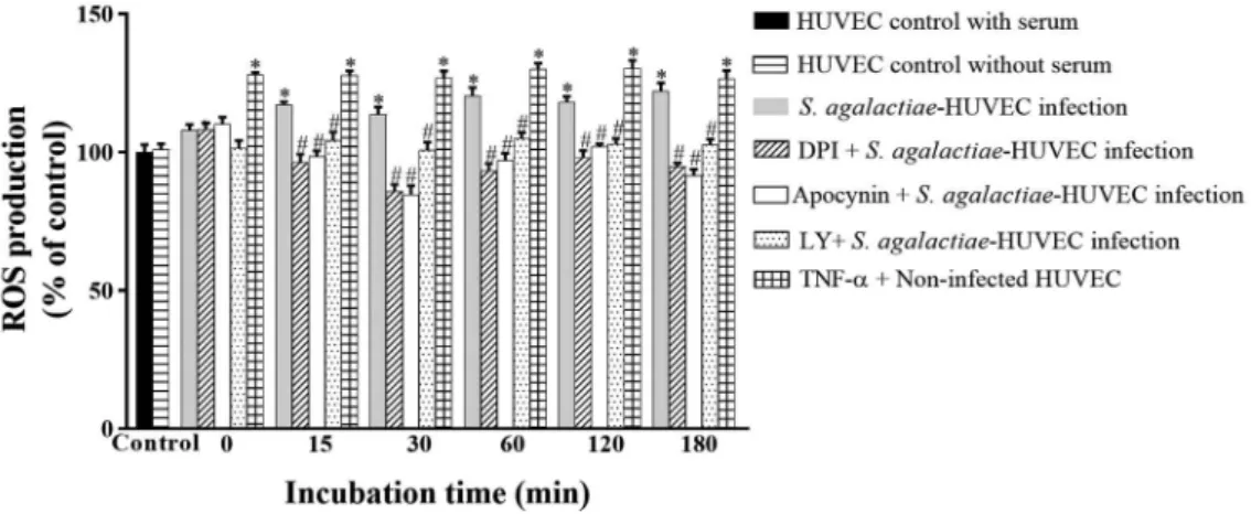
ROS production during S. agalactiae-HUVEC in-teraction - Levels of ROS generation were evaluated during S. agalactiae-HUVEC interaction using inhibi-tors of NADPH oxidase (DPI and apocynin) and PI3K (LY294002). Non-infected HUVEC monolayers treated
with TNF-α were used as positive controls. Increased
ROS production was observed in HUVECs after 15 min of infection (Fig. 1; p < 0.02). Moreover, pre-treatment of HUVEC with all inhibitors decreased ROS production during all time periods (p < 0.03). HUVECs grown in
Fig. 1: reactive oxygen species (ROS) generation by NADPH oxidase activity during infection of human primary endothelial cells (HU-VECs) by Streptococcus agalactiae. Experiments were performed at different periods of incubation using the GBS90356 type III strain of S. agalactiae and HUVECs in the presence or absence of NADPH inhibitors (10 μΜ DPI, 10 μΜ apocynin) or a PI3K inhibitor (5 μΜ
LY294002). Non-infected HUVEC monolayers with and without serum were used as negative controls. Non-infected HUVEC monolayers
with serum and 10 ng/mL TNF-α were used as positive controls. Data are expressed as mean ± SD of three independent experiments; *p≤
serum starvation conditions for 18-24 h to exclude se-rum-dependent effects were used as a negative control.
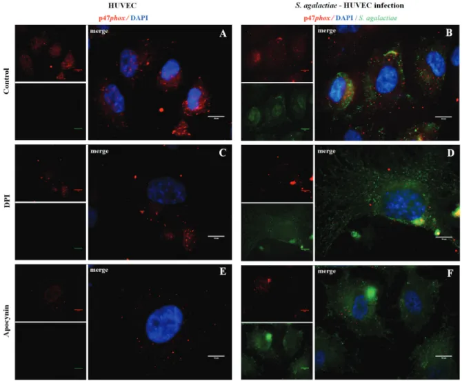
Activation of p47phox NADPH oxidase subunit dur-ing S. agalactiae-HUVEC interaction - Results of im-munofluorescence assays of p47phox activity during ROS generation by S. agalactiae infection of HUVEC were demonstrated in Fig. 2A-F. The NADPH oxidase p47phox subunit was mostly observed near the nucleus of uninfected host cells (Fig. 2A). HUVECs infected with S. agalactiae showed changes in subcellular localisation of the p47phox subunit, which was previously shown to be central for even distribution in the cell, reflecting activa-tion of p47phox in HUVECs (Fig. 2B). Additional assays are required to elucidate mechanisms involved in p47phox activation by S. agalactiae. DPI and apocynin inhibition assays (Fig. 2C-F) were performed at 60 min post-infec-tion when the highest adherence level to HUVECs by S. agalactiae was observed. Treatment with both inhibitors resulted in higher adherence levels of S. agalactiae 60 min post-infection of in HUVECs (Fig. 2D, F).
NADPH oxidase p47phox expression during S. aga-lactiae-HUVEC - Data displayed in Fig. 3A show an in-crease of p47phox mRNA expression at 30 min and 60 min post-infection, as measured by Real Time qRT-PCR (p < 0.03). Immunoblotting assay results confirmed high-er p47phox activities at upon 15 min and 60 min incuba-tion of S. agalactiae-HUVEC (p < 0.04) (Fig. 3B, C).
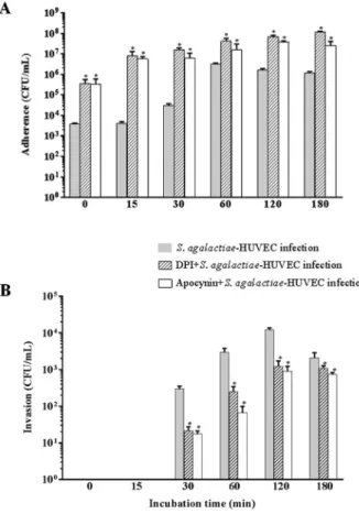
Effect of DPI and Apocynin on activation of the p47phox NADPH oxidase subunit during S. agalactiae-HUVEC interaction - The influence of the NADPH oxidase p47phox subunit on S. agalactiae adherence to and intracellular viability of HUVEC using inhibitors of p47phox (DPI or apocynin) are displayed in Fig. 4. A time-dependent increase in adherence to HUVECs was observed upon treatment with both inhibitors. A higher number of adherent bacteria was observed in HUVECs pre-treated with DPI or apocynin, compared to that of untreated HUVECs (p < 0.03) (Fig. 4A). A significant decrease in the number of internalised bacteria oc-curred in HUVECs treated with DPI and apocynin
af-Fig. 2: immunofluorescence assays of NADPH oxidase p47phox subunit activity during infection of human primary endothelial cells (HU-VECs) by Streptococcus agalactiae. (A, C, E) Uninfected HUVECs and (B, D, F) HUVECs infected by S. agalactiae in the presence or absence
of NADPH inhibitors (10 μΜ DPI, 10 μΜ apocynin). Anti-p47phox was labelled with Alexa 546 (red), S. agalactiae with
ter the first 30 min of infection compared to the number in untreated HUVECs (Fig. 4B).
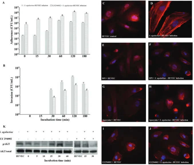
Involvement of the PI3K/Akt pathway during S. agalactiae-HUVEC interaction - Data shown in Fig. 5A-B demonstrate the effect of the PI3K/Akt pathway on the interaction between S. agalactiae and HUVECs pre-treated with the PI3K inhibitor, LY294002. A time-dependent increase in bacterial adherence was observed when HUVECs were treated with LY294002, similarly to results achieved with p47phox (DPI and apocynin) in-hibitors. The highest adherence level was observed at 120 min post-infection (p < 0.02) (Fig. 5A). Conversely, bac-terialinternalisation was detected at lower levels from 30 min to 180 min post-infection (p < 0.01) (Fig. 5B).
Immunofluorescence staining assays illustrated the involvement of PI3K/Akt pathway activation in the ac-tin stress fibre assembly of endothelial cells upon by S. agalactiae infection (Fig. 5C-D). Similar to non-infected HUVECs, pre-treatment with NADPH oxidase (DPI and
apocynin) or PI3K (LY294002) inhibitors in the presence (Fig. 5F, H, J) or absence of S. agalactiae infection (Fig. 5E, G, I) did not induce actin stress fibre arrangement. As shown in Fig. 5K, immunoblotting results revealed higher levels of phosphorylated Akt expression at 15 min and 30 min post-infection of HUVECs by S. agalactiae. Inhibition assays with LY294002 (15 min and 30 min post-infection) confirmed the involvement of the PI3K/ AKT pathway during S. agalactiae internalisation in en-dothelial cells. Results were confirmed by densitometry analysis (data not shown).
DISCUSSION
ROS have been classically regarded as host defence molecules related to destroying exogenous human patho-gens. Also, it has long been recognised that increased ROS levels modify cell signalling of host proteins, lead-ing to pathological processes such as inflammation and bacterial infections. Nevertheless, accumulated evi-dence indicates that ROS are second messengers and cell signalling modifiers (Zhang et al. 2016).
Only a few studies concerning NADPH oxidase ac-tivation during Streptococcus spp. infection were found in the available literature. For Streptococcus pyogenes, the production of O2- was found as the result of NADPH oxidase induction during bacterial infection in human
Fig. 3: expression of NADPH oxidase p47phox subunit during infection of HUVECs with Streptococcus agalactiae. Data were analysed at dif-ferent periods of time by (A) real time quantitative reverse transcription polymerase chain reaction (real-time qRT-PCR), (B) immunoblotting, and (C) densitometry assays. Actin was used as a control for protein
load-ing. Data are expressed as mean ± SD of three experiments; *p< 0.05.
Fig. 5: influence of the PI3K/Akt pathway on the Streptococcus agalactiae-human primary endothelial cells (HUVECs) interaction. (A) Ad-herence to and (B) invasion of S. agalactiae in HUVECs pre-treated with LY294002, an inhibitor of PI3K. Micrographic images of immuno-fluorescence staining indicated: (C) non-infected HUVEC monolayers as a negative control; (D) generation of actin stress fibres in HUVECs infected with S. agalactiae; (E, G, I) HUVECs pre-treated with DPI (10 µM), apocynin (10 µM), and LY294002 (5 µM), respectively; (F, H, J) HUVECs pre-treated with inhibitors (DPI, apocynin and LY294002, respectively) and infected with S. agalactiae. (K) Immunoblotting assay of phosphorylated Akt in infected HUVECs pre-treated with LY294002. Data are expressed as mean ± SD of three experiments. *p < 0.05.
primary keratinocytes (Regnier et al. 2016). The effect of NADPH oxidase inhibitor (DPI) treatment on intra-cellular survival of S. pyogenes during the early stage of phagocytosis by HL-60 cells was also verified (Nor-denfelt et al. 2009). Moreover, the ability of S. agalacti-ae totrigger oxidative burst in murine macrophages by NADPH oxidase activation was also demonstrated (Teix-eira et al. 2001). In addition, the role of p47phox in func-tionally active NADPH oxidase activity was evidenced by using p47phox-/- mice assays (Li and Shah 2002, Touyz et al. 2005). Similar effects were demonstrated for endothe-lial cells: the major source of ROS was also described as NADPH oxidase, whose activation and regulation were controlled by the phosphorylation of its cytosolic compo-nent p47phox (Schuett et al. 2017). However, physiological substrates, regulation, and control of signalling networks, especially of endothelial cells, need further investigation.
Presently, the involvement of p47phox subunit in S. agalactiae-HUVEC interaction was demonstrated by Real Time qRT-PCR, western blotting, and immuno-fluorescence assays. ROS production by activation of the p47phox NADPH oxidase subunit during S. agalactiae -HUVEC interaction was demonstrated. Thus, our results agree with those of Zhang et al. (2016) showing that DPI and apocynin reduce p47phox translocation and expres-sion, thereby suppressing ROS production in peripheral blood mononuclear cells from premature infants. Similar-ly, Streptococcus pneumoniae induces a higher oxidative burst in neutrophils by triggering neutrophil NADPH oxi-dase to produce more reactive oxygen intermediates and by activating p47phox by S. pneumoniae in the plasma membrane fraction of neutrophils (Barbuti et al. 2010).
et al. 2012). Interestingly, previous studies have also reported the involvement of ROS in cytoskeletal re-organisation accompanying O2- formation acting as a scaffold for the p47phox subunit (Touyz et al. 2005). A close relationship between actin polymerisation and ROS generation has also been observed after wound-ing endothelial cell monolayers (Li and Shah 2002).
The PI3K/Akt signalling pathway was found to be associated with cytoskeletal regulation, vesicle traffick-ing, and the balance between cellular survival and regu-lated cell death (Burnham et al. 2007). PI3K activation has been specifically implicated in modification and manipulation of the actin cytoskeleton, essentially in S. agalactiae invasion of host cells (Burnham et al. 2007). The involvement of the PI3K/Akt pathway during the S. agalactiae-HUVEC interaction was verified in the pres-ent study. Burnham et al. (2007) showed an inhibition of S. agalactiae invasion in HeLa cells treated with the LY294002 inhibitor of PI3K. Similar to previous results of these authors, current data concerning the inhibition of ROS production showed a decrease in invasion and an in-crease in the adhesion levels of S. agalactiae on HUVEC induced by LY294002. Data indicated that S. agalactiae -HUVEC interaction was partially due to alterations in the organisation of microfilaments of the host cells through the PI3K/Akt pathway. As soon as S. agalactiae began to establish physical contact with the host cell surface, the formation of stress fibres was observed in HUVECs. Additional results demonstrated loss of stress fibres due to inhibition of PI3K, reinforcing the participation of the PI3K/Akt pathway in human endothelial cells.
The ability of Akt to regulate multiple activities of host cells makes it an essential target for invading bac-terial pathogens, including S. agalactiae. A previous study in HeLa cells indicated that Akt was required for efficient S. agalactiae invasion, since Akt was rapidly phosphorylated in response to S. agalactiae (Burnham et al. 2007). In our work using HUVECs, a significant increase in Akt phosphorylation occurred at 15-30 min post-infection of S. agalactiae, suggesting the involve-ment of PI3K/Akt in this process.
In conclusion, ROS generation via the NADPH oxi-dase pathway in human endothelial cells accompanied by cytoskeletal reorganisation through the PI3K/Akt pathway occurred during invasion of the S. agalactiae GBS90356 strain isolated from a fatal case of meningi-tis. These results may contribute to the understanding of virulence mechanisms involved in the translocation of S. agalactiae across the blood-brain barrier.
AUTHORS’ CONTRIBUTION
JSSO, GSS and PEN - Conception of the general project and experimental design; JAM and AMS performed molecu-lar techniques and reviewed the experimental results; JSSO and GSS - performed immunoblot analysis and immunofluo-rescence assays; TCBF - provided reagents and assistance in drafting the manuscript; PEN and ALMG - analysis and dis-cussion of the results, assistance in creating the figures, litera-ture review, and preparation of the manuscript.
REFERENCES
Abid MR, Spokes KC, Shih SC, Aird WC. NADPH oxidase activity selectively modulates vascular endothelial growth factor signal-ing pathways. J Biol Chem. 2007; 282(48): 35373-85.
Awasthi YC, Ramana KV, Chaudhary P, Srivastava SK, Awasthi S. Regulatory roles of glutathione-S-transferases and 4-hy-droxynonenal in stress-mediated signaling and toxicity. Free Radic Biol Med. 2017; 111: 235-43.
Barbuti G, Moschioni M, Fumarulo R, Censini S, Montemurro P.
Streptococcus pneumoniae modulates the respiratory burst re-sponse in human neutrophils. FEMS Immunol Med Microbiol. 2010; 60(1): 57-62.
Boone TJ, Tyrrell GJ. Identification of the actin and plasminogen binding regions of group B streptococcal phosphoglycerate ki-nase. J Biol Chem. 2012; 287(34): 29035-44.
Bülbül N, Pala E, İğci YZ, Göğebakan B, Öztuzcu S, Cengiz B, et al. NADPH oxidase p22phox gene expression in ulcerative colitis. Turk J Gastroenterol. 2014; 25(6): 634-8.
Burnham CD, Shokoples SE, Tyrrell GJ. Invasion of HeLa cells by group B Streptococcus requires the phosphoinositide-3-kinase signaling pathway and modulates phosphorylation of host cell Akt and gly-cogen synthase kinase-3. Microbiology. 2007; 153(Pt 12): 4240-52.
Buscetta M, Firon A, Pietrocola G, Biondo C, Mancuso G, Midiri A, et al. PbsP, a cell wall-anchored protein that binds plasminogen to promote hematogenous dissemination of Group B Streptococcus. Mol Microbiol. 2016; 101(1): 27-41.
Costa AF, Moraes JA, de Oliveira JS, dos Santos MH, Santos GS, Barja-Fidalgo C, et al. Reactive oxygen species involved in apop-tosis induction of human respiratory epithelial (A549) cells by
Streptococcus agalactiae. Microbiology. 2016; 162(1): 94-9.
Costa AF, Pereira CS, Santos GS, Carvalho TM, Hirata Jr R, de Mat-tos-Guaraldi AL, et al. Group B Streptococcus serotypes III and V induce apoptosis and necrosis of human epithelial A549 cells. Int J Mol Med. 2011; 27(5): 739-44.
Dowd GC, Bhalla M, Kean B, Thomas R, Ireton K. Role of Host Type IA phosphoinositide 3-kinase pathway components in invasin-mediated internalization of Yersinia enterocolitica. Infect Im-mun. 2016; 84(6): 1826-41.
Jaffe EA, Nacchman RL, Becker CG, Minick CR. Culture of human endothelial cells of derivate from umbilical cords veins. Identi-fication on by morphologic and immunologic criteria. J Clin In-vest. 1973; 52(11): 2745-56.
Lambeth JD, Kawahara T, Diebold B. Regulation of Nox and Duox enzymatic activity and expression. Free Radic Biol Med. 2007; 43(3): 319-31.
Li J, Shah AM. Intracellular localization and preassembly of the NADPH oxidase complex in cultured endothelial cells. J Biol Chem. 2002; 277(22): 19952-60.
Li JM, Shah AM. Mechanism of endothelial cell NADPH oxidase ac-tivation by angiotensin II. Role of the p47phox subunit. J Biol Chem. 2003; 278(14): 12094-100.
Meijles DN, Fan LM, Howlin BJ, Li JM. Molecular insights of p47phox phosphorylation dynamics in the regulation of NADPH oxidase activation and superoxide production. J Biol Chem. 2014; 289(33): 22759-70.
Nordenfelt P, Bauer S, Lönnbro P, Tapper H. Phagocytosis of
Pimentel BA, Martins CA, Mendonça JC, Miranda PS, Sanches GF, Mattos-Guaraldi AL, et al. Streptococcus agalactiae infection in cancer patients: a five-year study. Eur J Clin Microbiol Infect Dis. 2016; 35(6): 927-33.
Regnier E, Grange PA, Ollagnier G, Crickx E, Elie L, Chouzenoux S, et al. Superoxide anions produced by Streptococcus pyogenes group A-stimulated keratinocytes are responsible for cellular necrosis and bacterial growth inhibition. Innate Immun. 2016; 22(2): 113-23.
Ribeiro-Pereira C, Moraes JA, Souza MJ, Laurindo FR, Arruda MA, Barja-Fidalgo C. Redox modulation of FAK controls melanoma survival: role of NOX4. PLoS One. 2014; 9(6): e99481.
Santos GS, Penha CL, Mattos-Guaraldi A, Attias M, Lopes-Bezerra LM, Silva-Filho FC, et al. Group B Streptococcus induces tyrosine phos-phorylation of annexin V and glutathione S-transferase in human umbilical vein endothelial cells. Int J Mol Med. 2009; 24(3): 393-9.
Santos MH, da Costa AF, Ferreira BJ, Souza SL, Lannes PS, Santos GS, et al. A phosphoramidon-sensitive metalloprotease induces apoptosis of human endothelial cells by Group B Streptococcus. Antonie van Leeuwenhoek. 2013; 104(6): 1125-33.
Schuett J, Schuett H, Oberoi R, Koch AK, Pretzer S, Luchtefeld M, et al. NADPH oxidase NOX2 mediates TLR2/6-dependent release of GM-CSF from endothelial cells. FASEB J. 2017; 31(6): 2612-24.
Shen Y, Fu W-Y, Cheng EYL, Fu AKY, Ip NY. Melanocortin-4 recep
-tor regulates hippocampal synaptic plasticity through a protein kinase A-dependent mechanism. J Neurosci. 2013; 33(2): 464-72.
Teixeira CF, Azevedo NL, Carvalho TM, Fuentes J, Nagao PE. Cy-tochemical study of Streptococcus agalactiae and macrophage interaction. Microsc Res Tech. 2001; 54(4): 254-9.
Touyz RM, Yao G, Quinn MT, Pagano PJ, Schiffrin EL. p47phox as-sociates with the cytoskeleton through cortactin in human vascular smooth muscle cells role in NAD(P)H oxidase regulation by angio-tensin II. Arterioscler Thromb Vasc Biol. 2005; 25(3): 512-8.
Ushio-Fukai M. Localizing NADPH oxidase-derived ROS. Sci STKE. 2006; 2006(349): re8.
Verani JR, McGee L, Schrag SJ, Division of Bacterial Diseases, Na-tional Center for Immunization and Respiratory Diseases, Cen-ters for Disease Control and Prevention (CDC). Prevention of perinatal group B streptococcal disease - revised guidelines from CDC, 2010. MMWR Recomm Rep. 2010; 59(RR-10): 1-36.
Watanabe H, Adachi R, Hirayama A, Kasahara T, Suzuki K. Triphe
-nyltin enhances the neutrophilic differentiation of promyelocytic HL-60 cells. Biochem Biophys Res Commun. 2003; 306(1): 26-31.
Yang K, Zhang H, Luo Y, Zhang J, Wang M, Liao P, et al. Gypenoside XVII prevents atherosclerosis by attenuating endothelial apopto-sis and oxidative stress: insight into the ERα-Mediated PI3K/Akt pathway. Int J Mol Sci. 2017; 18(2): pii: E77.